
BIOL-214 · Lesson 9 of 12 · 30 min
The cytoskeleton II: microtubules, intermediate filaments and motors
Actin filaments shape the cell surface and drive crawling and contraction. Microtubules do a different job: they are the long-distance highways of the cell, the scaffold that positions organelles, the tracks along which cargo travels from one end of a neuron to the other, and the machine that separates chromosomes at every cell division. Intermediate filaments, the third system, give cells and tissues mechanical strength. This lesson looks at how microtubules are built and organized, the remarkable behaviour called dynamic instability, the kinesin and dynein motors that move along them, cilia and flagella, the drugs that target microtubules in cancer treatment, and the intermediate filaments whose defects cause blistering skin diseases and premature ageing.
Tubulin and the structure of microtubules
Microtubules are hollow tubes about 25 nm in diameter, built from heterodimers of α-tubulin and β-tubulin, each about 50 kDa. Both subunits bind GTP. The GTP on α-tubulin is buried at the interface within the dimer and is never hydrolysed, but the GTP on β-tubulin is exposed at the end of the microtubule and is hydrolysed to GDP shortly after the dimer is added. Dimers line up head to tail into protofilaments, and 13 protofilaments in most cells associate side by side to form the wall of the tube. As with actin, every subunit points the same way, so the microtubule has a fast-growing plus end, where β-tubulin is exposed, and a slower minus end, where α-tubulin is exposed.
In animal cells, most microtubules grow from a microtubule-organizing centre, usually the centrosome near the nucleus. The centrosome contains a pair of centrioles, short cylinders of nine triplet microtubules, surrounded by pericentriolar material that contains many copies of the γ-tubulin ring complex. This ring, made of γ-tubulin and partner proteins, acts as a template that nucleates new microtubules and caps their minus ends. Microtubules therefore radiate from the centrosome with their minus ends anchored at the centre and their plus ends growing toward the cell periphery. Plants, which lack centrosomes, and many differentiated animal cells such as neurons and epithelial cells nucleate microtubules from other sites, including the Golgi apparatus, the nuclear envelope and the sides of existing microtubules.
Dynamic instability
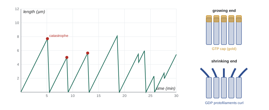
When Tim Mitchison and Marc Kirschner watched individual microtubules in 1984, they found something unexpected. A population of microtubules at steady state did not consist of filaments slowly treadmilling. Instead, each microtubule switched between long periods of steady growth and episodes of rapid shrinkage. The switch from growth to shrinkage is called catastrophe; the switch back to growth is called rescue. This behaviour, dynamic instability, lets microtubules explore the cytoplasm and be captured where they are needed.
The explanation lies in GTP hydrolysis. A growing microtubule has a cap of GTP-tubulin at its plus end, because new dimers add faster than their GTP is hydrolysed. GTP-tubulin dimers are straight and fit snugly into the lattice. GDP-tubulin dimers prefer a curved shape and are held straight only by the lattice around them. If growth slows and the GTP cap is lost, the protofilaments at the end peel apart and curl outward, like the frayed end of a rope, and the microtubule rapidly depolymerizes. Rescue occurs when the shrinking end regains a GTP cap, perhaps at islands of GTP-tubulin left within the lattice.
Four parameters describe the behaviour: growth speed v_g, shrinkage speed v_s, catastrophe frequency f_cat and rescue frequency f_res. When catastrophes dominate, microtubules have a stable average length given by the formula above; when growth dominates, they grow without limit until they hit the cell edge. Cells tune these parameters with dozens of microtubule-associated proteins (MAPs). Stabilizing MAPs such as tau in axons and MAP2 in dendrites bind along the lattice. Plus-end tracking proteins such as EB1 bind the growing tip; fluorescent EB1 is used to visualize microtubule growth as moving comets. Destabilizers such as kinesin-13 family members, which actively peel protofilaments, and stathmin, which sequesters tubulin dimers, increase catastrophes.
import numpy as np
rng = np.random.default_rng(214)

# Simulate dynamic instability of single microtubules (one-second steps)
vg, vs = 1.5, 25.0                  # um/min growth and shrinkage speeds
def simulate(fcat, fres, minutes=60, dt=1/60):
    L, growing, lengths, cats = 0.0, True, [], 0
    for _ in range(int(minutes/dt)):
        if growing:
            L += vg*dt
            if rng.random() < fcat*dt: growing, cats = False, cats + 1
        else:
            L -= vs*dt
            if L <= 0: L, growing = 0.0, True          # regrows from the centrosome
            elif rng.random() < fres*dt: growing = True
        lengths.append(L)
    return np.array(lengths), cats

for label, fcat, fres in [("interphase-like", 0.3, 1.5), ("mitosis-like", 2.0, 0.5)]:
    runs = [simulate(fcat, fres) for _ in range(40)]
    mean_len = np.mean([r[0][1800:].mean() for r in runs])       # last 30 minutes
    theory = vg*vs/(vs*fcat - vg*fres)
    print(f"{label:15s} f_cat {fcat}/min f_res {fres}/min: mean length {mean_len:5.1f} um "
f"(formula {theory:4.1f} um), catastrophes per hour {np.mean([r[1] for r in runs]):4.1f}")interphase-like f_cat 0.3/min f_res 1.5/min: mean length 6.4 um (formula 7.1 um), catastrophes per hour 16.8 mitosis-like f_cat 2.0/min f_res 0.5/min: mean length 0.7 um (formula 0.8 um), catastrophes per hour 112.5
The simulation shows how changing the catastrophe and rescue frequencies changes the length of microtubules in a population. The simulated averages come close to the formula; the small differences come from random variation over a finite simulated time. During mitosis, cells raise the catastrophe frequency several-fold. The long interphase microtubules are replaced by many short, highly dynamic ones that search the cytoplasm until their plus ends are captured by the kinetochores of chromosomes. This search-and-capture mechanism, together with microtubules nucleated near the chromosomes themselves, builds the mitotic spindle (lesson 12).
Drugs that target microtubules
Because dividing cells depend on a dynamic spindle, drugs that interfere with microtubules are among the most widely used cancer treatments. Some destabilize microtubules: colchicine, from the autumn crocus, and the vinca alkaloids vinblastine and vincristine, from the Madagascar periwinkle, bind tubulin and prevent polymerization. Others stabilize them: paclitaxel (Taxol), first isolated from the bark of the Pacific yew, binds β-tubulin inside the lattice and suppresses depolymerization. Both kinds of drug arrest cells in mitosis, because the spindle cannot work properly unless its microtubules are dynamic. The checkpoint that detects unattached chromosomes holds the cell in mitosis, and many cells eventually die (lesson 12). Their side effects include nerve damage, because neurons rely on microtubule transport. Colchicine is used at low doses to treat gout, where it reduces inflammation by blocking microtubule-dependent movement of white blood cells.
Motor proteins on microtubules
Two families of motors move along microtubules. Most kinesins move toward the plus end, carrying cargo outward from the cell centre; cytoplasmic dynein moves toward the minus end, carrying cargo inward. Kinesin-1, the conventional kinesin discovered in 1985 in squid axons by Ronald Vale and colleagues, has two motor heads that walk hand over hand along a single protofilament, taking 8-nm steps, the length of one tubulin dimer, and hydrolysing one ATP per step. It can take about 100 steps before falling off, a property called processivity, and moves at about 0.8 µm per second. Humans have about 45 kinesin genes; some carry cargo, some slide microtubules apart in the spindle, and some depolymerize microtubules.
Cytoplasmic dynein is a huge complex of over 1.4 MDa. Its motor domain is a ring of six AAA+ ATPase modules, unrelated to kinesin and myosin, with a long stalk that binds the microtubule. Dynein needs an activating complex called dynactin and a cargo adaptor to move processively. It carries endosomes, lysosomes and Golgi membranes toward the centre, transports virus particles toward the nucleus, positions the spindle, and pulls on kinetochores. A separate axonemal dynein drives the beating of cilia.
| Property | Kinesin-1 | Cytoplasmic dynein | Myosin V |
|---|---|---|---|
| Track | Microtubule | Microtubule | Actin filament |
| Direction | Plus end (outward) | Minus end (inward) | Barbed end |
| Step size | 8 nm | 8 nm on average (variable 4–32 nm) | 36 nm |
| Typical speed in cells | 0.5–1 µm/s | 0.5–1 µm/s | About 0.3–0.5 µm/s |
| Motor domain | Kinesin fold (related to myosin) | Ring of six AAA+ modules | Myosin fold |
| Examples of cargo | Synaptic vesicle precursors, mitochondria, mRNA granules | Endosomes, lysosomes, viruses | Melanosomes, secretory vesicles near the cell cortex |
Organelles often carry several motors at once, kinesin and dynein together, sometimes with myosin. Their net movement depends on which motors are active, controlled by adaptors, phosphorylation and the small GTPases that mark organelle identity (lessons 4 and 5). The result is a tug of war that can reverse direction in seconds. The melanosomes of fish and frog skin cells are a striking example: hormones that raise or lower cyclic AMP switch the pigment granules between dispersal by kinesin and aggregation by dynein, darkening or lightening the skin.
Axonal transport
Neurons pose an extreme transport problem. A motor neuron supplying a muscle in the foot has its cell body in the spinal cord, and an axon about one metre long. Nearly all proteins are made in the cell body and must be carried down the axon. In the axon, microtubules are arranged with their plus ends pointing away from the cell body, so kinesins carry cargo outward (anterograde transport) and dynein carries cargo back (retrograde transport), including signals from growth factors at the nerve terminal and worn-out organelles bound for lysosomes.
# How long does it take to reach the end of an axon?
lengths_m = {"cortical interneuron": 0.001, "spinal motor neuron (leg)": 1.0,
             "giraffe sciatic nerve": 4.5}
rates = {"fast transport (kinesin, 1 um/s)": 1.0e-6,           # m/s
         "slow transport (about 1 mm/day)": 1.0e-3/86400,
         "diffusion of a protein, D = 10 um^2/s": None}

D = 10e-12                                    # m^2/s
for cell, L in lengths_m.items():
    print(cell)
    for name, v in rates.items():
        t = L*L/(2*D) if v is None else L/v       # 1-D diffusion time ~ L^2/2D
        days = t/86400
print(f" {name:38s} {t:12.3g} s = {days:10.3g} days")cortical interneuron fast transport (kinesin, 1 um/s) 1e+03 s = 0.0116 days slow transport (about 1 mm/day) 8.64e+04 s = 1 days diffusion of a protein, D = 10 um^2/s 5e+04 s = 0.579 days spinal motor neuron (leg) fast transport (kinesin, 1 um/s) 1e+06 s = 11.6 days slow transport (about 1 mm/day) 8.64e+07 s = 1e+03 days diffusion of a protein, D = 10 um^2/s 5e+10 s = 5.79e+05 days giraffe sciatic nerve fast transport (kinesin, 1 um/s) 4.5e+06 s = 52.1 days slow transport (about 1 mm/day) 3.89e+08 s = 4.5e+03 days diffusion of a protein, D = 10 um^2/s 1.01e+12 s = 1.17e+07 days
The calculation shows why motors are essential. Fast kinesin transport delivers vesicles down a metre-long axon in under two weeks, while diffusion would take more than 1,500 years. Slow axonal transport, which moves cytoskeletal proteins and many soluble enzymes, is not a slower motor: cargo is carried in brief fast runs separated by long pauses. Defects in axonal transport contribute to several neurological diseases. Mutations in a kinesin, KIF5A, and in the dynactin subunit p150Glued cause forms of hereditary spastic paraplegia, amyotrophic lateral sclerosis and motor neuropathy, and the longest axons, which supply the legs, are usually affected first. In Alzheimer disease, tau detaches from microtubules and forms tangles; BIOL-420 discusses neurodegeneration in more detail.
Worked problem
Counting kinesin steps
A vesicle is carried 40 µm along an axon by a single kinesin-1 molecule that hydrolyses one ATP per 8-nm step. How many ATP molecules are used, and how long does the trip take at 0.8 µm/s?
Show solution
- Number of steps = 40 µm / 0.008 µm = 5,000 steps.
- One ATP per step, so 5,000 ATP molecules.
- Time = 40 µm / 0.8 µm/s = 50 s.
- Since kinesin-1 detaches after roughly 100 steps (0.8 µm), a real vesicle relies on several motors and rebinding to make the journey.
Answer: 5,000 ATP and about 50 s (in reality several motors share the work).
Cilia and flagella
Cilia and flagella are hair-like extensions of the cell surface built around a microtubule core called the axoneme. In motile cilia, the axoneme has nine doublet microtubules arranged around a central pair, the famous 9 + 2 arrangement. Axonemal dyneins attached to each doublet walk along the neighbouring doublet. Because the doublets are linked to one another by nexin links and anchored at the base, the sliding cannot happen freely and instead bends the axoneme. Coordinated waves of bending produce the whip-like beat that sweeps mucus out of the airways, moves eggs along the oviduct and propels sperm. Each cilium grows from a basal body, a modified centriole, and is built by intraflagellar transport, in which kinesin-2 carries trains of building blocks to the tip and dynein-2 returns them.
Almost every human cell also has a single non-motile primary cilium, with a 9 + 0 axoneme, that acts as an antenna for signals. Components of the Hedgehog pathway, important in development (BIOL-360), move into and out of the primary cilium as the pathway turns on. Defects in cilia cause ciliopathies, a diverse group of disorders. Primary ciliary dyskinesia causes chronic respiratory infections and infertility, and about half of patients have their internal organs mirrored left to right (situs inversus), because beating cilia in the embryo create the leftward fluid flow that establishes body asymmetry. Polycystic kidney disease and Bardet–Biedl syndrome involve primary cilia.
Intermediate filaments
Intermediate filaments, about 10 nm in diameter, are named because their width falls between that of actin filaments and microtubules. Unlike the other two, they have no polarity, do not bind nucleotides and are not used as tracks by motors. Their subunits are long fibrous proteins with a central α-helical rod domain. Two subunits wind together into a coiled-coil dimer, two dimers associate in a staggered antiparallel tetramer, and eight tetramers pack into a unit-length filament that joins end to end with others. Because the tetramers are antiparallel, both ends of the filament look alike. The result is a rope-like filament that can be stretched to several times its length without breaking, ideal for resisting mechanical stress.
| Type | Example proteins | Location | Disease when mutated |
|---|---|---|---|
| Keratins (types I and II) | Keratin 5 and 14, keratin 8 and 18 | Epithelial cells; hair and nails | Epidermolysis bullosa simplex (skin blisters from minor friction) |
| Type III | Vimentin, desmin, GFAP | Fibroblasts and white blood cells; muscle; astrocytes | Desmin myopathies; Alexander disease (GFAP) |
| Type IV (neurofilaments) | NF-L, NF-M, NF-H | Neurons, especially large axons | Charcot–Marie–Tooth neuropathy (some forms) |
| Type V (nuclear lamins) | Lamins A, B and C | Inner surface of the nuclear envelope | Hutchinson–Gilford progeria; muscular dystrophies |
Keratins form a network across epithelial cells and connect at desmosomes and hemidesmosomes to neighbouring cells and the underlying matrix (lesson 10). In epidermolysis bullosa simplex, a mutation in keratin 5 or 14 weakens the network in the basal layer of the skin, and cells rupture under minor friction, producing blisters. In a study reported in 2017, a boy in Germany with junctional epidermolysis bullosa, a related blistering disease caused by a defective anchoring protein, was treated with sheets of his own skin grown from genetically corrected stem cells; the grafts covered about 80 per cent of his body and remained healthy in follow-up, a dramatic demonstration of stem-cell gene therapy (lesson 12). The nuclear lamins support the nuclear envelope (lesson 2). A point mutation in lamin A that creates an abnormal farnesylated protein, progerin, causes Hutchinson–Gilford progeria syndrome, in which children age rapidly. A farnesyltransferase inhibitor, lonafarnib, was approved in the United States in 2020 and extends life expectancy in children with progeria.
Coordinating the three systems
The three cytoskeletal systems are not independent. Proteins called plakins, such as plectin, cross-link intermediate filaments to microtubules and actin. Microtubule plus ends deliver signals that regulate Rho GTPases and focal adhesions at the leading edge of migrating cells, and in neurons, actin in the growth cone and microtubules in the axon cooperate to steer the growing tip. During cell division, microtubules position the contractile ring of actin. The cytoskeleton is best thought of as one integrated, dynamic system in which each component has its own strengths.
Common mistakes
MistakeMicrotubules grow and shrink by treadmilling only.
InsteadIndividual microtubules show dynamic instability, switching between growth and rapid shrinkage.
MistakeThe GTP on α-tubulin is hydrolysed during polymerization.
InsteadOnly the GTP on β-tubulin is hydrolysed; α-tubulin’s GTP is buried and stable.
MistakeKinesins and dyneins both move toward the plus end.
InsteadMost kinesins move toward the plus end, while dynein moves toward the minus end.
MistakeTaxol kills cancer cells by preventing microtubule assembly.
InsteadTaxol stabilizes microtubules; it arrests mitosis by suppressing dynamics.
MistakeIntermediate filaments have plus and minus ends like microtubules.
InsteadTheir antiparallel tetramers make them non-polar, and motors do not walk on them.
Summary and key terms
- Microtubules are hollow tubes of α/β-tubulin dimers with a plus end and a minus end, usually nucleated by γ-tubulin at the centrosome.
- Dynamic instability, caused by GTP hydrolysis and loss of the GTP cap, lets microtubules switch between growth and shrinkage.
- MAPs and plus-end proteins tune growth, catastrophe and rescue; mitosis shifts microtubules toward short, dynamic forms.
- Microtubule drugs such as vinca alkaloids and taxanes block mitosis and are used against cancer.
- Kinesins mostly move toward plus ends, dynein toward minus ends; together they drive long-range transport such as axonal transport.
- Motile cilia beat by dynein-driven sliding of axonemal doublets; primary cilia act as signalling antennae.
- Intermediate filaments are non-polar rope-like fibres that give mechanical strength; lamins support the nucleus.
- tubulin
- The α/β heterodimer that is the building block of microtubules.
- centrosome
- The main microtubule-organizing centre of animal cells, containing centrioles and γ-tubulin ring complexes.
- dynamic instability
- The switching of individual microtubules between growth and rapid shrinkage.
- catastrophe
- The switch of a microtubule from growth to shrinkage.
- GTP cap
- A region of GTP-tubulin at a growing microtubule end that stabilizes it.
- kinesin
- A family of motor proteins that mostly move toward microtubule plus ends.
- dynein
- A large motor protein complex that moves toward microtubule minus ends.
- axoneme
- The microtubule core of a cilium or flagellum.
- processivity
- The ability of a motor to take many steps along a track before detaching.
- nuclear lamina
- The network of lamin intermediate filaments lining the inner nuclear membrane.
Practice problems
Work each problem on paper first, then reveal the answer to check.
Which tubulin subunit hydrolyses its GTP?
Show answer
β-tubulin.
What causes a microtubule catastrophe?
Show answer
Loss of the GTP cap, so that curved GDP-tubulin protofilaments peel apart.
Name a protein that tracks growing microtubule plus ends.
Show answer
EB1.
Calculate the mean microtubule length for v_g = 2, v_s = 20 µm/min, f_cat = 0.5 and f_res = 1 per minute.
Show answer
2 × 20 / (20 × 0.5 − 2 × 1) = 40/8 = 5 µm.
How do vinblastine and paclitaxel differ in their effect on microtubules?
Show answer
Vinblastine prevents polymerization; paclitaxel stabilizes microtubules.
In which direction does kinesin-1 carry cargo in an axon?
Show answer
Away from the cell body, toward the plus ends at the nerve terminal.
How many ATP does kinesin use to move 1 µm?
Show answer
1 µm / 8 nm = 125 steps, so about 125 ATP.
Why do some patients with primary ciliary dyskinesia have situs inversus?
Show answer
Motile cilia in the embryo create the flow that sets left–right asymmetry; without it, sidedness is random.
What is the structural defect in epidermolysis bullosa simplex?
Show answer
Mutant keratin 5 or 14 weakens the keratin network of basal skin cells.
Which protein is abnormal in Hutchinson–Gilford progeria?
Show answer
Lamin A, which is made as the permanently farnesylated progerin.
Lesson quiz · 5 questions
Score 60% or more to complete this lesson.
Create a free account to save your progress, take quizzes and earn a certificate.