
BIOL-214 · Lesson 5 of 12 · 31 min
Endocytosis, lysosomes and autophagy
Cells constantly take in material from outside and break down their own worn-out parts. Nutrients such as cholesterol and iron enter by binding receptors that are then pulled inside; immune cells engulf bacteria; and receptors, once they have done their job, are internalized to stop signalling. Inside, a network of endosomes sorts incoming material: some is returned to the surface, some is passed to lysosomes for destruction. Lysosomes are also the destination of autophagy, the process by which cells wrap up portions of their own cytoplasm, including whole organelles, and digest them. Together these pathways control nutrient supply, signalling, immunity and the quality of the cell’s contents. Their failure causes dozens of inherited disorders and contributes to neurodegeneration, infection and cancer. This lesson builds on lesson 4, which described the coats and SNAREs that also drive endocytic traffic.
Ways into the cell
Endocytosis is the uptake of material by inward budding of the plasma membrane. Several mechanisms differ in the size of the vesicle and the selectivity of uptake.
| Mechanism | Vesicle size | Selectivity | Examples |
|---|---|---|---|
| Phagocytosis | Over 250 nm, up to the size of the particle | Triggered by receptors for the particle | Macrophages and neutrophils engulfing bacteria and dead cells |
| Macropinocytosis | 0.2–5 µm | Low: bulk fluid | Antigen sampling by dendritic cells; nutrient scavenging by some cancer cells |
| Clathrin-mediated endocytosis | About 100 nm | High: receptor-bound cargo concentrated in coated pits | LDL, transferrin, many signalling receptors |
| Caveolae | 60–80 nm flask-shaped pits | Moderate | Abundant in endothelial cells, fat cells and muscle; also buffer membrane tension |
| Clathrin-independent pathways | Varied | Varied | Some toxins, glycosylphosphatidylinositol (GPI)-anchored proteins |
Clathrin-mediated endocytosis is the best understood. The adaptor complex AP-2 binds the lipid PIP₂ in the plasma membrane and short sorting motifs in the cytoplasmic tails of receptors, such as the NPxY sequence of the LDL receptor and the YxxΦ motif of the transferrin receptor (where Φ is a bulky hydrophobic residue). Clathrin assembles over the adaptors into a coated pit, which deepens into a bud; dynamin pinches it off, and the coat is quickly removed with the help of the ATPase Hsc70 and a phosphatase that converts PIP₂, so the naked vesicle can fuse with an early endosome. A coated pit forms and buds in about one minute, and a fibroblast may internalize the equivalent of its whole plasma membrane area every hour or so, most of which returns by recycling.
Receptor-mediated endocytosis of LDL
Cholesterol travels in the blood mainly as low-density lipoprotein (LDL), particles about 22 nm across, each containing roughly 1,500 cholesterol ester molecules in a core surrounded by phospholipid, free cholesterol and a single large protein, apolipoprotein B-100. Michael Brown and Joseph Goldstein discovered in the 1970s that cells take up LDL by receptor-mediated endocytosis. LDL receptors cluster in coated pits, bind LDL, and are internalized. In the acidic early endosome, LDL dissociates from its receptor; the receptor recycles to the surface, and LDL travels on to lysosomes, where the cholesterol esters are hydrolysed and free cholesterol is released for the cell to use. Rising cholesterol in the cell then shuts down both cholesterol synthesis and production of LDL receptors through the SREBP pathway, a feedback system. Brown and Goldstein received the 1985 Nobel Prize in Physiology or Medicine.
Their discovery came from studying familial hypercholesterolemia (FH), an inherited disorder in which mutations in the LDL receptor gene prevent cells from taking up LDL, so it accumulates in the blood and causes early heart disease. Heterozygous FH affects roughly 1 in 250 people and causes LDL levels about twice normal; the rare homozygous form causes levels four or more times normal and heart attacks in childhood without treatment. Different mutations disrupt different steps: some prevent receptor synthesis, some block transport from the ER, some prevent LDL binding, and one famous class, found in the patient known as J. D., produced receptors that bind LDL but fail to cluster in coated pits because of a single change in the NPxY internalization signal. Today FH is treated with statins, which block cholesterol synthesis and so increase LDL receptor numbers, and with antibodies or small interfering RNAs against PCSK9, a protein that sends LDL receptors to lysosomes for degradation instead of letting them recycle.
import numpy as np
from scipy.integrate import solve_ivp

# Uptake of a ligand by receptor-mediated endocytosis: surface binding, internalization,
# and recycling of empty receptors. Units: fraction of total receptors; rates per minute.
k_on_L = 0.5     # binding rate (ligand concentration x k_on), per min
k_int = 0.2      # internalization of occupied receptors (half-time ~3.5 min)
k_rec = 0.1      # recycling of internalized receptors to the surface

def model(t, y):
    free, bound, inside, delivered = y
    return [-k_on_L*free + k_rec*inside,
            k_on_L*free - k_int*bound,
            k_int*bound - k_rec*inside,
            k_int*bound]                      # ligand molecules delivered (cumulative)

t = np.linspace(0, 60, 61)
sol = solve_ivp(model, (0, 60), [1, 0, 0, 0], t_eval=t)
free, bound, inside, delivered = sol.y
for i in (0, 5, 10, 20, 40, 60):
    print(f"t={i:2d} min  free {free[i]:.2f}  ligand-bound {bound[i]:.2f}  internal {inside[i]:.2f}  "
          f"ligand delivered per receptor {delivered[i]:.1f}")
print(f"internalization half-time = {np.log(2)/k_int:.1f} min")
# A receptor that cannot be internalized (like the J.D. mutation) delivers nothing
print("mutant with k_int = 0 delivers 0 ligand per receptor")t= 0 min free 1.00 ligand-bound 0.00 internal 0.00 ligand delivered per receptor 0.0 t= 5 min free 0.13 ligand-bound 0.51 internal 0.37 ligand delivered per receptor 0.4 t=10 min free 0.10 ligand-bound 0.35 internal 0.55 ligand delivered per receptor 0.9 t=20 min free 0.12 ligand-bound 0.30 internal 0.59 ligand delivered per receptor 1.5 t=40 min free 0.12 ligand-bound 0.29 internal 0.59 ligand delivered per receptor 2.7 t=60 min free 0.12 ligand-bound 0.29 internal 0.59 ligand delivered per receptor 3.8 internalization half-time = 3.5 min mutant with k_int = 0 delivers 0 ligand per receptor
Because receptors recycle, each one can deliver many ligand molecules: in this model each receptor carries about four ligands into the cell in an hour, even though most receptors at any moment are inside the cell. LDL receptors in cultured cells make a round trip roughly every 10 minutes and can cycle more than a hundred times before they are degraded. The model also shows why a receptor that binds but cannot be internalized is useless for uptake, which is exactly the defect in the J. D. mutation.
Endosomes: sorting stations
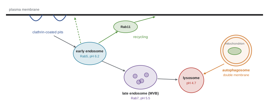
Endocytic vesicles fuse with early endosomes, tubular and vesicular compartments near the cell periphery marked by Rab5 and the lipid PI(3)P. The lumen of early endosomes is mildly acidic, about pH 6.0 to 6.5, maintained by V-type proton ATPases. The acidity releases many ligands from their receptors. Receptors and membrane are sorted into narrow tubules that return to the plasma membrane, either directly or through recycling endosomes marked by Rab11, while released cargo stays in the vacuolar part of the endosome.
The transferrin cycle illustrates how pH can be used cleverly. Transferrin carries two iron ions in the blood and binds its receptor at neutral pH. In the acidic endosome, iron is released, but iron-free transferrin, apotransferrin, remains bound to the receptor at low pH. Receptor and apotransferrin recycle to the surface together, where, at neutral pH, apotransferrin is released to pick up more iron. Nothing is wasted.
Over 10 to 15 minutes, early endosomes mature into late endosomes: they move toward the cell centre along microtubules, exchange Rab5 for Rab7, become more acidic (pH 5.0 to 6.0) and receive newly made lysosomal enzymes from the trans-Golgi network (lesson 4). Membrane proteins destined for destruction, such as activated growth factor receptors marked by ubiquitin, are sorted into small vesicles that bud into the lumen of the endosome. This inward budding is driven by the ESCRT machinery (endosomal sorting complexes required for transport), and the resulting structures are called multivesicular bodies. When late endosomes fuse with lysosomes, the internal vesicles and their cargo are digested. Some multivesicular bodies instead fuse with the plasma membrane and release their internal vesicles as exosomes, which carry proteins and RNAs to other cells and are being studied as disease biomarkers. ESCRT proteins also help close the nuclear envelope after mitosis, cut the bridge between daughter cells at the end of cell division and allow viruses such as HIV to bud from cells.
Lysosomes
Lysosomes, discovered by Christian de Duve in the 1950s through cell fractionation (he shared the 1974 Nobel Prize with Palade and Albert Claude), are the cell’s main digestive compartments. They contain about 60 kinds of acid hydrolases, including proteases (cathepsins), nucleases, lipases, glycosidases, phosphatases and sulfatases, which together can break down every kind of biological macromolecule. These enzymes work best at the low pH of the lysosome, about 4.5 to 5.0, maintained by V-type ATPases pumping protons in. The low optimum pH is a safety feature: if a lysosome leaks, its enzymes are much less active at the neutral pH of the cytosol. The lysosomal membrane is protected from its own enzymes by heavily glycosylated membrane proteins, such as LAMP1 and LAMP2, which form a sugar coat on the inner surface, and it contains transporters that export the products of digestion, amino acids, sugars and nucleotides, back to the cytosol.
Worked problem
How many free protons are in a lysosome?
A spherical lysosome has a diameter of 0.5 µm and a luminal pH of 4.7. How many free hydrogen ions does it contain? Compare with the same volume of cytosol at pH 7.2.
Show solution
- Volume = (4/3)πr³ = 4.19 × (0.25 × 10⁻⁶ m)³ ≈ 6.5 × 10⁻²⁰ m³ = 6.5 × 10⁻¹⁷ L.
- [H⁺] = 10⁻⁴·⁷ M ≈ 2.0 × 10⁻⁵ mol/L.
- Free H⁺ = 2.0 × 10⁻⁵ × 6.5 × 10⁻¹⁷ × 6.02 × 10²³ ≈ 790 ions.
- At pH 7.2: 10⁻⁷·² × 6.5 × 10⁻¹⁷ × 6.02 × 10²³ ≈ 2.5 ions.
Answer: About 800 free protons, compared with only 2 or 3 in the same volume of cytosol. Most protons pumped in are bound by buffers, so the pumps must move many more than this to keep the pH low.
Lysosomes are now also recognized as signalling hubs. The master growth regulator mTORC1, a kinase complex, is activated on the lysosomal surface when amino acids are plentiful; there it promotes protein synthesis and blocks autophagy. When nutrients are scarce, mTORC1 is released and inactive, and the transcription factor TFEB, no longer phosphorylated by mTORC1, enters the nucleus and switches on genes for lysosome biogenesis and autophagy. In this way the digestive organelle reports on the cell’s nutrient status.
Lysosomal storage diseases
More than 70 inherited disorders, collectively called lysosomal storage diseases, result from loss of a lysosomal enzyme, a membrane transporter or an activator protein. Undigested material accumulates and lysosomes swell, damaging cells, particularly in the brain, liver, spleen and bones. Individually rare, together they affect roughly 1 in 5,000 to 1 in 8,000 births.
| Disease | Missing protein | Stored material | Notes |
|---|---|---|---|
| Tay–Sachs disease | β-hexosaminidase A | GM2 ganglioside | Neurodegeneration in infancy; higher carrier frequency in some populations, including Ashkenazi Jewish, French Canadian and Cajun communities |
| Gaucher disease | Glucocerebrosidase | Glucocerebroside | Enlarged spleen and liver, bone disease; treated with enzyme replacement therapy |
| Pompe disease | Acid α-glucosidase | Glycogen | Muscle weakness and heart failure; enzyme replacement available |
| Niemann–Pick disease type C | NPC1 or NPC2 cholesterol transporters | Cholesterol and other lipids | Shows lysosomes export cholesterol from LDL |
| Fabry disease | α-galactosidase A | Globotriaosylceramide | X-linked; kidney, heart and nerve damage |
| I-cell disease | Enzyme adding the M6P tag | Many substrates | Enzymes are secreted instead of delivered (lesson 4) |
Enzyme replacement therapy for Gaucher disease, introduced in the early 1990s, works because recombinant enzyme injected into the blood is modified so that its glycans end in mannose, which is recognized by mannose receptors on macrophages, the cells most affected; the enzyme is taken up by endocytosis and delivered to lysosomes. Replacement enzymes for diseases affecting the brain are less effective because they cannot cross the blood–brain barrier, which has prompted work on gene therapies and on drugs that reduce the production of the stored substrate.
Autophagy: self-eating
Autophagy delivers portions of the cell’s own cytoplasm to lysosomes. In macroautophagy, the main form, a flat membrane cisterna called the phagophore appears, often at contact sites with the ER, grows around a region of cytoplasm and closes to form a double-membraned vesicle, the autophagosome. The autophagosome then fuses with a lysosome, and its contents and inner membrane are digested. The process is controlled by about 40 ATG (autophagy-related) genes, first identified in yeast by Yoshinori Ohsumi, who received the 2016 Nobel Prize in Physiology or Medicine. A key component is LC3 (Atg8 in yeast), a small protein attached to the phagophore membrane by a lipid; fluorescent LC3 forms bright dots wherever autophagosomes form and is the standard marker used to measure autophagy.
Autophagy rises sharply when cells are starved, when mTORC1 is inactive, providing amino acids and energy from recycled material. It also clears protein aggregates, damaged organelles and invading bacteria. Selective autophagy uses receptor proteins, such as p62, that bind both ubiquitinated cargo and LC3, linking the cargo to the growing phagophore. In mitophagy, damaged mitochondria that have lost their membrane potential accumulate the kinase PINK1 on their surface, which recruits the ubiquitin ligase Parkin; ubiquitin chains then mark the mitochondrion for engulfment. Mutations in PINK1 and Parkin cause inherited forms of Parkinson disease, supporting the idea that clearing damaged mitochondria protects neurons. Other forms of autophagy include microautophagy, in which the lysosome itself engulfs material, and chaperone-mediated autophagy, in which proteins with a specific motif are unfolded and pulled directly across the lysosomal membrane.
import numpy as np
rng = np.random.default_rng(214)

# Counting LC3 puncta (autophagosomes) per cell under three conditions.
# Simulated counts drawn from Poisson distributions (illustrative means).
conditions = {"fed": 4, "starved 2 h": 14, "starved + lysosome inhibitor": 27}
data = {k: rng.poisson(m, 60) for k, m in conditions.items()}
for k, v in data.items():
    print(f"{k:30s} mean {v.mean():5.1f} puncta/cell (SD {v.std(ddof=1):.1f}, n={v.size})")

# Autophagic flux: extra puncta when degradation is blocked
flux = data["starved + lysosome inhibitor"].mean() - data["starved 2 h"].mean()
print(f"\nPuncta accumulating when degradation is blocked: {flux:.1f} per cell")
print("A higher count alone cannot distinguish more formation from slower clearance;")
print("comparing with and without a lysosome inhibitor measures flux.")fed mean 4.2 puncta/cell (SD 2.1, n=60) starved 2 h mean 13.8 puncta/cell (SD 3.3, n=60) starved + lysosome inhibitor mean 26.8 puncta/cell (SD 5.3, n=60) Puncta accumulating when degradation is blocked: 13.0 per cell A higher count alone cannot distinguish more formation from slower clearance; comparing with and without a lysosome inhibitor measures flux.
This example illustrates an important experimental point. More autophagosomes could mean that cells are making more of them, or that they are failing to clear them. Blocking lysosomal degradation, for example with a V-ATPase inhibitor, and seeing how many more accumulate measures the flux through the pathway, the true rate of autophagy.
Worked problem
Recycling amino acids by autophagy
A starving hepatocyte degrades about 4 per cent of its protein per hour by autophagy. If it contains 0.5 ng of protein, and the average amino acid residue weighs 110 Da, how many amino acids are released per second?
Show solution
- Protein degraded per hour = 0.04 × 0.5 ng = 0.02 ng = 2 × 10⁻¹¹ g.
- Moles of residues = 2 × 10⁻¹¹ / 110 ≈ 1.8 × 10⁻¹³ mol per hour.
- Molecules = 1.8 × 10⁻¹³ × 6.02 × 10²³ ≈ 1.1 × 10¹¹ per hour.
- Per second ≈ 1.1 × 10¹¹ / 3,600 ≈ 3 × 10⁷ amino acids.
Answer: About 30 million amino acids per second, available for new protein synthesis and energy (illustrative values).
Common mistakes
MistakeAll endocytosis uses clathrin.
InsteadPhagocytosis, macropinocytosis, caveolae and other clathrin-independent routes also take material in.
MistakeLDL receptors are destroyed after delivering one LDL particle.
InsteadThey release LDL in acidic endosomes and recycle many times; PCSK9 diverts them to lysosomes.
MistakeLysosomal enzymes work best at neutral pH.
InsteadThey are acid hydrolases with optima near pH 4.5–5, which protects the cytosol if they leak.
MistakeMore LC3 dots always mean more autophagy.
InsteadThey could reflect slower clearance; flux must be measured with lysosomal inhibitors.
MistakeAutophagy only happens in starving cells.
InsteadIt also continually clears aggregates, damaged organelles and microbes.
Summary and key terms
- Cells take up material by phagocytosis, macropinocytosis, clathrin-mediated endocytosis, caveolae and other routes.
- AP-2 recognizes sorting motifs; dynamin pinches off clathrin-coated vesicles.
- LDL enters by receptor-mediated endocytosis; defects cause familial hypercholesterolemia, treated with statins and PCSK9 inhibitors.
- Early endosomes (Rab5) sort receptors for recycling; late endosomes (Rab7) form multivesicular bodies with ESCRT and fuse with lysosomes.
- Lysosomes hold about 60 acid hydrolases at pH 4.5–5 and act as nutrient-signalling hubs through mTORC1 and TFEB.
- Lysosomal storage diseases such as Tay–Sachs and Gaucher result from missing enzymes or transporters.
- Autophagy, governed by ATG genes, wraps cytoplasm in autophagosomes; mitophagy uses PINK1 and Parkin.
- endocytosis
- Uptake of material by inward budding of the plasma membrane.
- receptor-mediated endocytosis
- Selective uptake of ligands bound to receptors concentrated in coated pits.
- familial hypercholesterolemia
- An inherited disorder of LDL receptor function causing high blood cholesterol.
- early endosome
- A mildly acidic sorting compartment that receives endocytic vesicles; marked by Rab5.
- multivesicular body
- A late endosome containing internal vesicles formed by the ESCRT machinery.
- lysosome
- An acidic organelle containing hydrolases that digest macromolecules.
- V-type ATPase
- A proton pump that acidifies endosomes and lysosomes.
- lysosomal storage disease
- An inherited disorder in which undigested material accumulates in lysosomes.
- autophagosome
- A double-membraned vesicle that engulfs cytoplasm and fuses with a lysosome.
- mitophagy
- Selective autophagy of damaged mitochondria.
Practice problems
Work each problem on paper first, then reveal the answer to check.
Which adaptor links cargo to clathrin at the plasma membrane?
Show answer
AP-2, which binds PIP₂ and sorting motifs.
What signal in the LDL receptor tail directs it into coated pits?
Show answer
The NPxY motif.
How do statins lower blood LDL?
Show answer
By blocking cholesterol synthesis, which raises LDL receptor numbers through the SREBP pathway.
Why does transferrin not lose its receptor in the endosome?
Show answer
Iron-free apotransferrin remains bound to the receptor at low pH and recycles with it.
What does ESCRT do in endosomes?
Show answer
It drives inward budding of membrane carrying ubiquitinated cargo, forming multivesicular bodies.
Why is the acid pH optimum of lysosomal enzymes a safety feature?
Show answer
Leaked enzymes are much less active at the neutral pH of the cytosol.
How is recombinant glucocerebrosidase targeted to macrophages?
Show answer
Its glycans end in mannose, recognized by macrophage mannose receptors.
What happens to TFEB when nutrients are scarce?
Show answer
It is no longer phosphorylated by mTORC1, enters the nucleus and activates lysosome and autophagy genes.
How do PINK1 and Parkin mark damaged mitochondria?
Show answer
PINK1 accumulates on depolarized mitochondria and recruits Parkin, which adds ubiquitin.
A lysosome of 1 µm diameter is at pH 5. How many free protons does it hold?
Show answer
Volume ≈ 5.2 × 10⁻¹⁶ L; 10⁻⁵ × 5.2 × 10⁻¹⁶ × 6.02 × 10²³ ≈ 3,100.
Lesson quiz · 5 questions
Score 60% or more to complete this lesson.
Create a free account to save your progress, take quizzes and earn a certificate.