
BIOL-214 · Lesson 2 of 12 · 31 min
The nucleus and nuclear transport
The nucleus is the defining feature of eukaryotic cells and their largest organelle, typically 5 to 10 µm across in animal cells. It holds the genome, and with it the machinery for replicating DNA, transcribing genes and processing RNA. Separating the genome from the cytoplasm gives eukaryotes a layer of control that bacteria lack: transcription and translation happen in different places, so RNA can be spliced and checked before it meets a ribosome, and proteins that regulate genes can be kept out of the nucleus until they are needed. But separation comes at a price. Every protein that works in the nucleus, from histones to DNA polymerase, is made in the cytoplasm and must be imported, and every messenger RNA, transfer RNA and ribosomal subunit made in the nucleus must be exported. This lesson describes the structure of the nuclear envelope, the nuclear pore complexes through which all this traffic passes, the energy-driven system that makes transport directional, and the internal organization of the nucleus. BIOL-207 covered transcription, RNA processing and chromatin; here the focus is the nucleus as a compartment.
The nuclear envelope
The nucleus is enclosed by the nuclear envelope, two concentric membranes separated by a space of 20 to 40 nm. The outer nuclear membrane is continuous with the endoplasmic reticulum and, like the rough ER, is studded with ribosomes; the space between the two membranes, the perinuclear space, is continuous with the ER lumen. The inner nuclear membrane has its own set of proteins, some of which bind chromatin and the nuclear lamina. The two membranes join at the nuclear pores, where the envelope is pierced by large protein complexes.
Lining the inner surface of the envelope is the nuclear lamina, a meshwork of intermediate filament proteins called lamins, about 15 nm thick in many cells. The lamina gives the nucleus mechanical strength, anchors nuclear pore complexes and helps organize chromatin: regions of the genome attached to the lamina, called lamina-associated domains, tend to be gene-poor and transcriptionally silent. Mutations in LMNA, the gene for lamins A and C, cause a group of diseases called laminopathies, including some muscular dystrophies, cardiomyopathies and Hutchinson–Gilford progeria syndrome, a very rare disorder of accelerated ageing in children. In progeria, a point mutation activates a cryptic splice site so that the lamin A precursor is not processed normally; the abnormal protein, progerin, stays attached to the membrane by a lipid anchor and distorts the nuclear envelope. A drug that blocks the lipid modification, lonafarnib, was approved in the United States in 2020 as the first treatment shown to extend survival.
Proteins called LINC complexes span both membranes of the envelope and connect the lamina inside to the cytoskeleton outside. They transmit mechanical forces from the cell surface to the nucleus and help position the nucleus within the cell, for example during cell migration and in muscle fibres.
The nuclear pore complex
Nuclear pore complexes (NPCs) are among the largest protein assemblies in the cell. In vertebrates each is about 110 to 120 MDa and about 100 to 120 nm across, built from roughly 30 different proteins called nucleoporins, each present in multiple copies, so that a complete pore contains around 1,000 protein molecules. The core has eight-fold rotational symmetry. Cytoplasmic filaments extend into the cytosol, and a basket of filaments projects into the nucleus. A typical mammalian nucleus has 2,000 to 5,000 pores, and a large frog oocyte nucleus tens of millions.
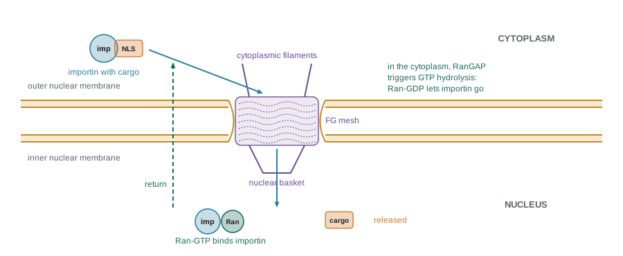
The central channel is filled with a dense, disordered mesh of protein chains rich in phenylalanine–glycine repeats, the FG repeats of a subset of nucleoporins. This mesh behaves like a selective gel. Small molecules, ions and small proteins diffuse through freely, but passage becomes progressively slower as size increases, and proteins larger than about 40 to 60 kDa pass very slowly unless they are bound to a transport receptor. Transport receptors, by contrast, contain many sites that bind FG repeats weakly and transiently; they can dissolve into the mesh and slide through, carrying their cargo with them. Cryo-electron tomography of NPCs in intact cells has shown that the pore is not a rigid ring: its diameter changes with the cell’s mechanical and energetic state.
Worked problem
How many pores, and how busy are they?
A spherical nucleus has a diameter of 10 µm and about 11 nuclear pores per square micrometre of envelope. A growing HeLa cell is estimated to import about 560,000 ribosomal proteins per minute and to export about 14,000 ribosomal subunits per minute (approximate textbook values). How many pores does the nucleus have, and how many import events per pore per second do ribosomal proteins alone require?
Show solution
- Surface area = 4πr² = 4 × 3.14 × 5² ≈ 314 µm².
- Pores ≈ 314 × 11 ≈ 3,500.
- Imports per second = 560,000 / 60 ≈ 9,300.
- Per pore ≈ 9,300 / 3,500 ≈ 2.7 ribosomal proteins per second.
- Single pores can handle on the order of 1,000 translocation events per second in total, so ribosome production uses only part of their capacity.
Answer: About 3,500 pores, each importing roughly 3 ribosomal proteins per second for ribosome assembly, alongside many other cargoes moving in both directions.
Signals for import and export
Proteins destined for the nucleus carry a nuclear localization signal (NLS). The classical NLS, first identified in the large T antigen of the virus SV40, is a short cluster of basic residues, PKKKRKV. Changing a single lysine in this sequence keeps the protein in the cytoplasm, and attaching the sequence to a cytoplasmic protein sends it into the nucleus. Bipartite NLSs, such as that of nucleoplasmin, have two short basic clusters separated by a spacer of about ten residues. Proteins that leave the nucleus carry nuclear export signals (NES), typically short leucine-rich motifs. Because NLSs and NESs are not cleaved off, proteins can shuttle in and out repeatedly, and the balance between import and export determines where they spend their time.
Signals can be hidden or revealed to regulate a protein’s location. The transcription factor NF-κB is held in the cytoplasm by a partner, IκB, that masks its NLS; when a cell senses infection or inflammation, IκB is phosphorylated, ubiquitinated and destroyed, and NF-κB enters the nucleus within minutes. Steroid hormone receptors (lesson 11) are released from cytoplasmic chaperones when they bind hormone, exposing their NLSs. Phosphorylation near an NLS or NES can also tip the balance; lesson 12 describes how the location of cyclin–CDK complexes is controlled in this way.
import re

# Search illustrative protein sequences for classical NLS patterns.
# Monopartite: K followed by K or R, any residue, then K or R (K-[KR]-x-[KR]),
# a commonly used simplified pattern. Bipartite: two basic pairs separated by 10-12 residues.
mono = re.compile(r"K[KR].[KR]")
bipartite = re.compile(r"[KR]{2}.{10,12}[KR]{3}")

proteins = {
    "SV40 large T fragment": "MDKAELIPEPPKKKRKVEDPKDF",
    "nucleoplasmin fragment": "AVKRPAATKKAGQAKKKKLDKED",
    "cytosolic enzyme fragment": "MSTAGLVDEAFGQPWLSNTEVGA",
}
for name, seq in proteins.items():
    m1 = [(m.start() + 1, m.group()) for m in mono.finditer(seq)]
    m2 = [(m.start() + 1, m.group()) for m in bipartite.finditer(seq)]
    print(f"{name:28s} monopartite: {m1 or 'none'}")
print(f"{'':28s} bipartite: {m2 or 'none'}")SV40 large T fragment monopartite: [(12, 'KKKR')]
bipartite: none
nucleoplasmin fragment monopartite: [(15, 'KKKK')]
bipartite: [(3, 'KRPAATKKAGQAKKKK')]
cytosolic enzyme fragment monopartite: none
bipartite: nonePattern searches like this are a first step only. They produce false positives, because many basic stretches are not NLSs, and they miss signals that do not fit the classical pattern. Experiments that move a signal onto a reporter protein, or mutate it in the original protein, remain the test of whether a sequence really works.
Transport receptors and the Ran cycle
Most nuclear transport is carried out by a family of related transport receptors called karyopherins: importins carry cargo into the nucleus and exportins carry it out. In classical import, the adaptor importin α binds the NLS and importin β binds both importin α and the FG repeats of the pore.
What makes transport directional is a small GTPase called Ran. Like other GTPases, Ran switches between a GTP-bound form and a GDP-bound form. Two enzymes are placed on opposite sides of the envelope. The guanine nucleotide exchange factor for Ran, RCC1, is bound to chromatin inside the nucleus, so Ran in the nucleus is loaded with GTP. The GTPase-activating protein, RanGAP, is in the cytoplasm, together with helpers, so Ran in the cytoplasm hydrolyses its GTP and ends up bound to GDP. The result is a steep gradient: Ran-GTP is high in the nucleus and low in the cytoplasm.
| Step | Import (importin) | Export (exportin) |
|---|---|---|
| Cargo binding | In the cytoplasm, where Ran-GTP is low | In the nucleus, together with Ran-GTP |
| Passage | Importin–cargo complex moves through FG mesh | Exportin–Ran-GTP–cargo complex moves through FG mesh |
| Cargo release | In the nucleus, Ran-GTP binds importin and releases cargo | In the cytoplasm, GTP hydrolysis releases cargo and Ran-GDP |
| Receptor recycling | Importin returns to cytoplasm bound to Ran-GTP | Exportin returns to nucleus empty |
| Ran recycling | Ran-GDP is imported by NTF2 and reloaded with GTP by RCC1 | Same |
The gradient is the energy source. The receptors themselves move through pores by diffusion, but because cargo binding and release depend on whether Ran-GTP is present, cargo accumulates on one side. Roughly one GTP is hydrolysed for each transport cycle. If the gradient is collapsed experimentally, for example by putting Ran-GTP into the cytoplasm, import stops. During mitosis the nuclear envelope breaks down in most animal cells, but the Ran-GTP gradient persists around the chromosomes, because RCC1 stays bound to them; there it helps assemble the mitotic spindle and later guides the reassembly of the envelope.
Worked problem
How much can a gradient concentrate a cargo?
Suppose import is driven so that a cargo reaches equilibrium when its nuclear concentration is 20 times its cytoplasmic concentration. What is the free-energy cost per molecule at 37 °C, and is one GTP enough?
Show solution
- ΔG = RT ln(ratio) = 8.314 × 310 × ln 20 ≈ 7.7 kJ/mol.
- GTP hydrolysis under cellular conditions releases roughly 50 kJ/mol.
- One GTP per cycle provides several times the energy needed for a 20-fold accumulation.
Answer: About 7.7 kJ/mol, well within what one GTP hydrolysis provides; the Ran system can drive large concentration differences.
Exporting RNA
RNA export uses both karyopherins and dedicated machinery. Transfer RNAs leave through exportin-t, and microRNA precursors through exportin-5. Ribosomal subunits, assembled in the nucleolus from rRNA and about 80 ribosomal proteins, are exported with the help of the exportin CRM1 and other factors. Messenger RNAs are exported differently: they leave as large ribonucleoprotein particles, coated with proteins that mark them as fully processed. A heterodimeric export receptor, NXF1 with its partner NXT1, binds the mRNA particle and interacts directly with FG repeats, and directionality comes not from Ran but from an RNA helicase on the cytoplasmic filaments that strips proteins off the emerging mRNA. Because export factors are loaded during splicing and 3′-end processing (BIOL-207), incompletely processed RNAs are retained in the nucleus and degraded, an important quality-control step.
Inside the nucleus
The nucleus has no internal membranes, but it is far from uniform. Each chromosome occupies its own region, a chromosome territory, with gene-rich chromosomes and active genes tending toward the interior and gene-poor regions near the lamina. Techniques such as Hi-C, which capture which parts of the genome lie close together, have shown that chromatin is folded into loops and domains, many of them formed by the protein complex cohesin extruding DNA loops until it meets boundary proteins such as CTCF.
The nucleus also contains membraneless compartments, or nuclear bodies. The largest is the nucleolus, where ribosomal RNA genes are transcribed by RNA polymerase I and ribosomal subunits are assembled; its size reflects how much protein the cell makes. Others include nuclear speckles, enriched in splicing factors, Cajal bodies, involved in assembling small nuclear ribonucleoproteins, and promyelocytic leukaemia (PML) bodies. Many of these bodies form by phase separation, in which proteins with flexible, multivalent interaction regions condense out of solution like oil droplets in water, concentrating the molecules needed for a task without enclosing them in a membrane. Phase separation also operates in the cytoplasm, for example in stress granules and processing bodies.
import math

# Passive diffusion through pores slows steeply with size.
# A simple illustrative model: equilibration half-time grows with molecular mass.
# Values are order-of-magnitude illustrations, not measurements.
def passive_half_time(kda):
    return 2.0 * math.exp(kda / 10.0)     # seconds

for kda in (5, 20, 40, 60, 90):
    t = passive_half_time(kda)
    unit = f"{t:8.0f} s" if t < 3600 else f"{t/3600:8.1f} h"
    print(f"{kda:3d} kDa protein: passive equilibration half-time ~ {unit}")

print("\nWith an importin, cargoes of several hundred kDa cross in milliseconds per event.")5 kDa protein: passive equilibration half-time ~ 3 s 20 kDa protein: passive equilibration half-time ~ 15 s 40 kDa protein: passive equilibration half-time ~ 109 s 60 kDa protein: passive equilibration half-time ~ 807 s 90 kDa protein: passive equilibration half-time ~ 4.5 h With an importin, cargoes of several hundred kDa cross in milliseconds per event.
The exact numbers depend on the protein’s shape and surface, but the trend is real: below about 30 kDa, proteins equilibrate within seconds to minutes, while larger proteins take hours or never equilibrate without help. This is why even small nuclear proteins carry NLSs: active import concentrates them in the nucleus rather than letting them spread evenly.
The nucleus in mitosis and disease
In most animal cells, the nuclear envelope breaks down at the start of mitosis. Phosphorylation of lamins by the mitotic cyclin–CDK (lesson 12) causes the lamina to disassemble, nuclear pores come apart into subcomplexes, and the nuclear membranes are absorbed into the ER. After the chromosomes separate, the process reverses: membranes coat the chromosomes, pores reassemble and lamins are dephosphorylated. Yeasts and some other organisms instead keep their envelope intact and build the spindle inside it, a closed mitosis.
Defects in nuclear transport contribute to several diseases. Mutations in nucleoporins cause rare developmental and kidney disorders, and some viruses, including HIV and influenza, have evolved ways to use or block nuclear transport. In some neurodegenerative diseases, including amyotrophic lateral sclerosis (ALS), aggregates and toxic repeat proteins disrupt the pores. Drugs that block CRM1-dependent export, such as selinexor, are used in some blood cancers, because they trap tumour-suppressor proteins in the nucleus where they can act.
Common mistakes
MistakeThe nuclear envelope is a single membrane.
InsteadIt is two membranes; the outer is continuous with the ER, and they join at nuclear pores.
MistakeNuclear pores are open holes that let anything through.
InsteadThe FG-repeat mesh lets small molecules pass but blocks large ones unless they are bound to transport receptors.
MistakeImportins burn ATP to push cargo through the pore.
InsteadReceptors move by diffusion; directionality comes from the Ran-GTP gradient and about one GTP per cycle.
MistakeRan-GTP is high in the cytoplasm because GTP is made there.
InsteadRan-GTP is high in the nucleus because the exchange factor RCC1 is bound to chromatin there.
MistakeThe nucleolus is surrounded by a membrane.
InsteadIt is a membraneless compartment formed by ribosomal RNA genes and associated proteins, partly by phase separation.
Summary and key terms
- The nuclear envelope has two membranes; the outer is continuous with the ER, and the lamina lines the inner surface.
- Lamin mutations cause laminopathies, including Hutchinson–Gilford progeria.
- Nuclear pore complexes of about 1,000 nucleoporins form a selective FG-repeat barrier.
- Classical NLSs are short basic clusters; leucine-rich NESs direct export; masking signals regulates location.
- Importins and exportins carry cargo; the Ran-GTP gradient, set by nuclear RCC1 and cytoplasmic RanGAP, makes transport directional.
- mRNA export uses NXF1–NXT1 and a cytoplasmic helicase, with quality control linked to processing.
- The nucleus contains chromosome territories, loops and domains, and membraneless bodies such as the nucleolus.
- nuclear envelope
- The double membrane surrounding the nucleus, pierced by nuclear pores.
- nuclear lamina
- A meshwork of lamin intermediate filaments lining the inner nuclear membrane.
- nuclear pore complex
- A large protein assembly that controls traffic between nucleus and cytoplasm.
- FG repeats
- Disordered phenylalanine–glycine-rich segments of nucleoporins forming the pore’s selective barrier.
- nuclear localization signal
- A sequence, often a short basic cluster, that directs a protein into the nucleus.
- nuclear export signal
- A sequence, often leucine-rich, that directs a protein out of the nucleus.
- importin
- A transport receptor that carries cargo into the nucleus.
- Ran
- A small GTPase whose GTP-bound form is concentrated in the nucleus, driving directional transport.
- nucleolus
- The nuclear body where rRNA genes are transcribed and ribosomal subunits assembled.
- phase separation
- Condensation of molecules into liquid-like droplets that form membraneless compartments.
Practice problems
Work each problem on paper first, then reveal the answer to check.
What is continuous with the outer nuclear membrane?
Show answer
The endoplasmic reticulum; the perinuclear space is continuous with the ER lumen.
What is the molecular defect in Hutchinson–Gilford progeria?
Show answer
A mutation in LMNA creates a cryptic splice site, producing progerin, which keeps a lipid anchor and distorts the envelope.
Roughly how many proteins make up one vertebrate nuclear pore complex?
Show answer
About 30 different nucleoporins in multiple copies, around 1,000 molecules in total.
Why can transport receptors pass through the pore when large proteins cannot?
Show answer
They bind FG repeats weakly and transiently, so they can dissolve into and move through the mesh.
Where is RCC1 located, and what does it do?
Show answer
It is bound to chromatin in the nucleus and loads Ran with GTP.
A cargo is bound to importin in the cytoplasm. What releases it in the nucleus?
Show answer
Binding of Ran-GTP to importin.
How does NF-κB reach the nucleus after an inflammatory signal?
Show answer
Its inhibitor IκB is phosphorylated and destroyed, unmasking its NLS.
A nucleus 8 µm across has 10 pores per µm². How many pores?
Show answer
4π × 4² × 10 ≈ 2,000.
How is directionality achieved for mRNA export?
Show answer
A helicase on the cytoplasmic filaments strips export proteins from the emerging mRNA.
Why do lamins disassemble at mitosis?
Show answer
They are phosphorylated by the mitotic cyclin–CDK.
Lesson quiz · 5 questions
Score 60% or more to complete this lesson.
Create a free account to save your progress, take quizzes and earn a certificate.