
BIOL-214 · Lesson 6 of 12 · 31 min
Mitochondria, chloroplasts and peroxisomes
Three organelles stand apart from the secretory and endocytic pathways described in lessons 3 to 5. Mitochondria and chloroplasts are the descendants of bacteria that were engulfed by ancestral cells more than a billion years ago; they still carry their own small genomes and ribosomes, divide by fission and are surrounded by two membranes. Peroxisomes have a single membrane and no genome, and they specialize in oxidative reactions that produce hydrogen peroxide. None of the three receives proteins in vesicles from the Golgi. Instead, almost all their proteins are made on cytosolic ribosomes and imported after translation through dedicated machines in their membranes. BIOL-107 introduced cellular respiration and photosynthesis, and BIOL-201 described maternal inheritance of mitochondrial DNA. This lesson focuses on how these organelles are built and maintained: their genomes, how proteins find their way in, how mitochondria fuse, divide and are cleared, and what goes wrong in diseases of these organelles.
Mitochondrial structure and the proton-motive force
A mitochondrion has an outer membrane, permeable to small molecules through large pores formed by the protein porin (VDAC), and an inner membrane, which is impermeable to most ions and folded into cristae. The inner membrane encloses the matrix, which contains the enzymes of the citric acid cycle and fatty acid oxidation, the mitochondrial genome and mitochondrial ribosomes. The respiratory chain complexes in the inner membrane pump protons from the matrix to the intermembrane space, creating an electrochemical gradient, the proton-motive force, which drives ATP synthase. Cryo-electron tomography has shown that ATP synthase dimers line the sharp edges of cristae, helping to bend the membrane, and that respiratory complexes cluster in supercomplexes on the flat faces.
The proton-motive force has two components: an electrical potential across the inner membrane, Δψ, typically about 150 to 180 mV with the matrix negative, and a pH difference, with the matrix about 0.5 to 0.8 units more alkaline than the cytosol.
import math

R, F, T = 8.314, 96485.0, 310.0          # J/(mol K), C/mol, K
dpsi = -0.160                              # V, matrix negative relative to outside
dpH = 0.7                                  # matrix pH minus outside pH

z = 2.303 * R * T / F                      # V per pH unit
dp = abs(dpsi) + z * dpH                   # magnitude of proton-motive force, V
dG_per_H = F * dp / 1000                   # kJ released per mol H+ moving into the matrix
print(f"2.303RT/F at 37 C = {z*1000:.1f} mV per pH unit")
print(f"proton-motive force = {dp*1000:.0f} mV "
      f"({abs(dpsi)*1000:.0f} mV electrical + {z*dpH*1000:.0f} mV chemical)")
print(f"free energy per mol of protons entering = {dG_per_H:.1f} kJ")

# ATP synthase: a c-ring of 8 subunits in mammals gives 8 protons per 3 ATP
h_per_atp = 8 / 3
dG_atp = 50.0                              # kJ/mol needed to make ATP in the cell (approx.)
print(f"protons per ATP = {h_per_atp:.2f}; energy supplied = {h_per_atp*dG_per_H:.1f} kJ/mol "
      f"vs about {dG_atp:.0f} kJ/mol needed")
# Add one more proton for importing phosphate and exchanging ATP for ADP
print(f"including transport: {(h_per_atp+1)*dG_per_H:.1f} kJ per ATP delivered to the cytosol")2.303RT/F at 37 C = 61.5 mV per pH unit proton-motive force = 203 mV (160 mV electrical + 43 mV chemical) free energy per mol of protons entering = 19.6 kJ protons per ATP = 2.67; energy supplied = 52.2 kJ/mol vs about 50 kJ/mol needed including transport: 71.8 kJ per ATP delivered to the cytosol
The calculation shows why ATP synthase needs more than two protons per ATP: each proton supplies about 20 kJ/mol, while making ATP under cellular conditions costs about 50 kJ/mol. The mammalian ATP synthase has a ring of eight c subunits, so one full rotation moves eight protons and makes three ATP, about 2.7 protons per ATP; exporting ATP and importing ADP and phosphate costs roughly one more proton. Organisms with larger c-rings, 10 to 15 subunits, need more protons per ATP.
The mitochondrial genome
Human mitochondrial DNA (mtDNA) is a circular molecule of 16,569 base pairs encoding 37 genes: 13 proteins, all subunits of the respiratory complexes and ATP synthase, plus 22 transfer RNAs and 2 ribosomal RNAs. Each cell contains hundreds to thousands of copies, packaged into nucleoids. The remaining roughly 1,100 to 1,500 mitochondrial proteins, about 99 per cent of the total, are encoded in the nucleus, made in the cytosol and imported. Mitochondria use a slightly different genetic code; in human mitochondria, for example, UGA codes for tryptophan instead of stop. Because the genes for some subunits of a complex are in the nucleus and others in mtDNA, the two genomes must be expressed in a coordinated way.
Mutations in mtDNA cause diseases such as Leber hereditary optic neuropathy and MELAS, and mutations in nuclear genes for mitochondrial proteins cause many more; together, mitochondrial diseases affect about 1 in 5,000 people. A cell can contain a mixture of normal and mutant mtDNA, a state called heteroplasmy, and symptoms appear only when the mutant fraction exceeds a threshold, often 60 to 90 per cent, which varies by tissue. BIOL-201 described how the mitochondrial bottleneck in egg development can shift this fraction between generations, and how mitochondrial donation techniques aim to prevent transmission.
Worked problem
Copies of mtDNA in a cell
A muscle cell contains about 3,000 copies of mtDNA. What length of mtDNA does it contain, and how does this compare with its nuclear genome of about 6.2 billion base pairs (diploid)?
Show solution
- Total mtDNA = 3,000 × 16,569 ≈ 5.0 × 10⁷ bp.
- Ratio = 5.0 × 10⁷ / 6.2 × 10⁹ ≈ 0.008.
- mtDNA is less than 1 per cent of the cell’s DNA, but its 13 protein genes are essential for oxidative phosphorylation.
Answer: About 50 million base pairs, under 1 per cent of total cellular DNA.
Importing proteins into mitochondria
Proteins destined for the mitochondrial matrix usually carry an N-terminal presequence of 15 to 50 residues that forms an amphipathic α helix: positively charged residues (arginine and lysine) cluster on one face and hydrophobic residues on the other, while acidic residues are nearly absent. The presequence is recognized by receptors on the outer membrane and guides the protein through two translocases.
| Complex | Location | Role |
|---|---|---|
| TOM (translocase of the outer membrane) | Outer membrane | General entry gate for almost all imported proteins |
| SAM (sorting and assembly machinery) | Outer membrane | Inserts β-barrel proteins such as porin into the outer membrane |
| TIM23 | Inner membrane | Imports presequence proteins into the matrix or inner membrane; needs membrane potential |
| PAM motor (mitochondrial Hsp70) | Matrix side of TIM23 | Uses ATP to pull the chain into the matrix |
| TIM22 | Inner membrane | Inserts multipass carrier proteins, such as the ADP/ATP carrier, into the inner membrane |
| MIA (Mia40 and Erv1) | Intermembrane space | Traps small cysteine-rich proteins by forming disulfide bonds |
| MPP (mitochondrial processing peptidase) | Matrix | Cleaves off the presequence |
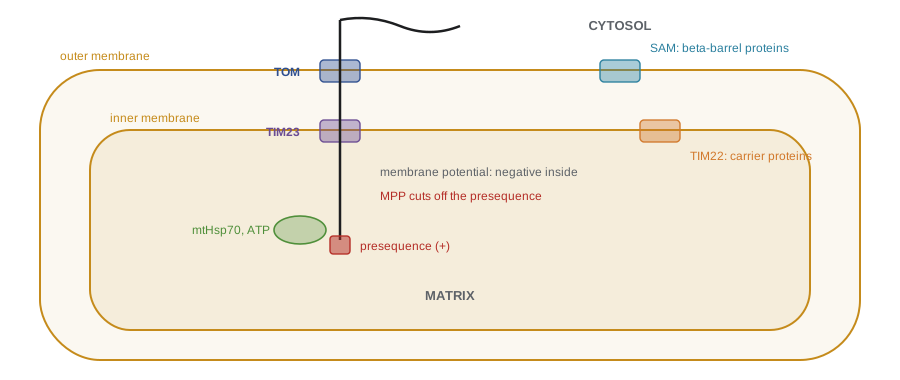
Import uses three sources of energy. The membrane potential across the inner membrane pulls the positively charged presequence toward the negative matrix. Mitochondrial Hsp70 in the matrix binds the incoming chain and, using ATP, pulls it inward, acting as a motor and a ratchet. And cytosolic chaperones, which keep precursors unfolded, use ATP. Because proteins must cross the membranes in an unfolded state, a tightly folded domain blocks import; classic experiments fused a matrix presequence to dihydrofolate reductase and showed that adding methotrexate, which stabilizes the folded enzyme, stalled the fusion protein in the import channels, spanning both membranes at contact sites.
import math, random, re

# Scoring an amphipathic presequence: net charge and hydrophobic moment
# (helical wheel with 100 degrees per residue). Sequences are illustrative.
KD = {"A": 1.8, "R": -4.5, "N": -3.5, "D": -3.5, "C": 2.5, "Q": -3.5, "E": -3.5, "G": -0.4,
      "H": -3.2, "I": 4.5, "L": 3.8, "K": -3.9, "M": 1.9, "F": 2.8, "P": -1.6, "S": -0.8,
      "T": -0.7, "W": -0.9, "Y": -1.3, "V": 4.2}
def charge(s): return sum(s.count(x) for x in "KR") - sum(s.count(x) for x in "DE")
def moment(s):
    x = sum(KD[a] * math.cos(math.radians(100 * i)) for i, a in enumerate(s))
    y = sum(KD[a] * math.sin(math.radians(100 * i)) for i, a in enumerate(s))
    return math.hypot(x, y) / len(s)

pre = "MLSLRQSIRFFKPATRTLCSSRYLL"        # yeast Cox4-like presequence
cyto = "MSDEETAKLQEAIDGLSEEVDEAA"        # N-terminus of a cytosolic protein (illustrative)
for name, p in (("matrix presequence", pre), ("cytosolic N-terminus", cyto)):
    print(f"{name:22s} net charge {charge(p):+d}, acidic residues {sum(p.count(x) for x in 'DE')}")

# Is the presequence more amphipathic than random arrangements of the same residues?
rng = random.Random(214)
shuffled = []
for _ in range(500):
    l = list(pre); rng.shuffle(l); shuffled.append(moment("".join(l)))
print(f"hydrophobic moment: presequence {moment(pre):.2f}, "
      f"shuffled mean {sum(shuffled)/len(shuffled):.2f}, "
      f"shuffles at least as high {sum(m >= moment(pre) for m in shuffled)/len(shuffled):.0%}")

# Peroxisomal targeting signal 1: C-terminal [SAC][KRH][LM]
for name, p in {"catalase-like C-terminus": "...GKNAKANL", "PTS1 protein": "...ADAPSKL",
                "cytosolic protein": "...GEEVKAG"}.items():
    ok = re.search(r"[SAC][KRH][LM]$", p) is not None
print(f"{name:26s} ends {p[-3:]}: PTS1 match = {ok}")matrix presequence net charge +5, acidic residues 0 cytosolic N-terminus net charge -8, acidic residues 9 hydrophobic moment: presequence 0.93, shuffled mean 0.56, shuffles at least as high 10% catalase-like C-terminus ends ANL: PTS1 match = False PTS1 protein ends SKL: PTS1 match = True cytosolic protein ends KAG: PTS1 match = False
The presequence carries a strong net positive charge and no acidic residues, while the cytosolic N-terminus is strongly acidic. Its hydrophobic moment is well above that of most random arrangements of the same residues, the signature of an amphipathic helix in which charged and hydrophobic residues sit on opposite faces. The PTS1 search shows a subtlety: human catalase ends in KANL, a weak, non-canonical signal that the receptor still recognizes, so simple pattern matching misses it. Prediction programs for both kinds of signal use machine learning trained on known examples, but experimental tests remain essential.
Mitochondrial dynamics and quality control
Mitochondria are not isolated bean-shaped organelles but a dynamic network that constantly fuses and divides. Fusion of the outer membranes is driven by the GTPases mitofusin 1 and 2, and of the inner membranes by OPA1. Fission is driven by Drp1, a dynamin-related GTPase recruited from the cytosol that forms spirals around the organelle and constricts it, usually at sites where ER tubules wrap around the mitochondrion and mark the division point. Fusion allows mitochondria to share contents, diluting damage and mixing mtDNA; fission allows them to be distributed to daughter cells, transported along microtubules to distant parts of neurons, and separated for quality control. Damaged fragments that lose their membrane potential are removed by mitophagy (lesson 5). Mutations in OPA1 cause dominant optic atrophy, and mutations in mitofusin 2 cause a form of Charcot–Marie–Tooth neuropathy, showing how much long nerve cells depend on mitochondrial dynamics.
Mitochondria also have roles beyond energy. They make the iron–sulfur clusters needed by many enzymes, synthesize heme and some phospholipids, buffer calcium taken up at contact sites with the ER, and act as signalling platforms in innate immunity. And they decide whether a cell lives or dies: release of cytochrome c from the intermembrane space is the key step in the intrinsic pathway of apoptosis (lesson 12).
Chloroplasts and other plastids
Chloroplasts have three membrane systems: an outer and an inner envelope membrane, and the internal thylakoid membranes, stacked into grana, which carry the photosystems, the electron transport chain and ATP synthase of photosynthesis. The space enclosed by thylakoids, the lumen, is separate from the stroma, which contains the enzymes of carbon fixation, including Rubisco, probably the most abundant protein on Earth. Chloroplast genomes are 120 to 160 kilobase pairs and encode about 100 genes; most of the roughly 3,000 chloroplast proteins are encoded in the nucleus. The large subunit of Rubisco is made in the chloroplast and the small subunit in the cytosol, another example of coordination between genomes. Plastids also exist as non-photosynthetic forms: amyloplasts store starch in roots and tubers, and chromoplasts give fruits and flowers their colours.
Nuclear-encoded chloroplast proteins carry N-terminal transit peptides recognized by the TOC and TIC translocons of the outer and inner envelope membranes; import is driven by GTP and ATP rather than by a membrane potential. Proteins bound for the thylakoid lumen have a bipartite signal: a transit peptide for import into the stroma, followed by a second, signal-peptide-like sequence that directs them across the thylakoid membrane, using pathways inherited from the bacterial ancestor. Some thylakoid proteins cross folded, using the twin-arginine translocation (Tat) system, which is powered by the proton gradient.
Peroxisomes
Peroxisomes are small (0.1 to 1 µm) single-membrane organelles found in nearly all eukaryotic cells. They take their name from hydrogen peroxide: their oxidases remove hydrogen atoms from substrates and transfer them to oxygen, producing H₂O₂, which the enzyme catalase then breaks down to water and oxygen or uses to oxidize other molecules, including toxins such as ethanol in liver cells. In animals, peroxisomes shorten very-long-chain fatty acids (those with more than 22 carbons) by β-oxidation before the products are completed in mitochondria, and they carry out the first steps in synthesizing plasmalogens, a class of phospholipids abundant in myelin. In plants, specialized peroxisomes called glyoxysomes convert stored fats to sugar in germinating seeds, and leaf peroxisomes take part in photorespiration.
Peroxisomal matrix proteins are imported fully folded, even as oligomers, which distinguishes this system from mitochondrial and chloroplast import. Most carry PTS1, a C-terminal tripeptide related to Ser–Lys–Leu (SKL); a minority carry an N-terminal PTS2. Cytosolic receptors, Pex5 for PTS1 and Pex7 for PTS2, bind the cargo, dock on the peroxisomal membrane and form a transient pore with other peroxins; the receptor is then ubiquitinated and pulled back out for reuse. New peroxisomes form both by growth and division of existing ones and, when necessary, from vesicles budding from the ER that carry peroxisomal membrane proteins.
| Disorder | Defect | Main features |
|---|---|---|
| Zellweger spectrum disorders | Mutations in PEX genes block peroxisome assembly | Severe neurological problems, liver disease; very-long-chain fatty acids accumulate |
| X-linked adrenoleukodystrophy | ABCD1 transporter that brings very-long-chain fatty acids into peroxisomes | Loss of myelin in the brain, adrenal failure; newborn screening in many US states and some provinces |
| Refsum disease | Enzyme for breaking down phytanic acid | Vision loss, neuropathy; managed with diet |
| Primary hyperoxaluria type 1 | A peroxisomal enzyme mistargeted to mitochondria in some patients | Kidney stones and kidney failure |
Worked problem
Catalase and hydrogen peroxide
Catalase is one of the fastest enzymes known, with a turnover number of about 4 × 10⁷ H₂O₂ molecules per second per enzyme. If a liver peroxisome contains 10⁵ catalase molecules working at 10 per cent of their maximum rate, how much H₂O₂ does it destroy per second?
Show solution
- Rate per enzyme at 10 per cent = 4 × 10⁶ molecules per second.
- Total = 10⁵ × 4 × 10⁶ = 4 × 10¹¹ molecules per second.
- In moles: 4 × 10¹¹ / 6.02 × 10²³ ≈ 6.6 × 10⁻¹³ mol per second.
Answer: About 4 × 10¹¹ molecules, or roughly 0.7 picomoles, of H₂O₂ per second per peroxisome (illustrative).
The endosymbiotic origin, revisited
The bacterial ancestry of mitochondria and chloroplasts, proposed by Lynn Margulis in 1967 and now firmly supported by genome sequencing, explains many of the features in this lesson: their double membranes, their circular genomes and bacterial-type ribosomes (which is why some antibiotics that target bacterial ribosomes, such as linezolid and chloramphenicol, can also affect mitochondrial protein synthesis), their division by fission, and machinery such as SAM and Tat that resembles bacterial systems. Over time most of the endosymbionts’ genes moved to the nucleus, which is why import machinery had to evolve. Peroxisomes, by contrast, are thought to have arisen from the endomembrane system, consistent with their ability to form from the ER.
Common mistakes
MistakeMitochondria make most of their own proteins.
InsteadmtDNA encodes only 13 proteins; about 99 per cent of mitochondrial proteins are imported from the cytosol.
MistakeMitochondrial import uses vesicles from the Golgi.
InsteadProteins are imported after translation through TOM, TIM and other translocases.
MistakeMitochondria are fixed, separate organelles.
InsteadThey form a dynamic network that fuses (mitofusins, OPA1) and divides (Drp1).
MistakePeroxisomal proteins must be unfolded to be imported.
InsteadPeroxisomes import folded and even oligomeric proteins.
MistakeEach ATP made needs exactly one proton.
InsteadMammalian ATP synthase needs about 2.7 protons per ATP, plus about one for transport.
Summary and key terms
- The proton-motive force combines about 150–180 mV of membrane potential with a pH difference; ATP synthase needs about 2.7 H⁺ per ATP in mammals.
- Human mtDNA (16,569 bp) encodes 13 proteins, 22 tRNAs and 2 rRNAs; heteroplasmy and thresholds shape mitochondrial disease.
- Matrix proteins carry amphipathic, positively charged presequences and enter through TOM and TIM23 using Δψ, Hsp70 and ATP.
- SAM, TIM22 and MIA insert β-barrel, carrier and small intermembrane-space proteins.
- Mitochondria fuse (mitofusins, OPA1) and divide (Drp1 at ER contacts); damaged parts are cleared by mitophagy.
- Chloroplast proteins use transit peptides and TOC/TIC; thylakoid proteins need a second signal.
- Peroxisomes import folded proteins via PTS1 (SKL-like) and Pex5; defects cause Zellweger disorders and X-ALD.
- proton-motive force
- The electrochemical gradient of protons across the inner mitochondrial membrane.
- heteroplasmy
- The presence of more than one mtDNA variant in a cell.
- presequence
- An N-terminal amphipathic, positively charged signal that targets proteins to the mitochondrial matrix.
- TOM complex
- The translocase of the outer mitochondrial membrane.
- TIM23 complex
- The inner membrane translocase for presequence proteins, driven by membrane potential.
- Drp1
- A dynamin-related GTPase that constricts and divides mitochondria.
- transit peptide
- An N-terminal signal that targets proteins into chloroplasts.
- peroxisome
- A single-membrane organelle that carries out oxidations producing hydrogen peroxide.
- PTS1
- A C-terminal SKL-like tripeptide that targets proteins to the peroxisomal matrix.
- catalase
- A peroxisomal enzyme that decomposes hydrogen peroxide.
Practice problems
Work each problem on paper first, then reveal the answer to check.
Give the two components of the proton-motive force.
Show answer
The membrane potential Δψ and the pH difference ΔpH across the inner membrane.
With Δψ = 170 mV and ΔpH = 0.5 at 37 °C, what is Δp?
Show answer
170 + 61.5 × 0.5 ≈ 201 mV.
Why does a c-ring of 8 subunits mean about 2.7 H⁺ per ATP?
Show answer
One rotation moves 8 protons and makes 3 ATP: 8/3 ≈ 2.7.
How many proteins does human mtDNA encode?
Show answer
13, all components of oxidative phosphorylation.
Describe a typical mitochondrial presequence.
Show answer
An N-terminal amphipathic helix rich in Arg and Lys, with hydrophobic residues on one face and few acidic residues.
What drives the presequence across the inner membrane?
Show answer
The membrane potential, negative inside, pulling the positive presequence, plus mitochondrial Hsp70 using ATP.
What did methotrexate-stabilized DHFR fusion proteins show?
Show answer
Proteins must unfold to be imported; a stabilized domain stalls the chain spanning both membranes.
Which protein divides mitochondria, and where?
Show answer
Drp1, usually at sites marked by ER tubules wrapping the mitochondrion.
What is unusual about peroxisomal protein import?
Show answer
Proteins are imported folded, even as oligomers.
What accumulates in X-linked adrenoleukodystrophy, and why?
Show answer
Very-long-chain fatty acids, because the ABCD1 transporter that brings them into peroxisomes is defective.
Lesson quiz · 5 questions
Score 60% or more to complete this lesson.
Create a free account to save your progress, take quizzes and earn a certificate.