
BIOL-214 · Lesson 4 of 12 · 31 min
The Golgi apparatus, vesicle traffic and secretion
Proteins that have folded correctly in the endoplasmic reticulum still have a long way to go. A digestive enzyme must be packaged into granules and released when a meal arrives; a receptor must reach the plasma membrane; a lysosomal hydrolase must be delivered to lysosomes and kept away from the cell surface. Moving cargo between compartments of the secretory and endocytic pathways is the job of vesicular transport: small membrane-bounded carriers bud from one compartment, travel and fuse with the next. At the centre of the pathway sits the Golgi apparatus, a stack of flattened membrane sacs that modifies proteins and lipids and sorts them for their destinations. This lesson describes how vesicles form, how they choose the right target, how the Golgi processes and sorts cargo, and how cells secrete. The mechanisms were worked out by combining yeast genetics, biochemistry in test tubes and electron microscopy, and they underlie the 2013 Nobel Prize in Physiology or Medicine shared by James Rothman, Randy Schekman and Thomas Südhof.
Tracing the pathway: pulse–chase
The route that secreted proteins follow was first mapped in the 1960s by George Palade and colleagues using pancreatic tissue slices. They gave the cells a short pulse of radioactive amino acids, so that only proteins made during those few minutes were labelled, then a chase with unlabelled amino acids, and fixed samples at intervals for autoradiography under the electron microscope. The labelled proteins appeared first in the rough ER, after about 10 minutes in the Golgi region, and later in secretory granules near the cell apex before being released. Palade shared the 1974 Nobel Prize for this and related work. Today the same logic is applied with fluorescent proteins whose release from the ER can be triggered on demand, so that a wave of cargo can be followed through living cells.
import numpy as np
from scipy.integrate import solve_ivp

# Pulse-chase as a chain of compartments: ER -> Golgi -> granules -> secreted
# First-order transfer rate constants per minute (illustrative values)
k_er, k_golgi, k_gran = 0.12, 0.08, 0.02

def rates(t, y):
    er, golgi, gran, sec = y
    return [-k_er*er, k_er*er - k_golgi*golgi, k_golgi*golgi - k_gran*gran, k_gran*gran]

t = np.linspace(0, 180, 181)
sol = solve_ivp(rates, (0, 180), [1, 0, 0, 0], t_eval=t)
er, golgi, gran, sec = sol.y
for tt in (0, 10, 20, 40, 60, 120, 180):
    i = tt
    print(f"t = {tt:3d} min  ER {er[i]:.2f}  Golgi {golgi[i]:.2f}  granules {gran[i]:.2f}  secreted {sec[i]:.2f}")
print(f"Golgi label peaks at about {t[np.argmax(golgi)]:.0f} min")
print(f"half of the label secreted by about {t[np.argmax(sec >= 0.5)]:.0f} min")t = 0 min ER 1.00 Golgi 0.00 granules 0.00 secreted 0.00 t = 10 min ER 0.30 Golgi 0.44 granules 0.24 secreted 0.02 t = 20 min ER 0.09 Golgi 0.33 granules 0.48 secreted 0.09 t = 40 min ER 0.01 Golgi 0.10 granules 0.58 secreted 0.32 t = 60 min ER 0.00 Golgi 0.02 granules 0.45 secreted 0.53 t = 120 min ER 0.00 Golgi 0.00 granules 0.14 secreted 0.85 t = 180 min ER 0.00 Golgi 0.00 granules 0.04 secreted 0.96 Golgi label peaks at about 10 min half of the label secreted by about 58 min
The model reproduces the pattern Palade saw: a wave of label moving from compartment to compartment, with each peak later and broader than the last. Real secretory cells store granules until stimulated, so release depends on signals rather than a constant rate, but kinetic models like this are used to estimate transit times from imaging data.
How vesicles form: coats
Transport vesicles bud from a donor membrane with the help of protein coats. A coat does two things: it bends the membrane into a bud, and it selects cargo by binding sorting signals on membrane proteins or on receptors that carry soluble cargo. Three main kinds of coated vesicle are known.
| Coat | Assembly GTPase | Route | Cargo selection |
|---|---|---|---|
| COPII | Sar1 | ER to Golgi (anterograde) | Sec24 binds export signals on cargo or cargo receptors |
| COPI | Arf1 | Golgi to ER (retrograde) and between Golgi cisternae | Binds C-terminal KKXX motifs; the KDEL receptor returns escaped ER proteins |
| Clathrin with adaptor AP-1 | Arf1 | Trans-Golgi network to endosomes | Mannose 6-phosphate receptors carrying lysosomal enzymes |
| Clathrin with adaptor AP-2 | None (recruited by PIP₂) | Plasma membrane to endosomes (endocytosis) | Tyrosine and dileucine motifs on receptors such as the LDL receptor |
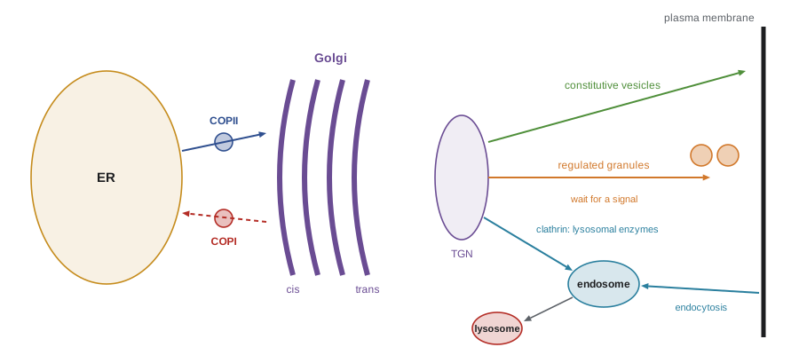
Coat assembly is controlled by small GTPases. COPII assembly begins when the ER membrane protein Sec12 loads Sar1 with GTP; Sar1-GTP inserts an amphipathic helix into the membrane, helping to bend it, and recruits the inner coat proteins Sec23 and Sec24, which bind cargo, and then the outer cage of Sec13 and Sec31. When Sar1 hydrolyses its GTP, the coat falls off, so the vesicle can fuse with its target. Arf1 plays the equivalent role for COPI and for clathrin at the Golgi. Clathrin itself is a three-legged protein, a triskelion, that assembles into a polyhedral lattice of hexagons and pentagons around the bud; adaptor proteins link it to cargo. Budding vesicles must then be pinched off; for clathrin-coated vesicles this is done by dynamin, a GTPase that assembles as a collar around the neck of the bud and constricts it.
Some cargoes are too large for standard COPII vesicles, which are 60 to 90 nm in diameter. Procollagen, the precursor of collagen, forms rigid rods about 300 nm long. Cells that secrete collagen, such as fibroblasts and chondrocytes, use the protein TANGO1 to build enlarged carriers or tunnels at ER exit sites. Mutations affecting this machinery cause skeletal disorders.
Worked problem
How much membrane do vesicles carry?
A COPII vesicle has a diameter of 70 nm. How much membrane area does it carry, and how many such vesicles would deliver a membrane area equal to the plasma membrane of a cell with 1,200 µm² of surface?
Show solution
- Area of a sphere = πd² = 3.14 × (0.070 µm)² ≈ 0.0154 µm².
- Number of vesicles = 1,200 / 0.0154 ≈ 78,000.
- Secretory cells cycle membrane rapidly; most of this membrane is retrieved again by retrograde and endocytic traffic.
Answer: About 0.015 µm² per vesicle, so roughly 78,000 vesicles would carry as much membrane as the whole plasma membrane.
Finding the right target: Rabs and tethers
A cell contains many membrane compartments, and a vesicle must fuse only with the correct one. Identity is conferred largely by Rab proteins, a family of more than 60 small GTPases in humans. Each compartment carries particular Rabs in their active, GTP-bound form: Rab1 on ER-to-Golgi carriers, Rab5 on early endosomes, Rab7 on late endosomes, Rab11 on recycling endosomes, Rab27 on secretory granules. Active Rabs recruit effector proteins, including motors that move vesicles along the cytoskeleton and tethering factors, long coiled-coil proteins or multi-subunit complexes that capture vesicles from a distance before fusion.
Like other GTPases, Rabs cycle between states. A guanine nucleotide exchange factor (GEF) on the target membrane loads a Rab with GTP and activates it; a GTPase-activating protein (GAP) stimulates GTP hydrolysis and inactivates it; and a GDP dissociation inhibitor (GDI) extracts inactive Rab-GDP from the membrane and keeps it soluble in the cytosol until it is delivered to a membrane again. Because the GEF for one Rab can be an effector of the previous Rab, cascades of Rab conversion can change a compartment’s identity over time, as when early endosomes carrying Rab5 mature into late endosomes carrying Rab7 (lesson 5).
import random
random.seed(214)

# A simple model of the Rab cycle on one compartment.
# States: "cytosol-GDP" (bound to GDI), "membrane-GDP", "membrane-GTP" (active).
# Per-step transition probabilities (illustrative):
P = {
    "cytosol-GDP":  {"membrane-GDP": 0.20},   # delivery to membrane (GDI displacement)
    "membrane-GDP": {"membrane-GTP": 0.30,    # GEF activation
                     "cytosol-GDP": 0.10},    # GDI extraction
    "membrane-GTP": {"membrane-GDP": 0.05},   # GAP-stimulated hydrolysis
}
def simulate(gap_rate, n=2000, steps=200):
    P["membrane-GTP"]["membrane-GDP"] = gap_rate
    counts = {s: 0 for s in P}
    for _ in range(n):
        s = "cytosol-GDP"
        for _ in range(steps):
            r, acc = random.random(), 0.0
            for nxt, p in P[s].items():
                acc += p
                if r < acc:
                    s = nxt; break
        counts[s] += 1
    return {k: v / n for k, v in counts.items()}

for gap in (0.05, 0.50):
    f = simulate(gap)
    print(f"GAP rate {gap:.2f}: active {f['membrane-GTP']:.2f}, "
f"inactive on membrane {f['membrane-GDP']:.2f}, in cytosol {f['cytosol-GDP']:.2f}")GAP rate 0.05: active 0.80, inactive on membrane 0.14, in cytosol 0.06 GAP rate 0.50: active 0.30, inactive on membrane 0.45, in cytosol 0.25
The model shows a general principle of GTPase switches: the fraction in the active state is set by the balance between activation (GEF) and inactivation (GAP). Localizing a GEF to one membrane and a GAP elsewhere, as cells do, creates domains where a Rab is active. The same logic applies to Ran in nuclear transport (lesson 2), Sar1 and Arf in coat assembly, and Ras and heterotrimeric G proteins in signalling (lesson 11).
Fusion: SNAREs
The final step, membrane fusion, is driven by SNARE proteins. A vesicle carries a v-SNARE (for vesicle; in neurons, synaptobrevin, also called VAMP), and the target membrane carries t-SNAREs (for target; in neurons, syntaxin and SNAP-25). Their helical domains zipper together into an extremely stable four-helix bundle, the trans-SNARE complex, which pulls the two membranes so close that they merge. Different compartments carry different SNAREs, adding a further layer of specificity. After fusion, the SNAREs sit together in the same membrane in a cis-complex, and an ATPase called NSF, with its adaptor α-SNAP, uses ATP to pry them apart so they can be reused; v-SNAREs are returned to their donor compartment by retrograde transport.
At synapses, fusion must happen within a millisecond of an electrical signal. Synaptic vesicles are docked and primed with partly zippered SNAREs, held in check by the proteins complexin and synaptotagmin. When an action potential opens calcium channels, calcium binds synaptotagmin, which triggers full zippering and fusion. Südhof’s work identified synaptotagmin as the calcium sensor. BIOL-420 discusses synaptic transmission in detail.
SNAREs are the targets of two of the most potent toxins known. Tetanus toxin and the seven types of botulinum toxin are proteases that enter neurons and cut specific SNAREs, blocking neurotransmitter release. Tetanus toxin acts in inhibitory neurons of the spinal cord, causing spastic paralysis; botulinum toxin acts at neuromuscular junctions, causing flaccid paralysis. Tiny doses of botulinum toxin type A are used medically to relax overactive muscles, treat chronic migraine and excessive sweating, and for cosmetic purposes.
The Golgi apparatus
The Golgi apparatus, discovered in nerve cells by Camillo Golgi in 1898 using a silver stain, consists in animal cells of stacks of four to eight flattened cisternae, often linked into a ribbon near the nucleus. Each stack has a direction. Cargo enters at the cis face, closest to the ER, through the ER–Golgi intermediate compartment, passes through the medial cisternae and leaves from the trans face and the trans-Golgi network (TGN), a tubular sorting station. Plant cells instead have hundreds of separate stacks, called dictyosomes, scattered through the cytoplasm.
How cargo moves through the stack has been debated for decades. In the vesicular transport model, cisternae are stable and cargo hops forward in COPI vesicles. In the cisternal maturation model, now widely supported, each cisterna itself moves forward, maturing from cis to trans as Golgi enzymes are carried backward in COPI vesicles to younger cisternae. Live imaging in yeast, where individual cisternae can be seen changing their resident proteins over time, gave strong evidence for maturation, and it explains how large cargoes such as procollagen aggregates and algal scales move through the stack without ever fitting into vesicles.
| Golgi region | Typical modifications | Example enzymes or events |
|---|---|---|
| cis-Golgi network | Phosphorylation of mannose on lysosomal enzymes; retrieval of ER proteins | GlcNAc-phosphotransferase; KDEL receptor |
| cis and medial cisternae | Trimming of mannose; addition of GlcNAc | Mannosidases, GlcNAc transferases |
| trans cisternae | Addition of galactose and sialic acid; O-linked glycosylation on Ser and Thr | Galactosyl- and sialyltransferases |
| trans-Golgi network | Sulfation; proteolytic processing begins; sorting into carriers | Sulfotransferases, furin, sorting receptors |
As N-linked glycans pass through the Golgi, they are trimmed and rebuilt. Some remain high-mannose glycans; most become complex glycans carrying N-acetylglucosamine, galactose, fucose and sialic acid, in arrangements that differ between cell types and species. The ABO blood group antigens are built in the Golgi: the A and B alleles encode glycosyltransferases that add N-acetylgalactosamine or galactose to a common precursor, the H antigen, while the O allele encodes an inactive enzyme. Glycosylation patterns also matter for therapeutic proteins; antibodies made in hamster cells for medical use are engineered and checked to have human-compatible glycans.
Sorting at the trans-Golgi network
The TGN directs cargo to three main destinations. Lysosomal hydrolases are tagged in the cis-Golgi with mannose 6-phosphate (M6P), recognized in the TGN by M6P receptors and carried in clathrin/AP-1 vesicles to endosomes, where the lower pH releases the enzymes and the receptors recycle. In I-cell disease (mucolipidosis II), the enzyme that adds the phosphate tag is missing; lysosomal enzymes are secreted instead of delivered, and undigested material accumulates in lysosomes as inclusions, causing severe developmental problems. Proteins for constitutive secretion and the plasma membrane leave in carriers that move continuously to the cell surface. Proteins for regulated secretion, such as hormones, digestive enzymes and neurotransmitters, aggregate in the acidic, calcium-rich environment of the TGN, are packed into dense-core secretory granules and stored until a signal, often a rise in cytosolic calcium, triggers fusion.
Worked problem
Concentrating cargo into secretory granules
In pancreatic acinar cells, digestive enzymes are concentrated about 200-fold between the ER lumen and mature zymogen granules. If the enzymes are at 0.5 mg/mL in the ER, what is their concentration in granules, and why is storage in an inactive form important?
Show solution
- Granule concentration ≈ 0.5 × 200 = 100 mg/mL.
- At such high concentrations, any active protease could digest the cell.
- The enzymes are stored as inactive precursors (zymogens), such as trypsinogen, and are activated only in the intestine.
- Premature activation inside the pancreas causes pancreatitis.
Answer: About 100 mg/mL; storing enzymes as zymogens prevents self-digestion.
In polarized epithelial cells, such as those lining the intestine and kidney tubules, the TGN and recycling endosomes also sort membrane proteins to the apical surface, facing the lumen, or the basolateral surface, facing the tissue. Tight junctions (lesson 10) stop proteins from mixing between the two domains once delivered.
Retrieval keeps compartments distinct
With so much membrane flowing forward, how do the ER and early Golgi keep their own proteins? Partly by retention, and partly by retrieval. Soluble ER residents such as BiP and PDI end with the tetrapeptide KDEL (HDEL in yeast). If they escape to the Golgi, KDEL receptors bind them, more strongly at the slightly acidic pH of the Golgi, and COPI vesicles carry them back. ER membrane proteins often carry C-terminal KKXX motifs that bind COPI directly. Golgi enzymes are kept in their cisternae partly by the length of their transmembrane segments, which match the thickness of the membranes in different regions, and partly by continuous COPI recycling.
Common mistakes
MistakeVesicles fuse with whichever membrane they bump into.
InsteadRabs, tethers and matching SNAREs ensure fusion only with the correct target.
MistakeCOPI vesicles carry cargo from the ER to the Golgi.
InsteadCOPII vesicles leave the ER; COPI vesicles carry material backward to the ER and between cisternae.
MistakeNSF drives membrane fusion by ATP hydrolysis.
InsteadSNARE zippering drives fusion; NSF uses ATP afterward to separate cis-SNARE complexes for reuse.
MistakeLysosomal enzymes reach lysosomes by default.
InsteadThey need the mannose 6-phosphate tag; without it, as in I-cell disease, they are secreted.
MistakeThe Golgi always moves cargo in small vesicles between fixed cisternae.
InsteadEvidence strongly supports cisternal maturation, in which cisternae themselves progress from cis to trans.
Summary and key terms
- Pulse–chase experiments showed secretory proteins moving from rough ER to Golgi to granules to the outside.
- COPII, COPI and clathrin coats bend membranes and select cargo; small GTPases Sar1 and Arf1 control coat assembly.
- Dynamin pinches off clathrin-coated vesicles; procollagen needs enlarged carriers built with TANGO1.
- Rab GTPases, controlled by GEFs, GAPs and GDI, give compartments identity and recruit motors and tethers.
- SNARE zippering fuses membranes; NSF recycles SNAREs; tetanus and botulinum toxins cut SNAREs.
- The Golgi modifies glycans and sorts cargo; cisternal maturation explains movement through the stack.
- The TGN sorts lysosomal (M6P), constitutive and regulated cargo; KDEL receptors retrieve escaped ER proteins.
- pulse–chase
- Labelling molecules briefly, then following the labelled cohort over time.
- COPII
- The coat of vesicles that carry cargo from the ER toward the Golgi.
- COPI
- The coat of vesicles that carry material from the Golgi back to the ER and between cisternae.
- clathrin
- A three-legged protein that forms the lattice coat of vesicles at the TGN and plasma membrane.
- dynamin
- A GTPase that constricts and pinches off the necks of budding vesicles.
- Rab GTPase
- A small GTPase that marks compartment identity and recruits effectors.
- SNARE
- Membrane proteins whose zippering into a four-helix bundle drives membrane fusion.
- cisternal maturation
- The model in which Golgi cisternae mature from cis to trans as enzymes recycle backward.
- trans-Golgi network
- The tubular network at the trans face of the Golgi where cargo is sorted.
- mannose 6-phosphate
- The sugar tag that directs soluble hydrolases to lysosomes.
Practice problems
Work each problem on paper first, then reveal the answer to check.
In Palade’s experiments, why was a short pulse followed by a chase?
Show answer
So that only proteins made during the pulse were labelled and could be followed as a cohort.
Which GTPase controls COPII coat assembly?
Show answer
Sar1, activated by Sec12 on the ER membrane.
What is the role of Sec24?
Show answer
It binds export signals on cargo or cargo receptors, selecting them into COPII vesicles.
A vesicle is 50 nm across. What membrane area does it carry?
Show answer
π × 0.05² ≈ 0.0079 µm².
How does a Rab GDI help the Rab cycle?
Show answer
It extracts inactive Rab-GDP from membranes and keeps it soluble until delivery.
Which SNAREs form the neuronal fusion complex?
Show answer
Synaptobrevin (VAMP) on the vesicle, syntaxin and SNAP-25 on the plasma membrane.
Why does botulinum toxin cause paralysis?
Show answer
It cleaves SNAREs at neuromuscular junctions, blocking acetylcholine release.
What evidence supports cisternal maturation?
Show answer
Live imaging of yeast cisternae changing resident proteins over time, and movement of cargoes too large for vesicles.
What happens to lysosomal enzymes in I-cell disease?
Show answer
They lack M6P tags and are secreted instead of delivered to lysosomes.
What signal returns BiP to the ER if it escapes?
Show answer
Its C-terminal KDEL sequence, bound by the KDEL receptor.
Lesson quiz · 5 questions
Score 60% or more to complete this lesson.
Create a free account to save your progress, take quizzes and earn a certificate.