
BIOL-214 · Lesson 12 of 12 · 33 min
The cell cycle, cell death and stem cells
Every cell in your body arose from a single fertilized egg through about 40 to 45 rounds of division, and in an adult, millions of cells divide every second to replace those lost from the skin, gut lining and blood. Just as important is the opposite process: cells die on purpose, removing the webbing between developing fingers, eliminating self-reactive immune cells and getting rid of damaged cells before they turn cancerous. Stem cells balance these losses by dividing to make both new stem cells and specialized cells. BIOL-107 described the phases of the cell cycle, the stages of mitosis and meiosis and how populations grow. This final lesson looks inside the control system: the cyclin-dependent kinases that drive the cycle, the checkpoints that halt it, the machinery of the mitotic spindle, the molecular pathways of programmed cell death and the biology and regulation of stem cells. It ends with an integrated investigation that draws on the whole course.
The cell-cycle control system
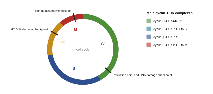
The cell cycle is driven by protein kinases called cyclin-dependent kinases (CDKs). A CDK is inactive on its own; it becomes active only when bound to a cyclin, a protein whose level rises and falls during the cycle. Different cyclin–CDK complexes trigger different events: G1/S cyclins commit the cell to a new cycle, S cyclins start DNA replication, and M cyclins drive entry into mitosis. Much of this was discovered in organisms as different as yeast, frogs and sea urchins. Lee Hartwell identified cell-division-cycle genes in budding yeast, Paul Nurse found the central CDK in fission yeast and Tim Hunt discovered cyclins as proteins that were destroyed at each division of sea urchin eggs; they shared the 2001 Nobel Prize in Physiology or Medicine.
| Complex (vertebrates) | Active in | Main role |
|---|---|---|
| Cyclin D–CDK4/6 | G1, in response to growth factors | Begins phosphorylation of Rb, linking mitogenic signals to the cycle |
| Cyclin E–CDK2 | Late G1 to early S | Completes Rb inactivation; passes the restriction point; starts replication |
| Cyclin A–CDK2 | S phase | Fires replication origins and prevents re-replication |
| Cyclin B–CDK1 | G2 to M | Drives nuclear envelope breakdown, chromosome condensation and spindle assembly |
Several layers of control make CDK activity switch on and off sharply. Inhibitory phosphorylation by the kinase Wee1 holds cyclin B–CDK1 inactive during G2 until the phosphatase Cdc25 removes the phosphate. Active CDK1 then activates Cdc25 and inhibits Wee1, a double positive feedback loop that turns on CDK1 abruptly and irreversibly. CDK inhibitor proteins, such as p21 and p27, bind and block cyclin–CDK complexes. Finally, cyclins are destroyed by ubiquitin-mediated proteolysis (lesson 5). Two ubiquitin ligases dominate: SCF targets G1/S proteins, and the anaphase-promoting complex or cyclosome (APC/C) targets M cyclins and securin, allowing the cell to exit mitosis.
A model of the cell-cycle oscillator
Early frog embryos divide every 30 minutes or so without growth phases or checkpoints, driven by a simple oscillator: cyclin B is made steadily, CDK1 becomes active once enough cyclin has built up, and active CDK1 triggers the APC/C, which destroys cyclin and resets the cycle. Because APC/C activation lags behind CDK1 activation, the system can overshoot and oscillate rather than settling at a steady state. A small set of equations, in which CDK1 activates APC/C through the intermediate kinase Plk1, captures this negative feedback with a delay.
import numpy as np
from scipy.integrate import solve_ivp

# A three-component negative feedback loop: CDK1 -> Plk1 -> APC/C -| CDK1
# (after Ferrell, Tsai and Yang, 2011; dimensionless, illustrative parameters)
def hill(x, K, n): return x**n / (K**n + x**n)

def make_rhs(n):
    def rhs(t, y):
        cdk, plk, apc = y
        d_cdk = 0.1 - 3.0*cdk*hill(apc, 0.5, n)          # cyclin made steadily, destroyed by APC/C
        d_plk = 3.0*(1 - plk)*hill(cdk, 0.5, n) - plk     # CDK1 activates Plk1
        d_apc = 3.0*(1 - apc)*hill(plk, 0.5, n) - apc     # Plk1 activates APC/C
        return [d_cdk, d_plk, d_apc]
    return rhs

for n in (8, 2):
    sol = solve_ivp(make_rhs(n), (0, 200), [0, 0, 0], max_step=0.05)
    t, cdk = sol.t, sol.y[0]
    late = t > 100
    lo, hi = cdk[late].min(), cdk[late].max()
    if hi - lo > 0.05:
        idx = [i for i in range(1, len(t)-1) if late[i] and cdk[i] > cdk[i-1] and cdk[i] >= cdk[i+1]]
        print(f"Hill n = {n}: oscillates, CDK1 between {lo:.2f} and {hi:.2f}, "
              f"period {np.diff(t[idx]).mean():.2f} time units")
    else:
print(f"Hill n = {n}: settles to a steady state, CDK1 = {cdk[-1]:.3f}")Hill n = 8: oscillates, CDK1 between 0.19 and 0.52, period 4.38 time units Hill n = 2: settles to a steady state, CDK1 = 0.145
With steep, switch-like responses at each step (a Hill coefficient of 8), CDK1 activity rises and falls in regular cycles, and the period does not depend on where the system starts. With shallow responses (a Hill coefficient of 2), the same loop damps down to a constant level of CDK1 activity and the cycle stops. Negative feedback alone is not enough: the loop also needs ultrasensitivity (lesson 11) and enough delay, which here comes from passing the signal through an intermediate, the kinase Plk1. In real embryos, the positive feedback loops around CDK1 provide additional steepness, and they make the oscillator robust to changes in the rate of cyclin synthesis.
In somatic cells, this core oscillator is embedded in a much larger network with checkpoints that can stop the cycle, and with links to growth signals and cell size. Even so, the logic is the same: positive feedback makes transitions switch-like, and negative feedback with a delay resets the system for the next round.
The restriction point and checkpoints
Mammalian cells decide in G1 whether to divide. Growth factors acting through the Ras–MAPK pathway (lesson 11) raise cyclin D, and cyclin D–CDK4/6 begins to phosphorylate the retinoblastoma protein, Rb. Unphosphorylated Rb binds and inhibits E2F transcription factors; phosphorylated Rb releases them, and E2F switches on genes for cyclin E, cyclin A and DNA replication enzymes. Cyclin E–CDK2 further phosphorylates Rb, another positive feedback loop. Once this loop has fired, the cell no longer needs growth factors to complete the cycle; this commitment is the restriction point. Most cancers disrupt this control, through loss of Rb, overproduction of cyclin D or loss of the CDK inhibitor p16. Palbociclib, ribociclib and abemaciclib, inhibitors of CDK4/6, are now standard treatments for the most common type of advanced breast cancer.
Checkpoints monitor whether earlier events have been completed correctly. The DNA damage checkpoints in G1 and G2 detect broken or unreplicated DNA through the kinases ATM and ATR, which activate Chk1 and Chk2. A key target is p53, a transcription factor normally kept at low levels by the ubiquitin ligase MDM2. After DNA damage, p53 is stabilized and switches on p21, which inhibits CDKs and halts the cycle while repairs are made; if damage is too severe, p53 triggers apoptosis or permanent arrest (senescence). Because of this role, p53 has been called the guardian of the genome, and its gene, TP53, is mutated in about half of human cancers. The spindle assembly checkpoint, described below, ensures that chromosomes are properly attached before anaphase. BIOL-450 discusses how cancers escape these controls.
The mitotic spindle and chromosome segregation
In mitosis, the microtubule cytoskeleton (lesson 9) is rebuilt into a bipolar spindle. The duplicated centrosomes separate, and kinesin-5 motors push antiparallel microtubules apart to form the two poles. Each replicated chromosome consists of two sister chromatids held together along their length by a ring-shaped protein complex, cohesin. At the centromere of each chromatid, a large protein structure called the kinetochore captures the plus ends of spindle microtubules. Correct, bi-oriented attachment, with sister kinetochores attached to opposite poles, puts the kinetochores under tension. Incorrect attachments are under low tension and are destabilized by the kinase Aurora B, giving the cell another chance.
Unattached kinetochores generate a signal that inhibits the APC/C through its activator Cdc20. This is the spindle assembly checkpoint. A single unattached kinetochore is enough to delay anaphase. When the last kinetochore attaches, the APC/C is released, and it ubiquitylates securin and cyclin B. Destruction of securin frees an enzyme called separase, which cuts cohesin, and the sister chromatids move to opposite poles as kinetochore microtubules shorten and the poles move apart. Destruction of cyclin B lowers CDK1 activity, allowing the nuclear envelope to reform and cytokinesis to begin. A contractile ring of actin and myosin II, positioned by signals from the spindle and assembled under the control of Rho (lesson 8), pinches the cell in two. Errors in segregation produce cells with abnormal chromosome numbers, aneuploidy, which is a hallmark of cancer.
Measuring the cell cycle by flow cytometry
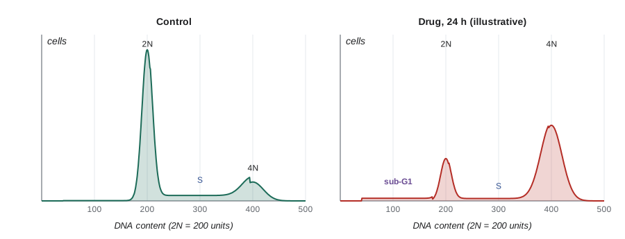
A flow cytometer measures fluorescence from thousands of single cells per second as they pass through a laser beam. If cells are stained with a dye that binds DNA stoichiometrically, such as propidium iodide, the brightness of each cell is proportional to its DNA content. Cells in G1 have two copies of the genome (2N), cells in G2 and M have 4N, and cells in S phase fall in between. Cells with less than 2N DNA, the sub-G1 population, are usually apoptotic cells whose DNA has been cut and partly lost.
import numpy as np
rng = np.random.default_rng(214)

# Simulated propidium iodide fluorescence for 20,000 cells per sample (2N peak at 200 units)
def sample(g1, s, g2m, sub, n=20000, cv=0.05):
    k = rng.multinomial(n, [g1, s, g2m, sub])
    return np.concatenate([
        rng.normal(200, 200*cv, k[0]),                 # G1: 2N
        rng.uniform(210, 390, k[1]),                   # S: between 2N and 4N
        rng.normal(400, 400*cv, k[2]),                 # G2/M: 4N
        rng.uniform(40, 175, k[3])])                   # sub-G1: fragmented DNA

def gate(x):
    bins = {"sub-G1": (0, 170), "G1": (170, 230), "S": (230, 370), "G2/M": (370, 460)}
    return {k: 100*np.mean((x >= lo) & (x < hi)) for k, (lo, hi) in bins.items()}

control = sample(0.58, 0.25, 0.15, 0.02)
drug = sample(0.15, 0.10, 0.60, 0.15)                 # invented: a microtubule-targeting drug, 24 h
for name, x in [("control", control), ("drug", drug)]:
    g = gate(x)
print(f"{name:8s}" + " ".join(f"{k} {v:5.1f}%" for k, v in g.items()))control sub-G1 2.0% G1 60.9% S 20.7% G2/M 16.5% drug sub-G1 14.5% G1 16.8% S 12.1% G2/M 56.5%
Simple rectangular gates like these slightly misassign cells at the edges of each peak, so commercial software fits curves to the G1 and G2/M peaks and models the S phase between them. Even so, the result is clear: the drug has caused cells to pile up with 4N DNA, as expected when the spindle checkpoint holds them in mitosis, and the sub-G1 fraction has risen, suggesting that some of the arrested cells are dying. Additional stains distinguish G2 from M: an antibody against histone H3 phosphorylated on serine 10 labels only mitotic cells, and pulse labelling with the nucleotide analogue EdU identifies cells actively replicating DNA.
Programmed cell death
Cells can die in two broad ways. Necrosis is accidental death from injury: cells swell and burst, spilling their contents and provoking inflammation. Apoptosis, by contrast, is an orderly, programmed process: the cell shrinks, its chromatin condenses, its DNA is cut into fragments and its membrane forms blebs that pinch off as apoptotic bodies. Phosphatidylserine, normally confined to the inner leaflet of the plasma membrane, flips to the outer leaflet and serves as an eat-me signal, so phagocytes engulf the dying cell before its contents can leak. Annexin V, a protein that binds phosphatidylserine, is used with a membrane-impermeant DNA dye to distinguish early apoptotic cells (annexin positive, dye negative) from late apoptotic or necrotic cells (both positive). The genetic basis of apoptosis was worked out in the nematode Caenorhabditis elegans, in which exactly 131 of the 1,090 somatic cells formed in development die; Sydney Brenner, Robert Horvitz and John Sulston shared the 2002 Nobel Prize for this work.
Apoptosis is carried out by caspases, proteases with a cysteine in their active site that cut after aspartate residues. They are made as inactive precursors. Initiator caspases, such as caspase-8 and caspase-9, are activated when brought together on adaptor platforms; they then cleave and activate executioner caspases, such as caspase-3 and caspase-7, which cut hundreds of cellular proteins, including nuclear lamins (lesson 9), and activate the nuclease that fragments DNA. Two pathways lead to caspase activation.
| Feature | Extrinsic pathway | Intrinsic (mitochondrial) pathway |
|---|---|---|
| Trigger | Death ligands such as Fas ligand or TNF bind death receptors | DNA damage, growth factor withdrawal, ER stress, loss of attachment |
| Platform | DISC (death receptor, FADD adaptor, procaspase-8) | Apoptosome (cytochrome c, Apaf1, procaspase-9) |
| Initiator caspase | Caspase-8 | Caspase-9 |
| Key regulators | FLIP (inhibits caspase-8 activation) | Bcl-2 family proteins: Bax and Bak (pro-death), Bcl-2 and Bcl-xL (pro-survival), BH3-only proteins (sensors) |
| Example | Cytotoxic T cells killing infected cells | p53-induced death after DNA damage |
In the intrinsic pathway, stress signals activate BH3-only proteins such as Puma, Noxa and Bim, which neutralize anti-apoptotic Bcl-2 proteins and activate Bax and Bak. Bax and Bak oligomerize in the outer mitochondrial membrane and make it permeable, so cytochrome c escapes from the intermembrane space (lesson 6) into the cytosol. There it binds Apaf1, which assembles into a wheel-shaped apoptosome that activates caspase-9. The point of no return is therefore a decision at the mitochondrial surface, set by the balance between pro-death and pro-survival Bcl-2 family proteins. Bcl-2 was first identified because a chromosomal translocation that overproduces it causes follicular lymphoma, by keeping cells alive that should die. Venetoclax, a drug that mimics BH3-only proteins and blocks Bcl-2, was approved in 2016 and is used for chronic lymphocytic leukaemia and acute myeloid leukaemia.
Other forms of regulated death have been described more recently. Necroptosis is a programmed form of necrosis triggered when caspase-8 is blocked, important in defence against viruses. Pyroptosis is an inflammatory death of immune cells, triggered by inflammasomes. Ferroptosis is an iron-dependent death caused by lipid peroxidation. Each is an active area of research for cancer, neurodegeneration and infection.
Stem cells
Stem cells are defined by two properties: they can renew themselves through division, and they can give rise to specialized cell types. Cells of the early embryo up to about the eight-cell stage are totipotent, able to form the whole organism and its placenta. Embryonic stem (ES) cells, derived from the inner cell mass of a blastocyst, are pluripotent: they can form any cell type of the body but not the placenta. Adult or tissue stem cells are usually multipotent, producing the cell types of one tissue. Hematopoietic stem cells in the bone marrow, for example, regenerate all blood cells and are the basis of bone marrow transplants, the oldest and most widely used stem cell therapy.
The lining of the small intestine is a striking example of a stem cell system. It is replaced every four to five days by stem cells at the base of crypts, identified by Hans Clevers and colleagues through their expression of the Wnt target gene Lgr5. Wnt signals from neighbouring Paneth cells keep the stem cells dividing (lesson 11); as their daughters move up the crypt, away from the Wnt source, they stop dividing and differentiate. Single Lgr5 stem cells can be grown in a three-dimensional matrix into organoids, miniature gut-like structures that are now widely used to study disease and test drugs.
In 2006, Shinya Yamanaka and Kazutoshi Takahashi showed that adult mouse cells could be reprogrammed to a pluripotent state by introducing four transcription factors, Oct4, Sox2, Klf4 and c-Myc. These induced pluripotent stem (iPS) cells, made from human cells the following year, behave much like ES cells but can be made from a patient’s own skin or blood cells. Yamanaka shared the 2012 Nobel Prize with John Gurdon, who had shown in 1962 that the nucleus of a frog intestinal cell could direct the development of a whole tadpole. iPS cells are used to model diseases in a dish and are in early clinical trials for conditions such as Parkinson disease and macular degeneration. Making and controlling stem cell therapies safely is challenging, because pluripotent cells that escape differentiation can form tumours, and BIOL-360 discusses how stem cells contribute to development and regeneration.
Regulation of stem cell research
In Canada, research with human embryos and embryonic stem cells is governed by federal law and by research funding policy. The Assisted Human Reproduction Act (2004) prohibits creating a human clone, creating embryos for research except to improve assisted reproduction procedures, and buying or selling embryos, and it requires informed consent for the use of embryos donated after fertility treatment. Researchers funded by the federal granting agencies (CIHR, NSERC and SSHRC) must also follow the Tri-Council Policy Statement, and research that derives or uses human pluripotent stem cell lines from embryos must be reviewed by the CIHR Stem Cell Oversight Committee as well as a local research ethics board. Research with iPS cells and adult stem cells raises fewer ethical issues but still needs ethics review when it involves human donors. For comparison, in the United States, federal funding policy is set by the National Institutes of Health, and in January 2026 NIH paused the addition of new lines to its registry of approved human embryonic stem cell lines while it sought public input on reducing reliance on them.
Capstone: an integrated cell biology investigation
To finish the course, consider how the methods and ideas of all twelve lessons come together in a single invented study. A research group has found a compound, which they call Z-17, in a screen for molecules that kill cancer cells. They want to know how it works.
| Experiment | Observation (invented data) | Lessons drawn on |
|---|---|---|
| Flow cytometry of DNA content after 24 h | Cells accumulate with 4N DNA; sub-G1 rises from 2% to 15% | 12 |
| Phospho-histone H3 staining | Most 4N cells are positive, so they are in mitosis, not G2 | 12 |
| Immunofluorescence of tubulin and DNA | Spindles are monopolar, with a single aster surrounded by chromosomes | 1, 9 |
| Live imaging of EB1 comets | Microtubule growth rate and catastrophe frequency are normal | 9 |
| Purified motor assay | Z-17 stops kinesin-5 from sliding microtubules apart but does not affect kinesin-1 | 9 |
| Annexin V staining and caspase activity at 48 h | Annexin V positive cells and caspase-3 activity increase; a pan-caspase inhibitor reduces death | 12 |
| Cells overexpressing Bcl-2 | Death after Z-17 is reduced by about three-quarters; arrest is unchanged | 6, 12 |
import numpy as np
rng = np.random.default_rng(2140)

# Invented counts from imaging: cells scored per condition (three experiments pooled)
data = {
    "DMSO":            dict(total=900, mitotic=38,  monopolar=2,   annexin=21),
    "Z-17":            dict(total=900, mitotic=412, monopolar=371, annexin=148),
    "Z-17 + Bcl-2":    dict(total=900, mitotic=405, monopolar=362, annexin=52),
}
for name, d in data.items():
    mi = d["mitotic"]/d["total"]
    mono = d["monopolar"]/max(d["mitotic"], 1)
    ann = d["annexin"]/d["total"]
    se = np.sqrt(ann*(1-ann)/d["total"])
    print(f"{name:13s} mitotic index {100*mi:5.1f}%, monopolar spindles {100*mono:5.1f}% of mitoses, "
          f"annexin V+ {100*ann:4.1f}% (95% CI {100*(ann-1.96*se):4.1f}-{100*(ann+1.96*se):4.1f})")

# How much of the Z-17-induced death does Bcl-2 prevent?
base = data["DMSO"]["annexin"]
rescue = 1 - (data["Z-17 + Bcl-2"]["annexin"] - base) / (data["Z-17"]["annexin"] - base)
print(f"Bcl-2 prevents {100*rescue:.0f}% of the extra death caused by Z-17")DMSO mitotic index 4.2%, monopolar spindles 5.3% of mitoses, annexin V+ 2.3% (95% CI 1.3- 3.3) Z-17 mitotic index 45.8%, monopolar spindles 90.0% of mitoses, annexin V+ 16.4% (95% CI 14.0-18.9) Z-17 + Bcl-2 mitotic index 45.0%, monopolar spindles 89.4% of mitoses, annexin V+ 5.8% (95% CI 4.3- 7.3) Bcl-2 prevents 76% of the extra death caused by Z-17
Putting the evidence together tells a coherent story. Z-17 inhibits kinesin-5, the motor that pushes spindle poles apart, so cells build monopolar spindles. Unattached or wrongly attached kinetochores keep the spindle assembly checkpoint active, and cells arrest in mitosis with 4N DNA. During the prolonged arrest, the balance of Bcl-2 family proteins shifts toward death, mitochondria release cytochrome c and caspases execute apoptosis, which is why Bcl-2 overexpression protects the cells without changing the arrest. Because Z-17 does not alter microtubule dynamics or kinesin-1, it might avoid the nerve damage caused by taxanes and vinca alkaloids, which disrupt axonal transport. Real kinesin-5 inhibitors, such as monastrol and ispinesib, were discovered and studied in just this way; their clinical results in solid tumours were disappointing, a reminder that a clear mechanism in cultured cells is only the first step toward a medicine.
This kind of reasoning, from microscopy and quantitative measurement (lesson 1) through organelles and transport (lessons 2 to 7), the cytoskeleton (lessons 8 and 9), adhesion (lesson 10), signalling (lesson 11) and the control of division and death (lesson 12), is the core of modern cell biology. The upper-year courses in cancer biology (BIOL-450), neurobiology (BIOL-420), immunology (BIOL-380), developmental biology (BIOL-360) and advanced biochemistry (BIOC-420) build directly on it.
Common mistakes
MistakeCyclins are kinases that drive the cell cycle.
InsteadCyclins are regulatory subunits; the kinases are the CDKs that cyclins activate.
MistakeThe restriction point is passed when mitosis begins.
InsteadIt is passed in late G1, when Rb phosphorylation and E2F feedback make the cell independent of growth factors.
MistakeSeparase cuts the spindle to start anaphase.
InsteadSeparase cuts cohesin, which holds sister chromatids together; it is released when APC/C destroys securin.
MistakeApoptosis and necrosis are the same process.
InsteadApoptosis is orderly and non-inflammatory; necrosis is uncontrolled swelling and bursting with inflammation.
MistakeBcl-2 is a caspase.
InsteadBcl-2 is a pro-survival protein that prevents mitochondrial outer membrane permeabilization.
MistakeiPS cells are the same as embryonic stem cells taken from an embryo.
InsteadiPS cells are made by reprogramming adult cells; they resemble ES cells but are not derived from embryos.
Summary and key terms
- Cyclin–CDK complexes drive the cycle; their activity is controlled by cyclin synthesis and destruction, phosphorylation and CDK inhibitors.
- Positive feedback makes transitions switch-like, and delayed negative feedback through APC/C resets the cycle.
- Growth factors raise cyclin D; Rb phosphorylation releases E2F and commits the cell at the restriction point.
- DNA damage checkpoints act through ATM/ATR and p53; the spindle assembly checkpoint delays anaphase until all kinetochores attach.
- Flow cytometry of DNA content measures cell-cycle distribution and apoptotic sub-G1 cells.
- Apoptosis is executed by caspases through extrinsic death receptor or intrinsic mitochondrial pathways controlled by the Bcl-2 family.
- Stem cells self-renew and differentiate; ES and iPS cells are pluripotent; research is regulated differently in Canada and the United States.
- cyclin-dependent kinase
- A kinase that is active only when bound to a cyclin and drives cell-cycle transitions.
- restriction point
- The point in late G1 after which a cell is committed to divide without growth factors.
- Rb
- The retinoblastoma protein, which inhibits E2F until phosphorylated by CDKs.
- p53
- A transcription factor that halts the cycle or triggers apoptosis after DNA damage.
- APC/C
- A ubiquitin ligase that targets securin and M cyclins for destruction.
- spindle assembly checkpoint
- A surveillance system that delays anaphase until all kinetochores are attached.
- caspase
- A cysteine protease that cleaves after aspartate and executes apoptosis.
- apoptosome
- A complex of cytochrome c, Apaf1 and caspase-9 that starts the intrinsic pathway.
- pluripotent
- Able to give rise to all cell types of the body.
- induced pluripotent stem cell
- A pluripotent cell made by reprogramming a differentiated cell.
Practice problems
Work each problem on paper first, then reveal the answer to check.
What is the function of Wee1 and Cdc25 in G2?
Show answer
Wee1 adds an inhibitory phosphate to CDK1; Cdc25 removes it to activate CDK1.
Why does a delay in APC/C activation matter for the embryonic oscillator?
Show answer
Delayed negative feedback lets CDK1 overshoot and oscillate rather than settle.
How do CDK4/6 inhibitors stop breast cancer cells dividing?
Show answer
They prevent Rb phosphorylation, so E2F remains inhibited and cells stay in G1.
What does p21 do after DNA damage?
Show answer
It is induced by p53 and inhibits cyclin–CDK complexes, halting the cycle.
What triggers the destruction of securin?
Show answer
Activation of APC/C–Cdc20 once all kinetochores are attached.
In a DNA histogram, cells with 4N DNA could be in which phases?
Show answer
G2 or M.
What does annexin V bind, and why is that useful?
Show answer
Phosphatidylserine exposed on the outer leaflet of apoptotic cells.
Which protein released from mitochondria triggers apoptosome assembly?
Show answer
Cytochrome c.
Name the four Yamanaka factors.
Show answer
Oct4, Sox2, Klf4 and c-Myc.
In the Z-17 study, why does Bcl-2 reduce death but not mitotic arrest?
Show answer
Bcl-2 acts downstream at the mitochondria, after the arrest, by blocking the intrinsic apoptosis pathway.
Lesson quiz · 5 questions
Score 60% or more to complete this lesson.
Create a free account to save your progress, take quizzes and earn a certificate.