
BIOL-360 · Lesson 9 of 12 · 29 min
Organogenesis: somites, limbs and organs
Once the germ layers and body axes are in place, the embryo builds its organs. Organogenesis uses the principles of earlier lessons, induction, morphogen gradients, lateral inhibition and changes in cell shape and adhesion, in new combinations. This lesson examines three systems in detail: the somites, which segment the vertebrate body; the limb, perhaps the best-studied example of pattern formation in three dimensions; and the heart, kidney and eye as examples of organs built by reciprocal interactions between tissues. Each illustrates a different strategy, and each has clinical relevance.
Mesoderm subdivided
After gastrulation, the vertebrate mesoderm on each side of the notochord is divided into strips from medial to lateral. The paraxial mesoderm, next to the neural tube, forms the somites. Intermediate mesoderm forms the kidneys and gonads. Lateral plate mesoderm splits into a layer that, with ectoderm, forms the body wall and limbs, and a layer that, with endoderm, forms the gut wall, heart and blood vessels. This medial-to-lateral pattern is set by BMP levels, which are high laterally and low medially, opposed by Noggin from the notochord and dorsal neural tube.
Somites and the segmentation clock
Somites are blocks of paraxial mesoderm that bud off in pairs from the anterior end of the presomitic mesoderm, at regular intervals, from head to tail. A chick forms a new pair every 90 minutes, a mouse every 2 hours, a zebrafish every 30 minutes and a human every 4 to 5 hours, ending with about 50 pairs in chicks, 65 in mice and 38 to 44 in humans. Somites give rise to the vertebrae and ribs (from the sclerotome), the skeletal muscles of the trunk and limbs (from the myotome), and the dermis of the back (from the dermatome). Their segmentation produces the segmental organization of vertebrae, muscles and spinal nerves.
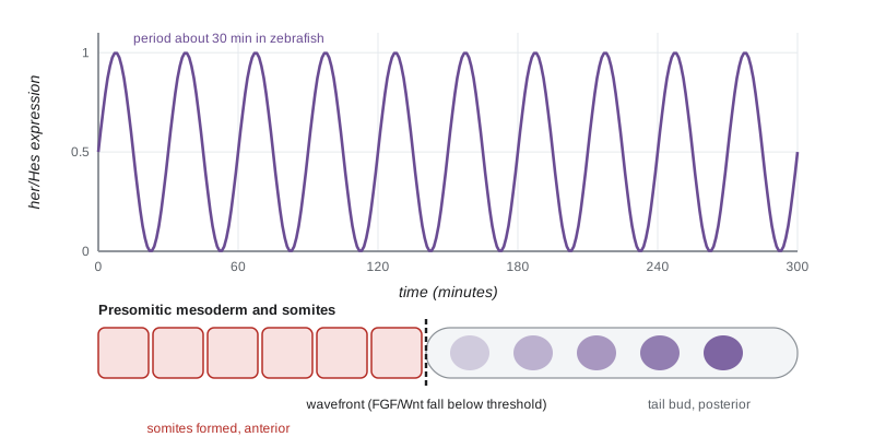
How is such regular timing achieved? In 1997 Olivier Pourquié's group discovered that the chick gene c-hairy1 is expressed in the presomitic mesoderm in waves that sweep from posterior to anterior once every 90 minutes, exactly the time taken to form one somite. This was the first molecular evidence for a segmentation clock, an idea proposed in the clock and wavefront model by Jonathan Cooke and Christopher Zeeman in 1976. The clock genes belong to the Hes and her families of transcriptional repressors, together with components of the Notch, Wnt and FGF pathways. The oscillation arises from delayed negative feedback: the Hes protein represses its own gene, but because transcription, splicing, export and translation take time, the protein accumulates and decays in cycles. Notch signalling synchronizes the oscillators of neighbouring cells.
# Delayed negative autoregulation (after Lewis 2003): Hes/her protein p represses its own mRNA m
# with a delay tau for transcription, splicing and export, giving sustained oscillations.
# dm/dt = k / (1 + (p(t - tau) / p0)^2) - c*m ;  dp/dt = a*m - b*p
k, p0, c, a, b = 33.0, 40.0, 0.23, 4.5, 0.23        # per-minute rates (zebrafish-like, Lewis 2003)
tau, dt, T = 12.0, 0.01, 600.0
n_delay = int(tau / dt)
m, p = 0.0, 0.0
hist = [0.0] * n_delay                            # past protein values
peaks, prev2, prev1 = [], 0.0, 0.0
for i in range(int(T / dt)):
    p_del = hist[i % n_delay]
    dm = k / (1 + (p_del / p0) ** 2) - c * m
    dp = a * m - b * p
    hist[i % n_delay] = p
    m += dt * dm; p += dt * dp
    t = i * dt
    if prev1 > prev2 and prev1 > p and t > 100: peaks.append(t)
    prev2, prev1 = prev1, p
periods = [round(peaks[j + 1] - peaks[j], 1) for j in range(len(peaks) - 1)]
print("protein peaks (min):", [round(x) for x in peaks[:6]])
print("period (min):", periods[:5])
mean_period = sum(periods) / len(periods)
# clock and wavefront: somite length = wavefront speed x clock period
v = 2.5                                            # wavefront regression, micrometres per minute (illustrative)
print(f"mean period {mean_period:.1f} min; somite length about {v * mean_period:.0f} micrometres")protein peaks (min): [103, 145, 187, 230, 272, 314] period (min): [42.3, 42.2, 42.1, 42.1, 42.1] mean period 42.1 min; somite length about 105 micrometres
The simple model, based on parameters estimated by Julian Lewis in 2003 for zebrafish her1 and her7, produces sustained oscillations with a period of about 40 minutes, the same order as the observed 30-minute rhythm, purely from the delay in feedback; slightly shorter delays bring it closer. The period depends mainly on the delay, so mutations that shorten the her gene by removing introns, reducing splicing time, should speed up the clock; experiments in mice in which introns were removed from Hes7 did accelerate oscillations. Recent work with stem cells has made it possible to study the human segmentation clock in a dish: human cells oscillate with a period of about 5 hours, compared with about 2 to 3 hours for mouse cells, and the difference appears to reflect a general slowing of biochemical reactions such as protein degradation in human cells.
The wavefront is a moving threshold. Fibroblast growth factor and Wnt are high in the posterior presomitic mesoderm, where cells are kept immature, and fall toward the anterior, opposed by retinoic acid from formed somites. As the embryo elongates, cells move relative to the gradient, and when a cell's FGF signal falls below a threshold it becomes competent to respond to the clock. Cells that pass the wavefront together during one clock cycle form one somite. The length of a somite is therefore the distance the wavefront moves during one period. Segmentation defects in humans, such as spondylocostal dysostosis, in which vertebrae and ribs are fused and malformed, are caused by mutations in clock and Notch pathway genes, including DLL3, MESP2, LFNG and HES7.
Snakes show how the clock can evolve. Corn snakes have more than 300 vertebrae. Céline Gomez and colleagues showed in 2008 that their segmentation clock runs about four times faster relative to their rate of growth than the clock in other vertebrates, so many more, smaller somites form before the presomitic mesoderm is used up. Changing the ratio of clock speed to growth rate is a simple route to evolving segment number.
The limb bud and its axes
The vertebrate limb is patterned along three axes: proximal to distal (shoulder to fingertip), anterior to posterior (thumb to little finger) and dorsal to ventral (back of the hand to palm). Each is controlled by a signalling centre. Limbs begin as buds of lateral plate mesoderm covered by ectoderm, at positions set by Hox genes and retinoic acid along the flank; Tbx5 specifies forelimb and Tbx4 with Pitx1 specifies hindlimb. FGF10 from the mesoderm induces the overlying ectoderm to form the apical ectodermal ridge (AER), a thickened ridge along the distal rim of the bud.
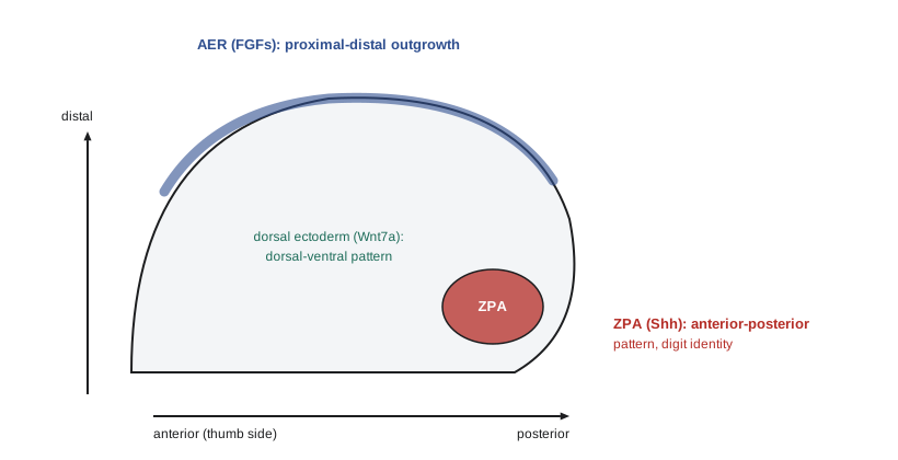
The AER secretes FGFs, especially FGF8, that keep the underlying mesenchyme proliferating and undifferentiated. Removing the AER from a chick wing bud stops outgrowth, producing truncated limbs; the later the removal, the more distal elements form. Beads soaked in FGF can replace the AER. How proximal–distal identity is specified is still debated. An older progress zone model held that cells acquire more distal identities the longer they remain under the AER. Current evidence favours a two-signal model in which proximal identity is specified early by retinoic acid from the flank and distal identity by FGFs from the AER, with intermediate identities between. The skeletal elements form in order: stylopod (humerus or femur), zeugopod (radius and ulna or tibia and fibula) and autopod (hand or foot), marked by expression of Meis, Hoxa11 and Hoxa13 respectively.
The anterior–posterior axis is controlled by the zone of polarizing activity (ZPA), a group of mesenchymal cells at the posterior margin of the bud. In 1968 John Saunders and Mary Gasseling grafted a ZPA to the anterior margin of a chick wing bud and obtained a mirror-image duplication of the digits. In 1993 Cliff Tabin's laboratory identified the ZPA signal as Sonic hedgehog. Shh beads placed anteriorly mimic a ZPA graft, and the extent of duplication depends on dose. The chick wing has three digits, conventionally numbered 2, 3 and 4; digit 4 forms nearest the ZPA, and the most anterior digit forms farthest from it, where Shh is lowest. Digit identity seems to depend on both the concentration of Shh and the duration of exposure: the posterior digits form from cells that were themselves Shh-expressing ZPA cells for longer periods. Shh also promotes growth of the bud by maintaining FGF expression in the AER through the BMP antagonist Gremlin, creating a feedback loop between the two signalling centres.
import math
# Toy model of digit identity from Sonic hedgehog exposure across the chick wing bud.
# Exposure = concentration x duration; digits form at positions where exposure crosses thresholds.
lam = 0.18          # decay length of Shh, as a fraction of bud width (illustrative)
def exposure(x, dose=1.0, hours=12.0):
    """x = distance from the ZPA (0 posterior, 1 anterior)."""
    return dose * math.exp(-x / lam) * hours / 12.0
thresholds = [(0.60, "digit 4"), (0.12, "digit 3"), (0.02, "digit 2")]
def digits(dose=1.0, hours=12.0, ectopic=False):
    out = []
    for i in range(0, 21):
        x = i / 20
        e = exposure(x, dose, hours)
        if ectopic:
            e = max(e, exposure(1 - x, dose, hours))          # second source at the anterior margin
        name = next((n for t, n in thresholds if e >= t), "no digit")
        if not out or out[-1] != name: out.append(name)
    return [d for d in out if d != "no digit"]
print("normal bud (posterior to anterior):", digits())
print("half the Shh dose                 :", digits(dose=0.5))
print("anterior ZPA graft :", digits(ectopic=True))normal bud (posterior to anterior): ['digit 4', 'digit 3', 'digit 2'] half the Shh dose : ['digit 3', 'digit 2'] anterior ZPA graft : ['digit 4', 'digit 3', 'digit 2', 'digit 3', 'digit 4']
The toy model reproduces three classic observations: normal buds form digits 4, 3 and 2 from posterior to anterior; reducing Shh loses the most posterior identities; and an anterior source produces a mirror-image duplication. Human limb malformations confirm the model. Point mutations in the ZRS limb enhancer of SHH, the enhancer lost in snakes, cause preaxial polydactyly, extra digits on the thumb side, because SHH becomes expressed ectopically in the anterior limb bud. Reaction–diffusion also plays a part: James Sharpe's group showed in 2014 that a Turing network involving Sox9, BMP and Wnt sets the periodic spacing of the digits, while Hox genes and Shh modulate its wavelength. Digits are separated by apoptosis of the tissue between them, driven by BMPs; in ducks, the BMP inhibitor Gremlin keeps the interdigital tissue alive, forming webbed feet.
The dorsal–ventral axis is controlled by the ectoderm. Wnt7a in the dorsal ectoderm induces the transcription factor Lmx1b in the underlying dorsal mesenchyme, and the ventral ectoderm expresses the transcription factor Engrailed-1, which represses Wnt7a ventrally. Mutations in human LMX1B cause nail–patella syndrome, with absent or underdeveloped nails and kneecaps, which are dorsal structures. The limb has also been a classic site of teratogenic damage: thalidomide, the subject of lesson 12, caused severe limb reductions.
The heart
The heart is the first functional organ in vertebrate embryos, beating by about day 22 in humans. It forms from cardiac mesoderm in the lateral plate, specified by signals from the adjacent endoderm, including BMPs and Wnt inhibitors. Progenitors expressing transcription factors such as Nkx2.5, Gata4 and Tbx5 form two fields: the first heart field forms a linear heart tube, mainly the future left ventricle, while the second heart field adds cells to both ends, forming the right ventricle, outflow tract and much of the atria. The tube then loops to the right, under the control of the left–right signals of lesson 5, and is divided by septa into four chambers, with contributions from cardiac neural crest to the outflow tract. Congenital heart defects are the most common birth defect, affecting nearly 1 per cent of births. Mutations in NKX2-5 cause atrial septal defects, mutations in TBX5 cause Holt–Oram syndrome with heart and upper limb defects, and deletion of a region of chromosome 22 including TBX1 causes DiGeorge syndrome, affecting the outflow tract, thymus and parathyroids.
The kidney and the eye: reciprocal induction
The permanent kidney, the metanephros, forms by reciprocal induction between two derivatives of intermediate mesoderm. The ureteric bud grows out of the nephric duct into the metanephric mesenchyme in response to GDNF secreted by the mesenchyme, acting on the RET receptor of the bud. The bud branches repeatedly to form the collecting system, and at each tip it induces the surrounding mesenchyme, through Wnt9b and then Wnt4, to condense and undergo a mesenchymal–epithelial transition to form nephrons. A human kidney contains on average about a million nephrons, though the number varies widely, and low nephron number at birth is associated with higher blood pressure later in life. The eye forms by interactions among the neural tube, which produces the optic vesicle, the surface ectoderm, which forms the lens and cornea, and neural crest mesenchyme. As described in lesson 1, the optic vesicle induces the lens placode, and the lens and optic cup then signal back and forth; Pax6, the subject of lesson 7, is required in both tissues.
| Organ | Interacting tissues | Key signals and factors | Example disorder |
|---|---|---|---|
| Somites and vertebrae | Presomitic mesoderm; notochord and neural tube | Hes/her clock, Notch, FGF, Wnt, retinoic acid | Spondylocostal dysostosis |
| Limb | Lateral plate mesoderm; ectoderm (AER) | FGF8, Shh, Wnt7a, Lmx1b, Hox genes | Preaxial polydactyly; nail–patella syndrome |
| Heart | Cardiac mesoderm; endoderm; neural crest | Nkx2.5, Gata4, Tbx5, BMP | Atrial septal defect; Holt–Oram syndrome |
| Kidney | Ureteric bud; metanephric mesenchyme | GDNF and RET, Wnt9b, Wnt4 | Renal agenesis |
| Eye | Optic vesicle; surface ectoderm | Pax6, BMP4, FGF | Aniridia |
Worked problem
Predicting limb phenotypes
Predict the result of each manipulation of a chick wing bud: (a) the AER is removed at an early stage; (b) the AER is removed at a later stage; (c) a bead soaked in Shh protein is implanted at the anterior margin; (d) dorsal ectoderm is removed and replaced with ventral ectoderm.
Show solution
- (a) Early AER removal stops outgrowth before most elements have been specified, so only a stylopod (humerus) forms.
- (b) Later removal allows proximal and middle elements to form, but distal elements (digits) are missing or truncated.
- (c) An anterior Shh source mimics a ZPA graft, producing a mirror-image duplication such as digits 4 3 2 2 3 4, depending on dose.
- (d) Without dorsal Wnt7a, the limb develops ventral characteristics on both surfaces, a double-ventral limb.
Answer: The AER controls proximal–distal outgrowth, the ZPA anterior–posterior pattern through Shh, and dorsal ectoderm the dorsal–ventral pattern through Wnt7a. These classic chick experiments established that limb pattern depends on three signalling centres.
Common mistakes
MistakeThinking somites form simultaneously.
InsteadThey bud off in sequence from head to tail at intervals set by the segmentation clock.
MistakeAssuming the segmentation clock alone sets somite boundaries.
InsteadThe clock interacts with a wavefront set by FGF, Wnt and retinoic acid gradients; somite size is wavefront speed times clock period.
MistakeSaying the ZPA forms the posterior digits only by producing more cells.
InsteadIt secretes Shh, which specifies digit identity through concentration and duration, and also promotes growth.
MistakeConfusing the AER and the ZPA.
InsteadThe AER is distal ectoderm controlling outgrowth through FGFs; the ZPA is posterior mesenchyme controlling anterior–posterior pattern through Shh.
MistakeBelieving digits are separated by cells moving apart.
InsteadInterdigital tissue is removed by BMP-driven apoptosis.
MistakeTreating the kidney as forming from a single tissue.
InsteadIt forms by reciprocal induction between the ureteric bud and the metanephric mesenchyme.
Summary and key terms
- Mesoderm is subdivided medial to lateral into paraxial, intermediate and lateral plate mesoderm by BMP levels.
- Somites form rhythmically under a segmentation clock based on delayed negative feedback of Hes/her genes, synchronized by Notch.
- A wavefront set by opposing FGF/Wnt and retinoic acid gradients converts the clock into somite boundaries.
- Limb outgrowth depends on FGFs from the AER; anterior–posterior pattern on Shh from the ZPA; dorsal–ventral pattern on Wnt7a and Lmx1b.
- Digit spacing involves a Turing-type network; interdigital apoptosis separates the digits.
- The heart forms from two heart fields and loops under left–right control; congenital heart defects affect nearly 1 per cent of births.
- The kidney and eye form by reciprocal induction between tissues.
- paraxial mesoderm
- Mesoderm beside the neural tube that forms the somites.
- somite
- A segmental block of paraxial mesoderm that forms vertebrae, skeletal muscle and dermis.
- segmentation clock
- An oscillator in presomitic mesoderm cells that times somite formation.
- clock and wavefront model
- A model in which an oscillator and a moving threshold together set somite boundaries.
- sclerotome
- The part of a somite that forms vertebrae and ribs.
- myotome
- The part of a somite that forms skeletal muscle.
- apical ectodermal ridge
- A thickened ridge of ectoderm at the distal rim of the limb bud that secretes FGFs.
- zone of polarizing activity
- Posterior limb bud mesenchyme that secretes Sonic hedgehog and patterns the anterior–posterior axis.
- polydactyly
- The presence of extra digits.
- heart field
- A population of cardiac progenitors contributing to particular parts of the heart.
- ureteric bud
- An outgrowth of the nephric duct that branches to form the kidney collecting system.
- metanephric mesenchyme
- Intermediate mesoderm that forms nephrons when induced by the ureteric bud.
Practice problems
Work each problem on paper first, then reveal the answer to check.
Name the three somite compartments and their derivatives.
Show answer
Sclerotome (vertebrae, ribs), myotome (skeletal muscle), dermatome (dermis of the back).
What evidence first revealed the segmentation clock?
Show answer
Waves of c-hairy1 expression sweeping through chick presomitic mesoderm every 90 minutes.
Why does delayed negative feedback produce oscillations?
Show answer
The protein represses its own gene only after a delay, so it overshoots and undershoots in cycles.
If the wavefront moves 3 µm per minute and the clock period is 90 minutes, how long is a somite?
Show answer
About 270 µm.
How do snakes produce so many vertebrae?
Show answer
Their segmentation clock runs fast relative to growth, forming more, smaller somites.
What happens if the AER is removed late in limb development?
Show answer
Proximal elements form but distal elements such as digits are missing.
Describe the Saunders and Gasseling experiment and its result.
Show answer
Grafting a ZPA to the anterior wing bud produced a mirror-image duplication of digits.
Why do ZRS mutations cause preaxial polydactyly?
Show answer
They cause ectopic Shh expression in the anterior limb bud, specifying extra digits.
What signals separate the digits, and how is this modified in ducks?
Show answer
BMPs drive interdigital apoptosis; Gremlin inhibits BMP in duck feet, preserving webbing.
Describe reciprocal induction in kidney development.
Show answer
Mesenchyme secretes GDNF to induce ureteric bud outgrowth and branching; bud tips secrete Wnt9b to induce nephron formation in the mesenchyme.
Lesson quiz · 5 questions
Score 60% or more to complete this lesson.
Create a free account to save your progress, take quizzes and earn a certificate.