
BIOL-360 · Lesson 3 of 12 · 27 min
Cleavage and gastrulation
After fertilization, the zygote divides rapidly without growing, partitioning the egg's large cytoplasm into many smaller cells. This is cleavage. It ends in a blastula, a ball or sheet of cells, often surrounding a fluid-filled cavity. Then comes gastrulation, the most dramatic reorganization in an animal's life, in which cells move into the interior to form the three germ layers and the body plan takes shape. As the embryologist Lewis Wolpert put it, "It is not birth, marriage or death, but gastrulation, which is truly the most important time in your life." This lesson compares how different animals cleave and gastrulate and asks what controls these processes.
The cell cycle in cleavage
Cleavage divisions are unusually fast because they skip the growth phases of the cell cycle. In Xenopus the first cleavage occurs about 90 minutes after fertilization and the next eleven follow at intervals of about 30 minutes, alternating S phase and M phase with almost no G1 or G2. A Drosophila embryo is faster still, with nuclear cycles of under 10 minutes. The driving engine is the oscillation of maturation-promoting factor, a complex of cyclin B and the kinase Cdk1, whose activity rises to trigger mitosis and falls as cyclin is destroyed. Because the egg is stocked with maternal cyclin mRNA, histones and nucleotides, the embryo can run many cycles without transcription.
The rapid cycles eventually slow. In Xenopus this happens at the twelfth cleavage, after about 4,000 cells have formed, an event called the mid-blastula transition. Cell cycles lengthen and become asynchronous, G phases appear, cells become motile and zygotic transcription begins on a large scale. A key trigger is the ratio of nuclear DNA to cytoplasm: each cleavage doubles the DNA while the total volume stays the same, and when enough DNA has accumulated to titrate out maternal factors that repress transcription or limit replication, the transition occurs. Embryos made with extra DNA reach the transition earlier. More generally, the handover from maternal to zygotic control, the maternal-to-zygotic transition, involves degradation of maternal mRNAs as well as activation of the zygotic genome; it occurs at the 2-cell stage in mice and over 4 to 8 cell stages in humans.
import math
# Cleavage without growth: cell number doubles each cycle while total volume is fixed.
egg_diameter_um = 1200.0     # Xenopus egg, about 1.2 mm
egg_volume = math.pi / 6 * egg_diameter_um ** 3
first_cycle_min, cycle_min = 90, 30
print("cycle  time(h)   cells    mean cell diameter (um)")
for n in range(0, 14):
    cells = 2 ** n
    t = 0 if n == 0 else (first_cycle_min + (n - 1) * cycle_min) / 60
    d = (6 * egg_volume / cells / math.pi) ** (1 / 3)
    flag = "  <- mid-blastula transition" if n == 12 else ""
    print(f"{n:5d}  {t:6.1f}  {cells:7d}   {d:8.1f}{flag}")
# nuclear-to-cytoplasmic ratio rises in proportion to cell number
print(f"DNA:cytoplasm ratio rises {2 ** 12}-fold between the zygote and cycle 12")cycle time(h) cells mean cell diameter (um)
0 0.0 1 1200.0
1 1.5 2 952.4
2 2.0 4 756.0
3 2.5 8 600.0
4 3.0 16 476.2
5 3.5 32 378.0
6 4.0 64 300.0
7 4.5 128 238.1
8 5.0 256 189.0
9 5.5 512 150.0
10 6.0 1024 119.1
11 6.5 2048 94.5
12 7.0 4096 75.0 <- mid-blastula transition
13 7.5 8192 59.5
DNA:cytoplasm ratio rises 4096-fold between the zygote and cycle 12The table of output shows why cleavage partitions rather than builds: by the mid-blastula transition, about 7 hours after fertilization, the embryo has thousands of cells, each only about 75 µm across, but the embryo as a whole is no bigger than the egg. The thousandfold increase in the ratio of DNA to cytoplasm is the timer for the transition.
Patterns of cleavage
The geometry of cleavage depends on two factors: the amount and distribution of yolk, which impedes division, and the orientation of the mitotic spindles, which is inherited from the egg's organization. In holoblastic (complete) cleavage the whole egg divides, as in sea urchins, frogs and mammals. In meroblastic (incomplete) cleavage only part of the egg divides, leaving a large undivided yolk mass, as in birds, reptiles, most fish and insects.
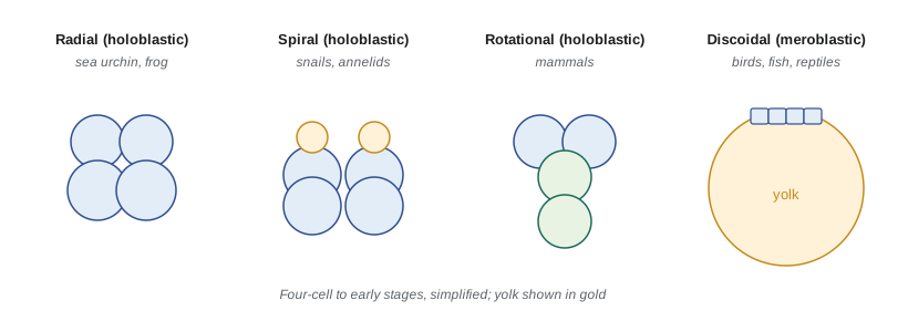
Radial cleavage, in sea urchins and with modification in frogs, divides the egg along planes parallel and perpendicular to the animal–vegetal axis, so tiers of cells sit directly above one another. In Xenopus, yolk concentrated in the vegetal hemisphere slows division there, so animal cells (micromeres) are smaller than vegetal cells (macromeres). Spiral cleavage, characteristic of molluscs, annelids and flatworms, the spiralians, tilts the spindles at an angle so that each tier is offset from the one below; it is usually accompanied by highly autonomous specification and a nearly invariant cell lineage. The direction of the spiral, left or right, is set by a maternal gene and determines the coiling of snail shells: Lymnaea snails with a mutation in the maternal formin gene Lsdia1 produce embryos with mirror-image cleavage and left-coiling shells.
Mammalian cleavage is rotational and slow. The first division is meridional, and in the second one cell divides meridionally while the other divides equatorially. Divisions take 12 to 24 hours and are asynchronous. At the 8-cell stage mouse blastomeres undergo compaction, flattening against each other and forming tight junctions, and the outer cells become polarized. Cells on the outside develop into the trophectoderm, which forms the embryonic part of the placenta, while the inside cells form the inner cell mass, from which the embryo proper arises. The trophectoderm pumps fluid into the interior to make a cavity, producing the blastocyst. This first fate decision depends on position and polarity: the Hippo signalling pathway is inactive in polarized outer cells, allowing the coactivator Yap to enter the nucleus and, with the transcription factor Tead4, activate trophectoderm genes such as Cdx2. Inner cells, lacking polarity, keep Hippo signalling active and express pluripotency factors such as Oct4. The inner cell mass then segregates into epiblast, which forms the embryo, and primitive endoderm, which forms the yolk sac.
Discoidal meroblastic cleavage in birds and fish confines division to a small disc of cytoplasm, the blastodisc, on top of the yolk. In the chick, by the time the egg is laid, cleavage has produced a blastoderm of some 20,000 cells. Insects show superficial cleavage: nuclei divide without cell membranes in the centre of the egg, migrate to the periphery to form a syncytial blastoderm, and only then are separated by membranes, forming the cellular blastoderm. This syncytial stage is what allows transcription factor gradients to pattern the Drosophila embryo, as lesson 4 explains.
Gastrulation: forming the germ layers
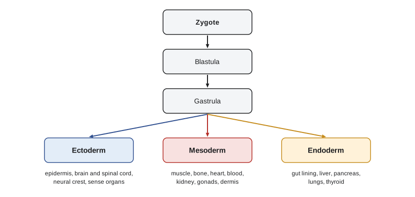
Gastrulation converts the blastula into an embryo with three germ layers. Ectoderm forms the outer layer and gives rise to the epidermis and the nervous system. Endoderm forms the innermost layer and gives rise to the lining of the gut and its derivatives. Mesoderm forms between them and gives rise to muscle, skeleton, connective tissue, the heart and blood, the kidneys and the gonads. Animals with three germ layers are called triploblasts; cnidarians such as sea anemones have only two and are diploblasts. The germ layers are not absolute commitments, and the neural crest, an ectodermal population that forms bone and cartilage in the head, is sometimes called a fourth germ layer.
Cell movements of gastrulation
Gastrulation uses a small repertoire of cell behaviours, combined differently in different species. Invagination is the infolding of a sheet of cells, like poking a finger into a balloon. Involution is the inturning of an expanding outer layer so that it spreads over the inner surface. Ingression is the migration of individual cells from the surface into the interior, often by epithelial–mesenchymal transition. Delamination is the splitting of one sheet into two. Epiboly is the spreading of an epithelial sheet to cover deeper layers. Convergent extension is the narrowing and lengthening of a tissue as cells intercalate between their neighbours, driven in vertebrates by the planar cell polarity pathway.
| Movement | Description | Example |
|---|---|---|
| Invagination | Infolding of a region of an epithelial sheet | Sea urchin archenteron; Drosophila ventral furrow |
| Involution | Inturning of an expanding outer layer over the inner surface | Xenopus mesoderm at the dorsal lip |
| Ingression | Individual cells leave an epithelium and migrate inward | Sea urchin primary mesenchyme; chick primitive streak |
| Delamination | Splitting of one cell sheet into two | Formation of the chick hypoblast |
| Epiboly | Spreading of an epithelial sheet over deeper layers | Xenopus animal cap ectoderm; zebrafish blastoderm over yolk |
| Convergent extension | Narrowing and lengthening by cell intercalation | Xenopus and zebrafish dorsal mesoderm forming the notochord |
Gastrulation in four embryos
In the sea urchin, about 32 small micromere descendants at the vegetal pole undergo epithelial–mesenchymal transition and ingress into the blastocoel as primary mesenchyme cells, which will build the larval skeleton. The vegetal plate then invaginates to form the archenteron, the primitive gut, which elongates by convergent extension and is pulled across the blastocoel by filopodia of secondary mesenchyme cells until it touches the opposite wall, where the mouth forms. The blastopore becomes the anus, the defining feature of deuterostomes. In protostomes, such as insects and molluscs, the blastopore typically gives rise to the mouth.
In Xenopus, gastrulation begins on the dorsal side with the formation of bottle cells, which constrict their apical surfaces and initiate invagination at the dorsal lip of the blastopore. Mesoderm then involutes over the lip and migrates along the inner surface of the blastocoel roof, using a fibronectin matrix, while animal cap ectoderm spreads by epiboly to cover the embryo. The blastopore lip extends laterally and ventrally to form a ring around a plug of yolky endoderm. The dorsal lip is the site of the Spemann–Mangold organizer, the subject of lesson 5.
In the chick, gastrulation proceeds through the primitive streak, a thickening that forms in the posterior epiblast and extends forward. Epiblast cells converge on the streak, undergo epithelial–mesenchymal transition and ingress through it. The first cells to ingress displace the hypoblast to form definitive endoderm; later cells spread between the layers as mesoderm. The anterior end of the streak, Hensen's node, is the bird equivalent of the amphibian organizer. As the streak regresses toward the posterior, it lays down the notochord and somites in its wake, so that the embryo develops from head to tail. Mammals, including humans, gastrulate through a primitive streak in the same way; in humans the streak appears around day 14 after fertilization, which is the developmental basis of the 14-day limit on human embryo research discussed in lesson 12.
Zebrafish gastrulation begins as the blastoderm spreads by epiboly over the yolk. At the margin, cells involute to form an inner layer, the hypoblast, and converge toward the dorsal side, where the embryonic shield, the fish organizer, forms. Convergent extension elongates the body axis. Throughout these embryos the same cell behaviours appear in different proportions, and the same signalling pathways, particularly Nodal, Wnt and fibroblast growth factor, specify the mesoderm and endoderm that move.
Mechanics and molecules of cell movement
Gastrulation is a problem in mechanics as well as in gene regulation. Apical constriction, driven by actomyosin contraction under the control of the small GTPase RhoA, changes cell shape from columnar to wedge-like and bends sheets. Changes in cell adhesion, especially of cadherins, allow cells to separate from or remain within epithelia; epithelial–mesenchymal transition involves loss of E-cadherin under the control of transcription factors such as Snail and Twist, the same factors that promote cancer metastasis. Mesodermal cells crawl on extracellular matrix proteins such as fibronectin. Differential adhesion and cortical tension can make tissues sort into layers, as Malcolm Steinberg showed by mixing dissociated cells from different tissues, which sorted out like immiscible liquids.
Worked problem
What does the yolk do?
A frog egg and a sea urchin egg are both fertilized and allowed to cleave. The sea urchin embryo forms tiers of similar-sized cells, while the frog embryo forms small cells near the animal pole and large cells near the vegetal pole, and its vegetal cells divide more slowly. Explain the difference, and predict what would happen to a frog egg centrifuged to redistribute its yolk evenly.
Show solution
- Sea urchin eggs contain little, evenly distributed yolk (isolecithal), so cleavage furrows pass through easily and cells are similar in size.
- Frog eggs are mesolecithal, with yolk concentrated in the vegetal hemisphere; yolk impedes cleavage furrows and slows division there.
- The animal–vegetal difference in cell size therefore reflects yolk distribution as well as spindle placement.
- If yolk were redistributed evenly, cleavage would be more uniform in cell size, although localized maternal determinants such as VegT mRNA might also be disturbed, with consequences for germ layer specification.
Answer: Yolk impedes cleavage, so its distribution shapes the pattern. Redistributing yolk would make cleavage more uniform but could also disrupt localized maternal determinants, illustrating how egg organization affects both mechanics and specification.
Common mistakes
MistakeThinking the embryo grows during cleavage.
InsteadCleavage partitions the egg into smaller cells; total volume stays roughly constant.
MistakeAssuming all embryos cleave completely.
InsteadYolky eggs of birds, reptiles and most fish cleave meroblastically; insects cleave superficially.
MistakeBelieving the inner cell mass and trophectoderm are specified by inherited determinants.
InsteadIn mammals this first decision depends on position and polarity through Hippo signalling.
MistakeTreating the blastopore as always forming the mouth.
InsteadIn deuterostomes, including vertebrates and sea urchins, it forms the anus.
MistakeSaying gastrulation is only about cells moving.
InsteadGastrulation also specifies germ layers through signals such as Nodal, Wnt and FGF and changes adhesion and shape.
MistakeThinking the mid-blastula transition is set by a clock counting hours.
InsteadIt is triggered mainly by the rising ratio of nuclear DNA to cytoplasm.
Summary and key terms
- Cleavage divides the egg rapidly without growth, driven by maternal cyclin B and Cdk1 oscillations.
- The mid-blastula transition, triggered by the DNA-to-cytoplasm ratio, lengthens cell cycles and starts zygotic transcription.
- Yolk and spindle orientation determine the cleavage pattern: radial, spiral, rotational, discoidal or superficial.
- In mammals, polarity and Hippo signalling separate trophectoderm from the inner cell mass.
- Gastrulation forms ectoderm, mesoderm and endoderm by invagination, involution, ingression, delamination, epiboly and convergent extension.
- Sea urchins invaginate an archenteron; frogs involute at the blastopore lip; birds and mammals ingress through a primitive streak; fish use epiboly and an embryonic shield.
- Apical constriction, changes in cadherin adhesion and epithelial–mesenchymal transition provide the mechanics.
- cleavage
- Rapid cell divisions of the early embryo without growth.
- blastula
- The embryonic stage at the end of cleavage, often a hollow ball of cells.
- mid-blastula transition
- The slowing and desynchronizing of cell cycles and onset of zygotic transcription in amphibian and fish embryos.
- holoblastic cleavage
- Cleavage in which the whole egg divides.
- meroblastic cleavage
- Cleavage restricted to part of the egg, leaving undivided yolk.
- compaction
- Flattening and tight adhesion of mammalian blastomeres at the 8-cell stage.
- inner cell mass
- The cells inside the blastocyst that form the embryo and some extraembryonic tissues.
- trophectoderm
- The outer layer of the blastocyst, which forms the embryonic part of the placenta.
- gastrulation
- The cell movements that form the three germ layers and establish the body plan.
- primitive streak
- The structure through which epiblast cells ingress during gastrulation in birds and mammals.
- convergent extension
- Narrowing and lengthening of a tissue by cell intercalation.
- epithelial–mesenchymal transition
- Conversion of epithelial cells into migratory mesenchymal cells, with loss of E-cadherin.
Practice problems
Work each problem on paper first, then reveal the answer to check.
Why are cleavage cell cycles so short?
Show answer
They lack G1 and G2 and use maternal stores, alternating S and M phases.
A Xenopus embryo made with twice the normal DNA reaches the mid-blastula transition one cycle early. Explain.
Show answer
The transition depends on the DNA-to-cytoplasm ratio; doubling DNA reaches the threshold one doubling sooner.
Distinguish holoblastic and meroblastic cleavage with an example of each.
Show answer
Holoblastic divides the whole egg (sea urchin, frog, mammal); meroblastic divides only part (chick, zebrafish).
What determines whether a mouse blastomere becomes trophectoderm?
Show answer
Being on the outside and polarized, which inactivates Hippo signalling so Yap and Tead4 activate Cdx2.
Name the three germ layers and one derivative of each.
Show answer
Ectoderm (brain), mesoderm (muscle), endoderm (gut lining).
What is the difference between invagination and ingression?
Show answer
Invagination folds a sheet; ingression moves individual cells inward.
What role do primary mesenchyme cells play in sea urchins?
Show answer
They ingress from the vegetal pole and build the larval skeleton.
What is the significance of Hensen's node?
Show answer
It is the anterior end of the primitive streak and acts as the bird organizer.
Why is the primitive streak relevant to policy on human embryo research?
Show answer
It appears around day 14, marking individuation and the start of gastrulation, which underlies the 14-day limit.
Which transcription factors drive epithelial–mesenchymal transition, and why are they relevant to cancer?
Show answer
Snail and Twist repress E-cadherin; reactivated in tumours, they promote invasion and metastasis.
Lesson quiz · 5 questions
Score 60% or more to complete this lesson.
Create a free account to save your progress, take quizzes and earn a certificate.