
BIOL-360 · Lesson 5 of 12 · 28 min
Axis formation in vertebrates and the organizer
In 1924 Hans Spemann and his doctoral student Hilde Mangold published one of the most famous experiments in biology. They took the dorsal lip of the blastopore from an early gastrula of a pale newt species and grafted it into the ventral side of a darkly pigmented newt embryo. The host developed a second body axis, a conjoined twin with its own neural tube, notochord and somites. Because the donor and host cells differed in pigment, Mangold could see that the graft formed mostly notochord, while the second neural tube and much of the second set of somites were made of host cells. The dorsal lip had induced host tissue to form a new axis, and Spemann called it the organizer. Mangold died in an accident before the paper appeared; Spemann received the Nobel Prize in 1935. This lesson explains what the organizer is, how it forms and how it works, using the frog Xenopus as the main example and comparing the chick, zebrafish and mouse.
The axes of a frog embryo
The Xenopus egg has an animal–vegetal axis built during oogenesis. The animal hemisphere is pigmented and will form ectoderm; the vegetal hemisphere is yolky and will form endoderm; mesoderm forms in a ring at the equator, the marginal zone. Maternal mRNAs localized in the vegetal cytoplasm, especially the transcription factor VegT and the signalling molecule Vg1, specify endoderm and help induce mesoderm. The dorsal–ventral axis, by contrast, is not fixed until fertilization. The sperm enters somewhere in the animal hemisphere, and the point of entry determines where the dorsal side will form: in most embryos, dorsal structures develop opposite the sperm entry point.
Cortical rotation and the Nieuwkoop centre
About 45 to 90 minutes after fertilization, the cortex of the egg, a thin layer of cytoplasm beneath the membrane, rotates about 30 degrees relative to the inner cytoplasm, moving away from the sperm entry point. The rotation is driven by a parallel array of microtubules organized by the sperm centriole. It carries dorsal determinants, including components of the Wnt signalling pathway such as Dishevelled and Wnt11 mRNA, from the vegetal pole toward the future dorsal side. Blocking the rotation with ultraviolet light applied to the vegetal pole, which destroys the microtubules, produces embryos with no dorsal structures, essentially a belly piece. Tilting the egg can rescue them, and treating eggs with lithium chloride, which inhibits the kinase GSK3 and so mimics Wnt signalling, produces embryos with excess dorsal structures.
The result of the rotation is the stabilization of β-catenin on the dorsal side. Normally a destruction complex containing GSK3 phosphorylates β-catenin and marks it for degradation; Wnt signalling inhibits the complex, allowing β-catenin to accumulate and enter nuclei. In Xenopus, dorsal β-catenin combines with the maternal VegT-dependent Nodal signals in the dorsal vegetal cells to create the Nieuwkoop centre, named after Pieter Nieuwkoop, who showed in the 1960s that vegetal cells can induce mesoderm in animal cap tissue. The Nieuwkoop centre itself does not become organizer; it induces the dorsal marginal zone cells above it to form the organizer by producing high levels of Nodal-related proteins.
Mesoderm induction
Nieuwkoop's experiments established that mesoderm arises by induction. When animal cap tissue, which normally forms epidermis, is cultured alone, it forms only ectoderm. When vegetal tissue, which normally forms endoderm, is cultured alone, it forms endoderm. When the two are placed together, the animal cells adjacent to the vegetal cells form mesoderm. Dorsal vegetal cells induce dorsal mesoderm such as notochord, and ventral vegetal cells induce ventral mesoderm such as blood. The main inducers are members of the TGF-β family, particularly the Nodal-related proteins Xnr1 to Xnr6, activated by maternal VegT, and the related protein Vg1. Activin, another TGF-β protein, can mimic the inducer in culture, and its effect is concentration dependent: low doses induce ventral mesoderm genes, higher doses induce muscle and the highest doses induce organizer genes such as goosecoid. This was one of the first demonstrations that a single signal can specify several fates by concentration, as the French flag model predicts.
What the organizer does
The organizer, in the dorsal lip of the blastopore, has several functions. Its cells are the first to involute and they become the prechordal plate mesoderm, which underlies the forebrain, and the notochord. It dorsalizes the adjacent mesoderm, so that ventral mesoderm that would otherwise form blood and mesenchyme forms muscle and kidney instead. It induces the overlying ectoderm to form the neural plate rather than epidermis. And it initiates the movements of gastrulation. Organizer cells express a characteristic set of transcription factors, including goosecoid, Siamois (induced directly by β-catenin), Xnot and Lim1, and secrete the signalling molecules that do the organizer's work.
The default model of neural induction
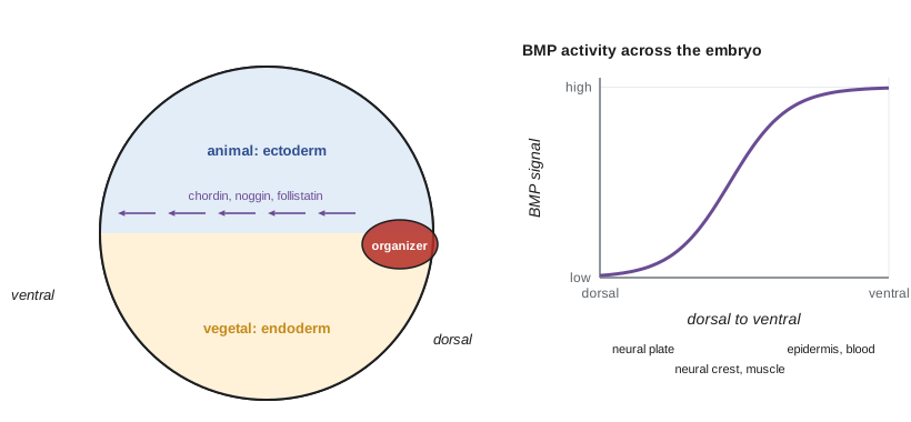
For decades embryologists searched for the neural-inducing signal from the organizer, assuming it would be an activator. The answer, found in the 1990s, was surprising. The ectoderm throughout the embryo is exposed to BMP4, which is expressed ventrally and laterally and instructs ectoderm to become epidermis. The organizer secretes proteins that bind BMPs and prevent them from reaching their receptors: Chordin, discovered by Edward De Robertis's group in 1994; Noggin, found by Richard Harland's group in 1992; and Follistatin. Where BMP is blocked, ectoderm becomes neural. Experiments with dissociated animal cap cells supported this default model: when cap cells are dispersed so that secreted BMP is diluted away, they become neural rather than epidermal, and adding BMP4 back converts them to epidermis.
The same antagonists dorsalize the mesoderm. BMP4 promotes ventral mesodermal fates such as blood; inhibiting it near the organizer allows dorsal fates such as muscle. The result is a gradient of BMP activity, low dorsally and high ventrally, that patterns both ectoderm and mesoderm across the dorsal–ventral axis. Intermediate BMP levels at the border between neural and epidermal ectoderm specify the neural crest and placodes. As lesson 4 showed, insects use the same system inverted. The default model is now known to be incomplete. In the chick, fibroblast growth factor signalling acts early to initiate neural development, and Wnt inhibition is also required, but BMP antagonism remains central to vertebrate neural induction.
import math
# BMP gradient across the dorsal-ventral axis, shaped by antagonists diffusing from the organizer.
# Free BMP = total BMP / (1 + Chordin / Kd), with Chordin decaying exponentially from the dorsal side.
L = 1.0                      # dorsal (0) to ventral (1), fraction of circumference
lam_chd = 0.25               # decay length of Chordin
chd0, Kd = 20.0, 1.0         # Chordin at the organizer, binding constant (relative units)
bmp_total = 1.0              # BMP assumed uniform here, for simplicity
thresholds = [(0.10, "neural plate"), (0.35, "neural crest / dorsal mesoderm"), (0.70, "lateral fates"), (1.01, "epidermis / blood")]
print(" position  Chordin   free BMP   fate")
for i in range(0, 11):
    x = i / 10
    chd = chd0 * math.exp(-x / lam_chd)
    bmp = bmp_total / (1 + chd / Kd)
    fate = next(name for t, name in thresholds if bmp < t)
    print(f"   {x:4.1f}   {chd:7.2f}   {bmp:7.3f}    {fate}")
# effect of doubling Chordin (as when an extra organizer is grafted or chordin mRNA injected)
x_np = next(i / 100 for i in range(101) if bmp_total / (1 + 2 * chd0 * math.exp(-(i / 100) / lam_chd) / Kd) >= 0.10)
x_n1 = next(i / 100 for i in range(101) if bmp_total / (1 + chd0 * math.exp(-(i / 100) / lam_chd) / Kd) >= 0.10)
print(f"neural plate extends to {x_n1:.2f} normally and {x_np:.2f} with twice the Chordin") position Chordin free BMP fate
0.0 20.00 0.048 neural plate
0.1 13.41 0.069 neural plate
0.2 8.99 0.100 neural crest / dorsal mesoderm
0.3 6.02 0.142 neural crest / dorsal mesoderm
0.4 4.04 0.198 neural crest / dorsal mesoderm
0.5 2.71 0.270 neural crest / dorsal mesoderm
0.6 1.81 0.355 lateral fates
0.7 1.22 0.451 lateral fates
0.8 0.82 0.551 lateral fates
0.9 0.55 0.647 lateral fates
1.0 0.37 0.732 epidermis / blood
neural plate extends to 0.20 normally and 0.38 with twice the ChordinThe toy model captures the logic: free BMP is lowest near the organizer, where Chordin is concentrated, and rises ventrally, so successive thresholds specify neural plate, neural crest and dorsal mesoderm, lateral fates and epidermis or blood. Doubling Chordin, as happens when chordin mRNA is injected, expands the neural plate ventrally, matching experiments. Real embryos are more complex. Chordin is cleaved by the protease Tolloid, and its complex with BMP is carried dorsally, actively shuttling BMP toward the ventral side, while a BMP-related protein called ADMP is made in the organizer itself and spreads ventrally, providing self-regulation. These mechanisms help explain how a frog embryo cut in half can regulate to form two smaller but well-proportioned embryos.
Head and trunk organizers
The organizer is not uniform. The first cells to involute, which form the anterior endoderm and prechordal mesoderm, induce head structures; later cells, which form notochord, induce trunk structures. Head induction requires inhibition of Wnt as well as BMP, and the anterior endoderm secretes Wnt inhibitors such as Dickkopf, Frzb and Cerberus. Cerberus inhibits Nodal, BMP and Wnt together, and injecting cerberus mRNA into a ventral cell can induce an ectopic head. Posteriorly, Wnt, FGF and retinoic acid transform neural tissue toward hindbrain and spinal cord identities. A two-step model, first proposed by Nieuwkoop, holds that BMP inhibition activates neural tissue with an anterior identity by default, and posteriorizing signals then transform it progressively. This explains why the forebrain needs protection from Wnt.
Organizers in other vertebrates
Every vertebrate studied has an organizer, though its form differs. In the chick it is Hensen's node at the anterior end of the primitive streak; grafting a node to the edge of a host embryo induces a second axis. In zebrafish it is the embryonic shield on the dorsal side of the blastoderm margin, and a maternal Wnt pathway again specifies the dorsal side. In the mouse it is the node, and an earlier signalling centre, the anterior visceral endoderm, located in an extraembryonic tissue, secretes Nodal and Wnt antagonists that specify the anterior. Its discovery showed that in mammals head development depends partly on tissues outside the embryo proper. The molecules are conserved across all these species: β-catenin, Nodal, BMP antagonists and Wnt inhibitors.
| Species | Organizer | Dorsal or anterior determinant | Key secreted factors |
|---|---|---|---|
| Xenopus (frog) | Dorsal lip of the blastopore | Maternal Wnt pathway after cortical rotation; Nieuwkoop centre | Chordin, Noggin, Follistatin, Cerberus, Dickkopf |
| Zebrafish | Embryonic shield | Maternal Wnt pathway; dorsal β-catenin | Chordin, Noggin, Dickkopf |
| Chick | Hensen's node | Posterior marginal zone; Vg1 and Wnt8c | Chordin, FGF8, Cerberus-like proteins |
| Mouse | Node (and anterior visceral endoderm) | Nodal signalling; position of distal visceral endoderm | Chordin, Noggin, Cerl, Lefty1, Dkk1 |
Left–right asymmetry
Vertebrates look bilaterally symmetrical from outside, but internal organs are asymmetric: the heart lies to the left, the liver mostly to the right, and the gut coils in a consistent direction. In the mouse, the left–right axis is established at the node, where several hundred cells each have a single motile cilium. The cilia rotate clockwise and, because they are tilted toward the posterior, they drive a leftward flow of fluid across the node, as Nobutaka Hirokawa's group showed in 1998 by tracking beads. Immotile crown cells around the node sense the flow, probably through the polycystin-2 calcium channel, and this leads to expression of Nodal on the left side of the lateral plate mesoderm. Nodal activates its own expression and that of its inhibitor Lefty; Lefty in the midline acts as a barrier keeping Nodal on the left. Nodal in turn activates the transcription factor Pitx2, which directs the left-sided development of organs.
Defects in cilia cause laterality disorders in people. In primary ciliary dyskinesia, mutations disable motile cilia, and about half of affected people have situs inversus, a mirror-image arrangement of internal organs, because the left–right decision becomes random. Other species use different early mechanisms: chicks rely on asymmetric cell movements around Hensen's node rather than ciliary flow, and frogs may use ion flows established very early. But asymmetric Nodal and Pitx2 expression in the left lateral plate is conserved across vertebrates.
Worked problem
Interpreting a lithium and ultraviolet experiment
Xenopus eggs are divided into three groups after fertilization. Group A is irradiated with ultraviolet light on the vegetal side before cortical rotation. Group B is treated with lithium chloride at the 32-cell stage. Group C receives both treatments. Predict the phenotypes and explain them in terms of β-catenin.
Show solution
- Ultraviolet light destroys the vegetal microtubule array, so cortical rotation fails and dorsal determinants are not moved. β-catenin is not stabilized dorsally, no Nieuwkoop centre or organizer forms, and group A embryos are ventralized, lacking axial structures.
- Lithium inhibits GSK3, so β-catenin is stabilized in all cells. Group B embryos are dorsalized, with excess dorsal and anterior structures, such as enlarged heads and a radial cement gland.
- In group C, lithium acts downstream of the defect caused by ultraviolet light, stabilizing β-catenin even though cortical rotation failed.
- Group C embryos are therefore dorsalized, like group B, showing that lithium bypasses the need for cortical rotation.
Answer: A: ventralized; B: dorsalized; C: dorsalized. Because lithium rescues ultraviolet-treated embryos, the dorsal signal moved by cortical rotation must act upstream of GSK3 and β-catenin, an epistasis argument that placed the Wnt pathway at the heart of dorsal axis formation.
Common mistakes
MistakeSaying the organizer grafted by Spemann and Mangold formed the entire second axis itself.
InsteadIt formed mainly notochord; it induced host cells to form the second neural tube and much of the somites.
MistakeThinking the neural inducer is an activating signal.
InsteadThe organizer mainly secretes BMP antagonists; inhibition of BMP allows ectoderm to become neural.
MistakeConfusing the Nieuwkoop centre with the organizer.
InsteadThe Nieuwkoop centre is in dorsal vegetal cells and induces the organizer in the overlying marginal zone.
MistakeBelieving the dorsal–ventral axis of Xenopus is fixed in the egg before fertilization.
InsteadIt is set by the sperm entry point and cortical rotation after fertilization.
MistakeAssuming all vertebrates break left–right symmetry with nodal cilia.
InsteadMice and fish use ciliary flow, but chicks rely on asymmetric cell movements; Nodal–Pitx2 expression is the conserved downstream step.
MistakeTreating activin as the natural mesoderm inducer in frogs.
InsteadActivin mimics the inducer in culture; Nodal-related proteins and Vg1 are the main endogenous inducers.
Summary and key terms
- Spemann and Mangold showed in 1924 that the dorsal blastopore lip, the organizer, induces a second axis.
- In Xenopus, cortical rotation moves Wnt pathway components dorsally and stabilizes β-catenin, forming the Nieuwkoop centre.
- Mesoderm is induced by Nodal-related proteins and Vg1 from vegetal cells; activin mimics this in a dose-dependent way.
- The organizer secretes Chordin, Noggin and Follistatin, which block BMP and allow ectoderm to become neural and mesoderm to become dorsal.
- Head induction needs Wnt inhibition (Dickkopf, Cerberus); posteriorizing signals transform anterior neural tissue.
- All vertebrates have organizers (shield, Hensen's node, node) using conserved molecules.
- Left–right asymmetry arises from leftward nodal flow in mice, leading to left-sided Nodal and Pitx2.
- organizer
- The dorsal region of the early gastrula that induces and patterns the body axis.
- cortical rotation
- Rotation of the egg cortex after fertilization that moves dorsal determinants in amphibians.
- β-catenin
- A protein that, when stabilized by Wnt signalling, enters the nucleus and activates target genes.
- Nieuwkoop centre
- Dorsal vegetal cells of the amphibian blastula that induce the organizer.
- mesoderm induction
- The specification of mesoderm in marginal cells by signals from vegetal cells.
- Chordin
- A BMP antagonist secreted by the organizer.
- default model
- The model in which ectoderm becomes neural unless BMP signalling directs it to epidermis.
- Cerberus
- A secreted inhibitor of Nodal, BMP and Wnt that promotes head formation.
- Hensen's node
- The organizer of the bird embryo at the anterior end of the primitive streak.
- anterior visceral endoderm
- An extraembryonic signalling centre in the mouse that specifies the anterior.
- situs inversus
- A mirror-image reversal of the left–right arrangement of internal organs.
- Pitx2
- A transcription factor expressed on the left side that directs asymmetric organ development.
Practice problems
Work each problem on paper first, then reveal the answer to check.
How did the pigment difference between newt species help Spemann and Mangold interpret their experiment?
Show answer
It showed which tissues came from the graft (mostly notochord) and which from the host (neural tube and much of the somites), proving induction.
What is the effect of lithium chloride on Xenopus embryos, and why?
Show answer
It inhibits GSK3, stabilizing β-catenin everywhere, which dorsalizes the embryo.
Describe Nieuwkoop's recombination experiment.
Show answer
Animal caps alone form ectoderm and vegetal tissue alone forms endoderm, but combined, the animal cells next to vegetal cells form mesoderm.
What is the evidence for the default model of neural induction?
Show answer
Dissociated animal cap cells become neural as BMP is diluted, and adding BMP4 makes them epidermal; BMP antagonists induce neural tissue.
Why are Wnt inhibitors needed for head development?
Show answer
Wnt posteriorizes neural tissue; blocking it in the anterior allows forebrain and head structures to form.
Name the organizer in the chick and in the zebrafish.
Show answer
Hensen's node; the embryonic shield.
What is the role of the anterior visceral endoderm?
Show answer
It secretes Nodal and Wnt antagonists that specify the anterior of the mouse embryo.
How do nodal cilia create left–right asymmetry?
Show answer
Their tilted clockwise rotation drives leftward fluid flow, sensed by crown cells, leading to left-sided Nodal.
Why do half of people with primary ciliary dyskinesia have situs inversus?
Show answer
Without ciliary flow the left–right decision is random, so half show reversed organ placement.
In the toy model, why does doubling Chordin expand the neural plate?
Show answer
More antagonist keeps free BMP below the neural threshold farther from the organizer.
Lesson quiz · 5 questions
Score 60% or more to complete this lesson.
Create a free account to save your progress, take quizzes and earn a certificate.