
BIOL-360 · Lesson 4 of 12 · 27 min
Axis formation in Drosophila
No animal has taught us more about how body plans are laid out than the fruit fly Drosophila melanogaster. In the late 1970s Christiane Nüsslein-Volhard and Eric Wieschaus, working at the European Molecular Biology Laboratory in Heidelberg, mutagenized flies and screened tens of thousands of lines for mutations that disrupted the pattern of the larval cuticle. They found about 600 mutations in some 120 genes and sorted them into a logical hierarchy. With Edward Lewis, who had studied homeotic genes since the 1940s, they received the 1995 Nobel Prize in Physiology or Medicine. Their screen revealed a cascade of transcription factors that subdivides the embryo step by step, from broad gradients to sharp stripes to the identity of each segment. Because many of these genes have counterparts in vertebrates, including humans, the fly cascade became a template for understanding development in general.
The fly embryo and its special features
The Drosophila egg is about 0.5 mm long and already polarized along its anterior–posterior and dorsal–ventral axes before fertilization, through interactions between the oocyte and surrounding follicle cells in the ovary. After fertilization, the zygote nucleus divides 13 times without cell membranes, forming a syncytium; after the ninth division most nuclei move to the periphery, forming the syncytial blastoderm, and at the end of division 13 membranes grow in from the surface to make about 6,000 cells, the cellular blastoderm, about 2.5 hours after fertilization. Because nuclei share cytoplasm during these early stages, transcription factors can diffuse among them and form gradients. Gastrulation follows, and the embryo forms a segmented larva that hatches after about a day at 25 °C.
The larva and adult are built from repeating units. In the embryo the basic unit is the parasegment, which is offset by half a segment from the visible segments of the larva. There are 14 parasegments: three in the head region, three thoracic and eight abdominal, plus terminal structures at each end. The pattern of denticle belts on the ventral cuticle of the larva provides a convenient readout of segmentation, and it was the cuticle that Nüsslein-Volhard and Wieschaus examined.
The hierarchy of segmentation genes
The genes controlling the anterior–posterior axis fall into five classes acting in sequence. Maternal effect genes set up gradients in the egg. Gap genes, expressed in broad domains, respond to these gradients. Pair-rule genes, expressed in seven stripes, respond to combinations of gap proteins. Segment polarity genes, expressed in 14 stripes, define the boundaries and polarity of each parasegment. Homeotic selector (Hox) genes then specify the identity of each segment. Each class was named for the phenotype of its mutants: gap mutants lack a contiguous block of segments, pair-rule mutants lack every other segment, and segment polarity mutants have part of each segment deleted and replaced by a mirror image of the remaining part.
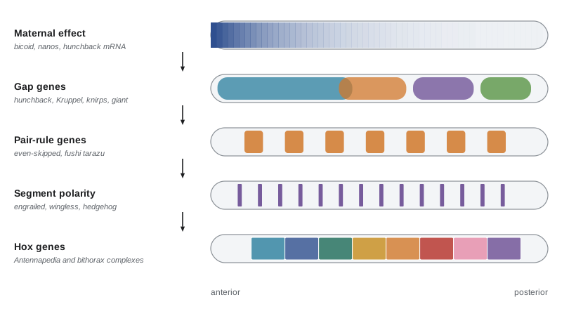
Maternal gradients: bicoid and nanos
The anterior of the embryo is organized by bicoid. During oogenesis, bicoid mRNA is transported along microtubules to the anterior pole and anchored there through its 3' untranslated region. After fertilization it is translated, and Bicoid protein spreads posteriorly through the syncytium, forming a gradient that falls roughly exponentially over the anterior half of the embryo. Embryos from mothers lacking bicoid have no head or thorax and instead develop posterior structures at both ends. Injecting bicoid mRNA into the posterior of such embryos makes a head there. Bicoid was the first morphogen to be identified molecularly, in 1988 by Wolfgang Driever and Nüsslein-Volhard, who showed that the level of Bicoid protein determines position along the axis: mothers with extra copies of bicoid produce embryos whose pattern shifts toward the posterior.
Bicoid acts in two ways. It is a homeodomain transcription factor that activates anterior genes, including the gap gene hunchback, in a concentration-dependent manner. It also binds the mRNA of caudal, a posterior determinant, and represses its translation, producing an opposing gradient of Caudal protein. The posterior is organized by nanos, whose mRNA is localized at the posterior pole along with the germ plasm. Nanos protein represses translation of maternal hunchback mRNA, so Hunchback protein is restricted to the anterior. The termini are specified separately by the Torso receptor tyrosine kinase, which is activated only at the two poles.
import math, random
random.seed(1)
# Bicoid gradient: C(x) = C0 exp(-x / lam), with x as a fraction of egg length (EL).
lam = 0.20                     # decay length about 20% of egg length (published estimates are near this)
threshold = math.exp(-0.5 / lam)   # set so the boundary lies at mid-embryo
def boundary(C0):
    return -lam * math.log(threshold / C0)
print("bicoid copies  relative C0   hunchback boundary (% EL from anterior)")
for copies, C0 in ((1, 0.5), (2, 1.0), (4, 2.0), (6, 3.0)):
    print(f"{copies:13d}  {C0:10.1f}   {100 * boundary(C0):6.1f}")
# positional error: if C0 varies 20% between embryos, how much does the boundary vary?
xs = [boundary(1.0 * random.gauss(1.0, 0.20)) for _ in range(5000)]
m = sum(xs) / len(xs)
sd = (sum((x - m) ** 2 for x in xs) / len(xs)) ** 0.5
print(f"20% embryo-to-embryo variation in C0 gives boundary SD = {100 * sd:.1f}% EL")bicoid copies relative C0 hunchback boundary (% EL from anterior)
1 0.5 36.1
2 1.0 50.0
4 2.0 63.9
6 3.0 72.0
20% embryo-to-embryo variation in C0 gives boundary SD = 4.2% ELThis simple model makes two testable predictions. Doubling bicoid dosage should shift the hunchback boundary posteriorly by λ ln 2, about 14 per cent of egg length, and real embryos from mothers with extra bicoid copies do shift, although by somewhat less than this simple model predicts, suggesting buffering by other inputs. Second, 20 per cent variation in Bicoid levels between embryos should produce about 4 per cent of egg length variation in the boundary. Measurements by Thomas Gregor and colleagues in 2007 found that the hunchback boundary is positioned to within about 1 per cent of egg length, roughly the width of a single nucleus. How embryos achieve such precision, through time averaging, cross-regulation among gap genes and input from the posterior system, has become a classic problem in quantitative biology.
Gap genes
The gap genes, including hunchback, Krüppel, knirps and giant, encode transcription factors expressed in broad, partly overlapping domains along the embryo. Hunchback is activated by high Bicoid in the anterior. Other gap genes are activated by Bicoid, Caudal or the terminal system and, crucially, repress one another. Krüppel, for instance, is expressed in the middle of the embryo where Hunchback is moderate; high Hunchback represses it anteriorly, and Knirps and Giant repress it posteriorly. Mutual repression between adjacent gap genes sharpens fuzzy maternal gradients into crisp domains, a general mechanism for converting graded inputs into discrete outputs.
Pair-rule genes and modular enhancers
The first periodic pattern appears with the pair-rule genes, expressed in seven transverse stripes. Primary pair-rule genes such as even-skipped (eve) and hairy are regulated directly by gap and maternal proteins. The eve gene has separate enhancers for different stripes. The stripe 2 enhancer, a stretch of about 500 base pairs, contains binding sites for activators (Bicoid and Hunchback) and repressors (Giant anteriorly and Krüppel posteriorly). Stripe 2 is expressed only where activators are present and both repressors are low. When Michael Levine and colleagues joined this enhancer to a reporter gene, the reporter was expressed in a single stripe. This modular organization, in which each enhancer reads a different combination of inputs, is a general feature of developmental genes and allows evolution to alter one expression domain without disturbing others.
| Gene class | Example genes | Expression pattern | Mutant phenotype |
|---|---|---|---|
| Maternal effect (anterior) | bicoid | Anterior gradient of protein from localized mRNA | No head or thorax; posterior structures at both ends |
| Maternal effect (posterior) | nanos, oskar | Posterior localization; represses hunchback translation | Loss of abdominal segments |
| Maternal effect (terminal) | torso | Receptor active at both poles | Loss of extreme anterior and posterior structures |
| Gap | hunchback, Krüppel, knirps, giant | Broad domains | Loss of a block of adjacent segments |
| Pair-rule | even-skipped, fushi tarazu, hairy | Seven stripes | Loss of alternate segments |
| Segment polarity | engrailed, wingless, hedgehog | Fourteen stripes | Part of each segment replaced by mirror image of the rest |
| Hox (homeotic) | Antennapedia, Ultrabithorax | Domains along the axis | One segment transformed into another |
Segment polarity genes and parasegment boundaries
After cellularization, cells can no longer share transcription factors, so the pattern must be maintained by cell–cell signalling. The segment polarity genes do this. Pair-rule proteins activate engrailed in a narrow stripe at the anterior of each parasegment and wingless in the adjacent row of cells just anterior to it. Engrailed-expressing cells secrete Hedgehog, which keeps neighbouring cells expressing wingless; Wingless, the fly Wnt protein, signals back to keep engrailed on. This positive feedback loop stabilizes the boundary between parasegments, and the gradients of Wingless and Hedgehog across each segment pattern the denticles and naked cuticle. The names hedgehog and wingless come from mutant phenotypes; the vertebrate versions of these pathways, Sonic hedgehog and the Wnts, appear throughout this course.
Homeotic genes and segment identity
Once segments exist, each must acquire a distinct identity: antennae on the head, legs on the thorax, wings only on the second thoracic segment, halteres (balancing organs) on the third. This is the job of the homeotic selector genes, which lie in two clusters on chromosome 3, the Antennapedia complex and the bithorax complex. Mutations in them cause homeotic transformations, in which one body part develops like another. Dominant Antennapedia mutations, which express the gene in the head, turn antennae into legs. Edward Lewis's triple mutants in the bithorax complex transformed the third thoracic segment into a second thoracic segment, producing four-winged flies. The products of these genes share a 60-amino-acid DNA-binding homeodomain, encoded by a 180-base-pair homeobox, and the discovery in 1984 that homeobox genes exist in vertebrates, the Hox genes, opened up comparative developmental biology. Lesson 7 examines Hox genes and their role in evolution.
The dorsal–ventral axis
The dorsal–ventral axis is set by a separate system. During oogenesis, the position of the oocyte nucleus determines where the follicle cells are signalled to adopt dorsal fates, and ventral follicle cells then generate a protease cascade in the perivitelline space that activates the Toll receptor on the ventral side of the embryo. Toll signalling causes the transcription factor Dorsal, initially present throughout the embryo's cytoplasm, to enter nuclei on the ventral side, forming a nuclear gradient highest ventrally. High nuclear Dorsal activates twist and snail, specifying mesoderm; intermediate levels activate genes of the neurogenic ectoderm; and where Dorsal is absent from nuclei, genes such as decapentaplegic (dpp) are expressed dorsally. Dpp, a BMP family protein, then patterns the dorsal ectoderm in a gradient shaped by its antagonist Short gastrulation. The Toll pathway, discovered through this developmental role, later turned out to be central to innate immunity in insects and mammals, earning Jules Hoffmann a share of the 2011 Nobel Prize.
Remarkably, the dorsal–ventral system of insects mirrors that of vertebrates, but inverted: in flies, the BMP Dpp is dorsal and the nervous system ventral, while in vertebrates BMP is ventral and the nervous system dorsal. The fly antagonist Short gastrulation corresponds to vertebrate Chordin. This observation revived an idea of the nineteenth-century naturalist Geoffroy Saint-Hilaire that the vertebrate body is an inverted arthropod, and it suggests that a BMP–Chordin system patterned the dorsal–ventral axis of the common ancestor of flies and vertebrates more than 550 million years ago.
Worked problem
Predicting a phenotype from the gene hierarchy
An embryo is homozygous for a loss-of-function mutation in Krüppel. Predict the effect on (a) the bicoid gradient, (b) the hunchback domain, (c) eve stripe 2, and (d) the larval cuticle.
Show solution
- Bicoid is a maternal effect gene upstream of Krüppel, so its gradient is unaffected.
- Krüppel represses hunchback at its posterior border, so the hunchback domain may expand somewhat posteriorly.
- Krüppel is the posterior repressor of eve stripe 2; without it, stripe 2 expands posteriorly into the region where Krüppel normally acts.
- Gap mutants lack a contiguous block of segments; Krüppel mutants lack thoracic and anterior abdominal segments, which are replaced by a mirror-image duplication of posterior abdominal segments.
Answer: The maternal gradient is normal, downstream patterns shift or expand where Krüppel normally represses, and the larva lacks a block of central segments. Working through the hierarchy, upstream genes are unaffected and downstream genes are changed, is the logic that let Nüsslein-Volhard and Wieschaus order the genes.
Common mistakes
MistakeThinking the segment polarity genes act in a syncytium like the gap genes.
InsteadThey act after cellularization, maintaining the pattern by cell–cell signalling through Wingless and Hedgehog.
MistakeAssuming bicoid mRNA forms a gradient by diffusing.
InsteadThe mRNA is anchored at the anterior pole; the protein forms the gradient.
MistakeConfusing pair-rule and segment polarity phenotypes.
InsteadPair-rule mutants lack alternate segments; segment polarity mutants lose part of every segment.
MistakeThinking each eve stripe needs a different gene.
InsteadOne gene with separate enhancers produces several stripes.
MistakeSaying homeotic mutations delete segments.
InsteadThey transform the identity of one segment into another, as in four-winged bithorax mutants.
MistakeTreating the fly and vertebrate dorsal–ventral systems as unrelated.
InsteadThey use the same BMP–Chordin logic with the axis inverted.
Summary and key terms
- Large mutant screens revealed a hierarchy of maternal, gap, pair-rule, segment polarity and Hox genes.
- Bicoid protein forms an anterior morphogen gradient from localized mRNA; Nanos represses hunchback translation posteriorly.
- Gap genes are expressed in broad domains and sharpen boundaries by mutual repression.
- Pair-rule genes form seven stripes, each driven by a modular enhancer reading gap and maternal inputs.
- Segment polarity genes maintain parasegment boundaries through a Wingless–Hedgehog feedback loop.
- Hox genes specify segment identity; mutations cause homeotic transformations.
- A Toll-driven nuclear gradient of Dorsal and a dorsal BMP gradient pattern the dorsal–ventral axis, inverted relative to vertebrates.
- syncytial blastoderm
- The stage at which many nuclei share a common cytoplasm at the periphery of the insect embryo.
- parasegment
- A unit of the Drosophila embryo offset by half a segment from the visible segments.
- bicoid
- A maternal effect gene whose protein forms an anterior gradient and organizes the head and thorax.
- nanos
- A maternal effect gene localized at the posterior that represses hunchback translation.
- gap gene
- A segmentation gene expressed in a broad domain; mutants lack a block of adjacent segments.
- pair-rule gene
- A segmentation gene expressed in seven stripes; mutants lack alternate segments.
- segment polarity gene
- A gene expressed in 14 stripes that patterns each parasegment.
- enhancer
- A regulatory DNA region that controls where and when a gene is transcribed.
- homeotic transformation
- Development of one body part with the identity of another.
- homeobox
- A 180-base-pair sequence encoding the DNA-binding homeodomain of homeotic and Hox proteins.
- Dorsal
- A transcription factor whose nuclear gradient specifies dorsal–ventral fates in the fly embryo.
- decapentaplegic
- The Drosophila BMP family protein that patterns the dorsal embryo.
Practice problems
Work each problem on paper first, then reveal the answer to check.
Why can transcription factors pattern the early Drosophila embryo directly?
Show answer
Nuclei share a common cytoplasm, so proteins can diffuse between them and form gradients.
Embryos from bicoid mutant mothers develop with posterior structures at both ends. Why are they called maternal effect mutants?
Show answer
The phenotype depends on the mother's genotype, because bicoid mRNA is supplied to the egg by the mother.
How does Bicoid repress the posterior determinant Caudal?
Show answer
It binds caudal mRNA and represses its translation.
How does mutual repression between gap genes help pattern?
Show answer
It sharpens graded inputs into discrete domains with crisp boundaries.
What are the activators and repressors of eve stripe 2?
Show answer
Activators: Bicoid and Hunchback; repressors: Giant (anterior) and Krüppel (posterior).
Why is the modular organization of enhancers important for evolution?
Show answer
One expression domain can change without disrupting others.
Describe the Wingless–Hedgehog feedback loop.
Show answer
Engrailed cells secrete Hedgehog, which maintains wingless in adjacent cells; Wingless maintains engrailed, stabilizing the boundary.
What would a bithorax triple mutant look like?
Show answer
A fly with the third thoracic segment transformed into a second, so it has four wings instead of two wings and two halteres.
If the bicoid decay length is 0.2 egg lengths, how far should doubling bicoid shift an anterior boundary?
Show answer
λ ln 2 ≈ 0.14 egg lengths.
Which cells form mesoderm in the fly, and why?
Show answer
Ventral cells with high nuclear Dorsal, which activates twist and snail.
Lesson quiz · 5 questions
Score 60% or more to complete this lesson.
Create a free account to save your progress, take quizzes and earn a certificate.