
BIOL-360 · Lesson 1 of 12 · 31 min
Principles of development
Every animal in this course began as a single cell. A fertilized human egg is about 0.1 mm across; some 38 weeks later it has become a baby of roughly two trillion cells, organized into more than 200 recognizable cell types arranged in tissues and organs of astonishing precision. A zebrafish embryo goes from one cell to a swimming larva with a beating heart in two days. Developmental biology asks how this happens: how cells become different from one another, how they come to be in the right place, how tissues acquire shape and size, and how the whole process is reproduced so reliably generation after generation. This lesson sets out the principles that recur in every later lesson, so that the details of fruit flies, frogs, chicks and mice can be read as variations on a small number of themes.
The questions are old. Aristotle opened chick eggs at intervals and argued that structures appear progressively rather than being present in miniature from the start, a view later called epigenesis. Its rival, preformationism, held that the embryo is a tiny but complete organism that merely grows. The debate was settled in favour of epigenesis by careful observation in the eighteenth and nineteenth centuries, but in a modern sense both ideas contain truth: the genome is inherited intact, yet the structures it encodes are built step by step through interactions among cells. The rest of this course is about those interactions.
Four processes make an embryo
Development can be broken into four overlapping processes. Cell proliferation, through cleavage divisions and later growth, generates the cells. Cell differentiation makes them different, so that a muscle cell expresses myosin and actin in a contractile arrangement while a neuron extends an axon and fires action potentials. Morphogenesis shapes the tissues: cells change shape, migrate, adhere, separate and die in organized patterns, folding sheets into tubes and condensing mesenchyme into cartilage. Pattern formation organizes the other three in space, so that differentiated cells appear in the right places relative to the body axes. Growth control then determines how large each part becomes. These processes are coupled. A signal that sets up a pattern often also controls proliferation and cell movement, and the same molecules reappear in many different contexts.
Differential gene expression
The central principle of developmental biology is that cells become different because they express different subsets of the same genome. Three lines of evidence established this. First, nuclear transplantation showed that nuclei from differentiated cells retain the information needed to make a whole animal. In 1962 John Gurdon transplanted nuclei from tadpole intestinal cells into enucleated frog eggs and obtained swimming tadpoles and, occasionally, adult frogs. In 1996 Dolly the sheep was cloned from the nucleus of an adult mammary cell. Second, molecular studies showed that the DNA content of different cell types is essentially the same, with rare exceptions such as the rearranged immunoglobulin genes of lymphocytes. Third, in 2006 Shinya Yamanaka showed that four transcription factors can reprogram adult mouse fibroblasts into pluripotent stem cells, a result we return to in lesson 11. Gurdon and Yamanaka shared the 2012 Nobel Prize in Physiology or Medicine.
If the genome is the same, differences must lie in which genes are transcribed, how transcripts are processed and translated, and how proteins are modified and localized. The mechanisms of gene regulation, including enhancers, transcription factors, chromatin modification and RNA processing, were covered in BIOL-207 lessons 9 and 10. In development the key point is combinatorial control. A gene's enhancer typically contains binding sites for several transcription factors, and the gene is expressed only in cells that contain the right combination. Because a modest number of regulators can be combined in many ways, a few hundred developmental transcription factors can specify thousands of distinct expression domains.
Specification, determination and differentiation
A cell's fate is what it will become in normal development. Fate can be mapped by labelling a cell and following its descendants, an approach that has produced fate maps for many embryos. But fate tells us nothing about how firmly a cell is committed. Developmental biologists distinguish two stages of commitment. A cell is specified when it will develop according to its fate if isolated and cultured in a neutral environment, but can still be redirected by other signals. It is determined when it will develop according to its fate even if moved to a different region of the embryo. Differentiation, the overt expression of cell-type-specific proteins, usually comes later still. Tests of commitment therefore involve isolating cells or transplanting them, and asking what they become.
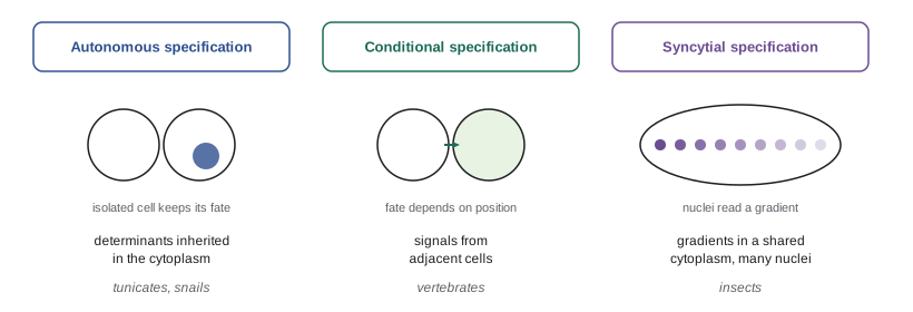
Embryos specify cells in three main ways. In autonomous specification, a cell inherits cytoplasmic determinants, such as mRNAs or proteins localized in the egg, that direct its fate. Remove the cell and it still forms the same tissue; remove the determinant and that tissue fails to form. The classic example is the tunicate embryo, in which the yellow-pigmented myoplasm of the egg, containing the mRNA for the transcription factor Macho-1, is partitioned into the cells that will form tail muscle. Embryos that rely heavily on autonomous specification show mosaic development: the embryo behaves like a mosaic of independently specified parts.
In conditional specification, a cell's fate depends on its position and on signals from other cells. Remove some cells and the remaining cells can often adjust to compensate, a property called regulative development. Hans Driesch showed in 1891 that each cell isolated from a two-cell sea urchin embryo could form a complete, if small, larva. Vertebrate embryos are highly regulative; separation of an early human embryo can give identical twins. In syncytial specification, found in insects, the early embryo is a syncytium in which many nuclei share one cytoplasm, so transcription factors can diffuse between nuclei and form gradients that nuclei interpret according to their position. Most embryos use all three modes to different degrees, and the distinction is a matter of emphasis rather than a strict classification.
Induction and competence
Induction is the process by which one group of cells changes the behaviour of an adjacent group. The inducing tissue sends a signal; the responding tissue must be competent, meaning it has the receptors, signal transduction machinery and transcription factors needed to respond. Competence is often restricted in time and space. Ectoderm from an amphibian gastrula can be induced to form neural tissue, but older ectoderm cannot. Induction can be instructive, when the signal directs the responding tissue toward one of several fates, or permissive, when the tissue is already committed and needs only a supportive environment to express that fate. Many organs form through reciprocal induction, in which two tissues signal back and forth. The vertebrate eye is a classic case: the optic vesicle induces lens formation in the overlying ectoderm, and the developing lens then signals back to shape the optic cup.
Signals act over different ranges. Juxtacrine signals pass between cells in contact, through membrane-bound ligands such as Delta acting on Notch receptors, or through gap junctions. Paracrine signals are secreted proteins that diffuse or are transported a few to many cell diameters; most developmental signals, including Hedgehog, Wnt, TGF-beta family members and fibroblast growth factors, belong here. Endocrine signals, such as the thyroid hormone that triggers amphibian metamorphosis or the ecdysone that controls insect moulting, act at a distance. The receptor biochemistry of these pathways was introduced in BIOL-214 lesson 11; lesson 6 of this course looks at how developing tissues use them.
Morphogens and positional information
A morphogen is a substance that forms a concentration gradient across a field of cells and specifies different fates at different concentrations. The concept was developed by Lewis Wolpert, who in 1969 proposed that cells acquire positional information, a value representing their position relative to a boundary, and then interpret that value according to their genetic programme. Wolpert's illustration, the French flag model, imagines a line of cells that must form blue, white and red stripes. A morphogen produced at one end decays with distance. Cells exposed to concentrations above a high threshold become blue, those between the high and low thresholds become white, and those below the low threshold become red. The flag forms correctly whatever the length of the line, if the gradient scales with tissue size, and the same mechanism can produce different patterns if cells interpret it differently.
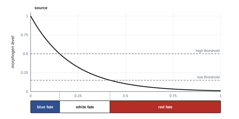
A simple and common way to form a gradient is for a substance to be produced at a localized source, to spread by diffusion and to be degraded everywhere at a constant rate. At steady state this gives an exponential profile, with concentration falling by a factor of e over a characteristic length λ equal to the square root of the diffusion coefficient divided by the degradation rate constant. With diffusion coefficients of a few square micrometres per second and lifetimes of tens of minutes, λ comes out at tens of micrometres, a few to a dozen cell diameters, which matches the range of many real morphogens.
import math
# Steady-state morphogen gradient C(x) = C0 exp(-x / lam), lam = sqrt(D / k),
# read by two thresholds (French flag model).
D = 3.0          # diffusion coefficient, square micrometres per second (illustrative)
k = 1 / 600      # degradation rate constant, per second (lifetime of 10 min)
lam = math.sqrt(D / k)
C0, high, low = 1.0, 0.5, 0.15
x_high = -lam * math.log(high / C0)
x_low = -lam * math.log(low / C0)
print(f"decay length lambda = {lam:.1f} micrometres")
print(f"blue/white boundary at {x_high:.1f} um, white/red boundary at {x_low:.1f} um")
# How far does a boundary move if the source doubles its output?
for factor in (0.5, 1.0, 2.0):
    xb = -lam * math.log(high / (C0 * factor)) + 0.0
    xb = max(xb, 0.0)
    print(f"source x{factor:<3}: blue/white boundary at {xb:6.1f} um")
cell = 8.0
print(f"a doubling shifts the boundary by {lam * math.log(2):.1f} um, about {lam * math.log(2) / cell:.1f} cell diameters")decay length lambda = 42.4 micrometres blue/white boundary at 29.4 um, white/red boundary at 80.5 um source x0.5: blue/white boundary at 0.0 um source x1.0: blue/white boundary at 29.4 um source x2.0: blue/white boundary at 58.8 um a doubling shifts the boundary by 29.4 um, about 3.7 cell diameters
The code shows an important property of exponential gradients. Changing the amount of morphogen made at the source moves every boundary by the same distance, λ multiplied by the natural log of the change. Doubling the source moves boundaries about 0.7λ, here roughly four cell diameters. Embryos are remarkably robust to such variation, and much current research asks how. Proposed mechanisms include self-enhanced degradation, in which the morphogen promotes its own breakdown so that the gradient becomes less sensitive to the source level, feedback between the morphogen and its receptors, and averaging of noisy signals over time. Sharpening of fuzzy boundaries by mutual repression between target genes, which we will see in the Drosophila gap genes in lesson 4, also helps.
Gradients need not be read only as concentrations. Cells may integrate signal over time, so that duration of exposure matters as well as level, and the vertebrate neural tube appears to use both: higher concentrations of Sonic hedgehog and longer exposure push progenitors toward more ventral fates. Some tissues read the slope of a gradient rather than its level, and others use opposing gradients from two ends. Positional information is a powerful idea, but it is one strategy among several.
Self-organization and reaction–diffusion
Not all patterns require a pre-existing source. In 1952 the mathematician Alan Turing showed that two interacting substances diffusing at different rates can spontaneously form periodic patterns from an almost uniform starting state. In the simplest version, a short-range activator stimulates its own production and that of a long-range inhibitor; the inhibitor spreads farther and suppresses activation nearby, so peaks of activator become separated by regular gaps. Turing's idea was ignored by most biologists for decades, but it is now supported in several systems: the spacing of hair follicles and feather buds, the ridges of the mammalian palate, the stripes of zebrafish skin, and the spacing of digits in the limb, discussed in lesson 9. Real systems often combine self-organization with positional cues, which orient the pattern and fix its scale.
Model organisms
Developmental biology has been built on a handful of model organisms chosen for practical advantages. Each answers some questions well and others poorly, and the general principles emerged by comparing them.
| Organism | Generation time | Main advantages | Main limitations |
|---|---|---|---|
| Fruit fly (Drosophila melanogaster) | about 10 days | Powerful genetics, large mutant screens, rapid embryogenesis, polytene chromosomes | Syncytial early embryo is unlike vertebrates; cells cannot be cultured easily |
| Nematode (Caenorhabditis elegans) | about 3.5 days | Transparent, invariant cell lineage of 959 somatic cells in the adult hermaphrodite, easy genetics and RNA interference | Highly derived and mostly autonomous development |
| Zebrafish (Danio rerio) | about 3 months | Transparent external embryos, vertebrate body plan, forward genetic screens, imaging of live development | Genome duplication complicates gene function studies |
| African clawed frog (Xenopus laevis) | 1 to 2 years | Large robust eggs, easy microinjection and grafting, classic embryology | Allotetraploid genome; slow to breed (X. tropicalis is diploid and faster) |
| Chick (Gallus gallus) | about 6 months | Accessible embryo in the egg, surgery and grafting, limb studies | Limited genetics until recent gene editing |
| Mouse (Mus musculus) | about 2 months | Mammal, targeted mutation and transgenics, close to human development | Embryo develops inside the mother; expensive |
| Human organoids and embryo models | not applicable | Human genome, human-specific processes, disease modelling | Incomplete organization; ethical oversight required (lesson 12) |
Plants have their own models, especially Arabidopsis thaliana, whose flower development and ABC model were covered in BIOL-340 lesson 10. Plant development differs in fundamental ways from animal development. Plant cells are encased in walls and do not migrate, so shape comes from oriented cell division and expansion; and plants continue to form new organs throughout life from meristems. Yet the logic of combinatorial transcription factors, signalling gradients and feedback is shared, a reminder that the principles of this lesson apply across kingdoms.
Robustness, evolution and medicine
Development is robust: embryos tolerate variation in temperature, nutrition and gene dosage and still produce a normal animal. Conrad Waddington called this canalization and pictured development as a ball rolling down a landscape of branching valleys, a metaphor we use in lesson 11. Robustness arises from redundancy, such as related genes with overlapping functions, from negative feedback, and from the buffering of protein-folding chaperones. Robustness is not unlimited, and failures of development cause congenital anomalies in about 3 per cent of births. Some are genetic; others result from environmental agents, the teratogens of lesson 12. Because the same pathways that build embryos also maintain adult tissues, developmental genes appear repeatedly in cancer, in regeneration and in stem cell therapies. And because changes in developmental genes produce changes in form, developmental biology is central to understanding evolution, a theme introduced in BIOL-321 lesson 10 and pursued here in lesson 7.
Worked problem
Interpreting a classic transplantation result
Cells from the region of an early amphibian gastrula fated to become epidermis are transplanted to the region fated to become neural plate. In one experiment, done at the early gastrula stage, the transplanted cells form neural tissue. In another, done at the late gastrula stage, they form epidermis in the neural plate region. What do these results show about the commitment of presumptive epidermis?
Show solution
- In the early experiment the cells adopted the fate of their new location, so they were not yet determined; their fate depended on signals in the neural plate region.
- In the late experiment the cells kept their original fate in a new environment, so by then they were determined as epidermis.
- Determination therefore occurs between early and late gastrulation for this tissue.
- The early cells must also have been competent to respond to neural-inducing signals; the late cells either became determined or lost competence, and further experiments would be needed to distinguish these possibilities.
Answer: Presumptive epidermis is not determined at the early gastrula stage but becomes determined by the late gastrula. This is a classic test of commitment and illustrates conditional specification in a regulative vertebrate embryo, studied in detail by Hans Spemann and colleagues in the early twentieth century.
Common mistakes
MistakeThinking cells become different because they lose genes they do not need.
InsteadWith rare exceptions, differentiated cells keep the whole genome; nuclear transfer and reprogramming prove this. They differ in which genes are expressed.
MistakeUsing fate and determination as synonyms.
InsteadFate is what a cell normally becomes; determination is a tested state of commitment that persists in a new environment.
MistakeAssuming a cell that differentiates correctly when isolated must have been determined.
InsteadThat shows specification. Determination requires the fate to persist in a different region of the embryo.
MistakeClassifying whole species as purely mosaic or purely regulative.
InsteadMost embryos combine autonomous, conditional and syncytial specification; the categories describe emphasis.
MistakeBelieving a morphogen must act through concentration alone.
InsteadCells may read duration of exposure, slope, or opposing gradients, and many patterns also involve self-organization.
MistakeThinking induction needs only a signal.
InsteadThe responding tissue must be competent, with receptors and transcription factors able to respond at that time.
Summary and key terms
- Development combines proliferation, differentiation, morphogenesis, pattern formation and growth control.
- Cells become different through differential gene expression of an identical genome, controlled combinatorially by transcription factors.
- Specification is reversible commitment shown in isolation; determination persists in a new environment; differentiation follows.
- Specification can be autonomous (cytoplasmic determinants), conditional (signals from other cells) or syncytial (gradients among nuclei sharing cytoplasm).
- Induction requires a signal and a competent responder; it may be instructive or permissive, and often reciprocal.
- Morphogens form gradients that convey positional information; an exponential gradient has decay length λ equal to the square root of D over k.
- Reaction–diffusion systems can generate periodic patterns without a pre-existing pattern.
- differential gene expression
- The expression of different subsets of the same genome in different cells.
- fate map
- A map of an embryo showing what each region will normally become.
- specification
- A labile state of commitment in which a cell follows its fate if isolated in a neutral environment.
- determination
- An irreversible state of commitment in which a cell follows its fate even when moved to another region.
- cytoplasmic determinant
- A molecule localized in the egg or early embryo that directs the fate of the cells inheriting it.
- mosaic development
- Development in which parts are specified largely independently, so removed parts are not replaced.
- regulative development
- Development in which remaining cells can compensate for removed or added cells.
- induction
- A signal from one tissue that changes the behaviour or fate of another.
- competence
- The ability of a tissue to respond to an inducing signal.
- morphogen
- A substance whose concentration gradient specifies different cell fates at different levels.
- positional information
- A value held by a cell that represents its position in a developing field.
- reaction–diffusion
- A mechanism in which interacting diffusing substances generate patterns spontaneously.
Practice problems
Work each problem on paper first, then reveal the answer to check.
State the evidence that differentiated cells retain a complete genome.
Show answer
Nuclear transfer from intestinal or adult mammary cells can produce complete animals; DNA content is the same across cell types; adult cells can be reprogrammed to pluripotency.
Distinguish specification and determination by the experiments used to test them.
Show answer
Specification is tested by isolation in a neutral medium; determination by transplantation to a different region.
Give an example of autonomous specification and the evidence for it.
Show answer
Tunicate tail muscle: the myoplasm containing macho-1 mRNA directs muscle fate, and cells inheriting it form muscle when isolated.
Why did Driesch's sea urchin experiment challenge mosaic theories?
Show answer
A single isolated cell from a two-cell embryo formed a complete small larva, showing that cells can regulate to replace missing parts.
What is competence, and why does it matter for induction?
Show answer
The ability to respond to a signal; without it, a tissue ignores the signal, so competence determines where and when induction can work.
A morphogen has D = 4 µm² per second and a lifetime of 25 min. Estimate λ.
Show answer
k = 1/1500 s⁻¹, so λ = √(4 × 1500) ≈ 77 µm.
In the French flag model, what happens to boundaries if the source makes three times as much morphogen?
Show answer
Every boundary shifts away from the source by λ ln 3, about 1.1λ.
How does a Turing system make spots or stripes?
Show answer
A short-range self-enhancing activator and a long-range inhibitor interact so that activation peaks suppress activation nearby, spacing peaks regularly.
Name one advantage and one limitation of C. elegans as a model.
Show answer
Invariant, fully mapped lineage and easy genetics; but development is highly autonomous and derived.
Give an example of reciprocal induction.
Show answer
The optic vesicle induces the lens, and the lens then signals to shape the optic cup.
Lesson quiz · 5 questions
Score 60% or more to complete this lesson.
Create a free account to save your progress, take quizzes and earn a certificate.