
BIOL-360 · Lesson 2 of 12 · 29 min
Gametes and fertilization
Development begins before fertilization. The egg and sperm are among the most specialized cells an animal makes, and the egg in particular is stocked with the mRNAs, proteins, nutrients and organelles that will run the first stages of development, often before the embryo's own genes are switched on. Fertilization then accomplishes two things at once: it combines the genetic material of two parents, and it activates the egg, triggering the metabolic and cell-cycle events that start embryogenesis. This lesson follows gametes from their formation to the fusion of the two pronuclei, using the sea urchin and the mouse as the main examples, with human applications where they illuminate the biology.
Making gametes
Gametes arise from primordial germ cells, a lineage set aside early in development and discussed in lesson 10. After migrating to the developing gonad, germ cells proliferate and then enter meiosis, the two divisions that halve the chromosome number and shuffle parental alleles, as described in BIOL-107 lesson 12 and BIOL-201 lesson 6. Spermatogenesis and oogenesis differ strikingly in timing and in what they produce.
In mammalian spermatogenesis, spermatogonial stem cells in the seminiferous tubules divide throughout adult life. Some daughters enter meiosis as primary spermatocytes, and each gives rise to four haploid spermatids. The spermatids then undergo spermiogenesis, a dramatic remodelling in which the nucleus condenses as histones are replaced by protamines, the Golgi forms the acrosome, a centriole organizes the flagellum, mitochondria wrap around the midpiece, and most of the cytoplasm is discarded. The cells remain joined by cytoplasmic bridges until release, which allows haploid cells to share gene products. In humans the process takes about 74 days from stem cell to sperm, and a man produces on the order of a hundred million sperm per day.
Oogenesis is slower and more conservative. In mammals, oogonia proliferate only before birth, then enter meiosis and arrest in prophase of meiosis I as primary oocytes within follicles. A human female is born with roughly one to two million oocytes, of which only a few hundred will ever be ovulated; most degenerate. At puberty, hormonal cycles recruit follicles each month; the selected oocyte completes meiosis I, extruding a small first polar body, and arrests again at metaphase of meiosis II. It completes meiosis only if fertilized. The asymmetric divisions conserve cytoplasm: one large egg and up to three tiny polar bodies are produced, rather than four equal cells. This long arrest, decades in humans, is thought to contribute to the rise in chromosomal errors such as trisomy 21 with maternal age.
| Feature | Spermatogenesis (mammal) | Oogenesis (mammal) |
|---|---|---|
| Stem cells in adult | Yes, spermatogonial stem cells divide throughout life | No; oogonia proliferate only before birth |
| Meiotic arrests | None; meiosis is continuous | Prophase I (from fetal life) and metaphase II (until fertilization) |
| Products per meiosis | Four equal spermatids | One egg and up to three polar bodies |
| Duration | About 74 days in humans | From fetal life to ovulation, up to five decades |
| Cytoplasm | Mostly discarded | Greatly enlarged and stocked with maternal products |
| Output | About 10⁸ sperm per day | Usually one egg per cycle |
What the egg contains
Eggs vary in size from about 0.1 mm in mammals to 1.2 mm in Xenopus and several centimetres in birds, where most of the volume is yolk. Beyond yolk, the egg contains the maternal contribution to development. Maternal mRNAs, transcribed during oogenesis and stored in a translationally repressed state, encode proteins for the early cell cycles, signalling components and transcription factors. Some are localized in specific regions of the egg, where they act as cytoplasmic determinants: bicoid mRNA at the anterior of the Drosophila egg, VegT mRNA in the vegetal cytoplasm of Xenopus, and germ plasm in many species. The egg also contains ribosomes, mitochondria (all inherited from the mother in most animals), cortical granules beneath the plasma membrane, and protective coats: the vitelline envelope of sea urchins and the zona pellucida of mammals. Genes expressed by the mother that affect the embryo through these products are called maternal effect genes, because the phenotype of the embryo depends on the mother's genotype rather than its own.
Getting gametes together
Species with external fertilization, such as sea urchins, many fish and frogs, release gametes into water, where sperm must find eggs of their own species among those of others. Many marine eggs release chemoattractant peptides. The sea urchin Arbacia punctulata releases resact, which binds a receptor guanylyl cyclase on the sperm tail and steers swimming up the concentration gradient. Chemoattraction is species-specific, the first of several recognition steps.
In mammals, sperm deposited in the female tract are not immediately able to fertilize. They must undergo capacitation, a set of changes that take hours in the female tract and include removal of cholesterol and glycoproteins from the sperm membrane, increased membrane fluidity, phosphorylation of proteins and a change to a vigorous, whip-like hyperactivated motility. Capacitation is why in vitro fertilization requires incubating sperm in specific media. Only a few hundred of the millions of sperm in an ejaculate reach the oviduct, where fertilization occurs. Guidance there probably combines rheotaxis (swimming against fluid flow), thermotaxis toward the slightly warmer site of fertilization, and possibly chemotaxis, mediated in part by the sperm-specific CatSper calcium channel.
Recognition and the acrosome reaction
When a sea urchin sperm meets the jelly coat of an egg, sugar polymers carrying sulphate groups in the jelly trigger the acrosome reaction: the acrosomal vesicle fuses with the sperm plasma membrane and releases enzymes that digest a path through the jelly, and actin polymerization extends an acrosomal process. The process carries the protein bindin, which binds species-specifically to a receptor on the vitelline envelope. Bindin from one sea urchin species binds poorly to the eggs of another, and bindin sequences evolve rapidly under selection, contributing to reproductive isolation between species.
In mammals the sperm must penetrate the cumulus cells surrounding the egg and then the zona pellucida. The mouse zona is made of three or four glycoproteins, ZP1 to ZP3 in mice and ZP1 to ZP4 in humans. Current evidence favours a model in which the N-terminal region of ZP2 is the main sperm-binding site, and the acrosome reaction may often begin before the sperm reaches the zona, in the cumulus. After passing through the zona, the sperm fuses with the egg membrane. In 2005 Masaru Okabe's group identified the sperm protein Izumo1 as essential for fusion, and in 2014 Gavin Wright's group identified its egg receptor, Juno; knockout mice lacking either protein are infertile. More proteins required for gamete fusion continue to be discovered.
Fusion and blocks to polyspermy
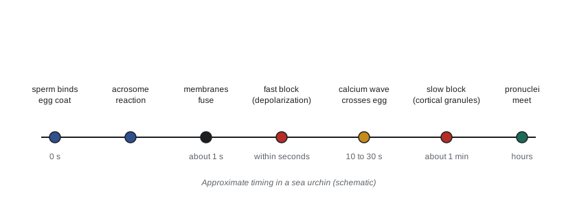
In most animals, fertilization by more than one sperm, polyspermy, is lethal, because the extra centrioles and chromosomes disrupt mitosis. Eggs therefore have mechanisms that prevent more than one sperm from entering. Sea urchins have two. The fast block acts within one to three seconds: sperm fusion opens sodium channels in the egg membrane, and the membrane potential shifts from about −70 mV to about +20 mV. Sperm cannot fuse with a depolarized egg. Laurinda Jaffe showed in 1976 that holding sea urchin eggs at a negative potential by passing current allowed polyspermy, while depolarizing them prevented fertilization. The fast block is transient and is not used by mammals.
The slow block lasts. About 20 to 60 seconds after fusion, cortical granules beneath the egg membrane fuse with it and release their contents. Enzymes cleave the links between the vitelline envelope and the membrane, mucopolysaccharides draw water into the space and lift the envelope away, and a peroxidase hardens it into a fertilization envelope that sperm cannot penetrate. In mammals, the cortical reaction releases the protease ovastacin, which cleaves ZP2 so that the zona no longer binds sperm; Juno is also rapidly shed from the egg membrane after fertilization, which may help block membrane fusion.
Egg activation and the calcium wave
The cortical reaction is triggered by a wave of calcium that sweeps across the egg from the point of sperm entry. Free calcium rises from about 0.1 µM to around 1 µM, released from the endoplasmic reticulum through inositol trisphosphate (IP3) receptors. The wave crosses a sea urchin egg in about 30 seconds. Calcium is both necessary and sufficient for activation: injecting a calcium chelator blocks activation by sperm, while calcium ionophores activate unfertilized eggs, a phenomenon called parthenogenetic activation. In mammals, the sperm delivers a phospholipase, PLCζ, into the egg cytoplasm; it generates IP3 and drives a series of calcium oscillations that continue for hours. Human sperm lacking functional PLCζ cause failure of fertilization even with intracytoplasmic sperm injection, and artificial activation with calcium ionophore is used in some fertility clinics, a practice still being evaluated.
Calcium triggers the events of egg activation. Early responses include the cortical reaction and, in mammals, the completion of meiosis II and extrusion of the second polar body, as calcium leads to destruction of cyclin B and inactivation of maturation-promoting factor. Later responses, over minutes to hours, include a burst of protein synthesis from stored maternal mRNAs, an increase in intracellular pH in sea urchins, resumption of DNA synthesis and entry into the first mitotic cell cycle.
import math, random
random.seed(7)
# A calcium wave crossing an egg: a wavefront moving at constant speed v from the sperm entry point.
# Each patch of cortex releases its cortical granules when the wave arrives.
diameter_um = 100.0      # sea urchin egg diameter (approximate)
v = 5.0                  # wave speed, micrometres per second (illustrative, within the reported range)
# arrival time at points around the circumference, measured from the entry point (angle 0)
for deg in (0, 45, 90, 135, 180):
    arc = math.pi * diameter_um * deg / 360          # distance along the surface
    print(f"angle {deg:3d} deg: wave arrives after {arc / v:5.1f} s")
# fraction of cortex activated over time (uniform points on the circle)
pts = [random.uniform(0, 180) for _ in range(20000)]
for t in (5, 10, 20, 30):
    reached = sum(1 for d in pts if math.pi * diameter_um * d / 360 / v <= t) / len(pts)
print(f"t = {t:2d} s: {100 * reached:5.1f} % of the cortex has released granules")angle 0 deg: wave arrives after 0.0 s angle 45 deg: wave arrives after 7.9 s angle 90 deg: wave arrives after 15.7 s angle 135 deg: wave arrives after 23.6 s angle 180 deg: wave arrives after 31.4 s t = 5 s: 16.0 % of the cortex has released granules t = 10 s: 32.2 % of the cortex has released granules t = 20 s: 63.9 % of the cortex has released granules t = 30 s: 95.4 % of the cortex has released granules
The simulation reproduces the order of magnitude of the real event: a wave travelling at a few micrometres per second crosses a 100 µm egg in about 30 seconds, and the cortical reaction follows it around the egg. In real eggs the wave is self-propagating, because calcium released at one site promotes release at neighbouring sites through calcium-sensitive IP3 receptors, so its speed stays roughly constant rather than slowing with distance as simple diffusion would.
The pronuclei unite
After fusion, the sperm nucleus decondenses as protamines are replaced by maternal histones, and a male pronucleus forms. In mammals the egg also finishes meiosis to form the female pronucleus. The sperm centriole, in most animals, organizes a microtubule aster that brings the pronuclei together. In sea urchins the pronuclei fuse to form a diploid zygote nucleus. In mammals they do not fuse directly; instead, both replicate their DNA, their envelopes break down, and the chromosomes come together on the first mitotic spindle, so the two parental genomes first share a nucleus at the two-cell stage. Recent imaging has shown that mammalian zygotes may assemble two separate spindles, one around each parental genome, which then align, offering one explanation for the frequent errors in early human embryos.
The parental genomes are not equivalent. Genomic imprinting marks certain genes, by DNA methylation laid down during gametogenesis, so that only the maternal or only the paternal copy is expressed. Mouse embryos made with two maternal or two paternal genomes fail to develop, which is why mammals cannot normally reproduce parthenogenetically. Imprinted genes such as Igf2 (expressed from the paternal copy) influence growth and placental development, and errors in imprinting cause disorders such as Prader–Willi and Angelman syndromes. After fertilization, most of the genome is demethylated and then remethylated as the embryo develops, but imprinted regions are protected; this epigenetic reprogramming was introduced in BIOL-201 lesson 12.
Fertilization technology and its limits
Knowledge of fertilization underlies assisted reproduction. In vitro fertilization (IVF), first successful in humans with the birth of Louise Brown in 1978, relies on capacitating sperm in culture and on the fact that the egg completes meiosis on activation. Intracytoplasmic sperm injection (ICSI), introduced in 1992, bypasses the zona and membrane fusion entirely and is now used in a majority of IVF cycles worldwide. Embryos produced by IVF beyond those transferred are often frozen and may later be donated for research, which is how most human embryonic stem cell lines were derived. The legal frameworks governing that research differ between Canada and the United States and are the subject of lesson 12.
Worked problem
Diagnosing a fertilization failure
In a laboratory study, mouse eggs are inseminated with sperm from a mutant male line. Sperm reach and penetrate the zona pellucida and accumulate in the perivitelline space, but no sperm fuse with the egg membrane and no calcium oscillations occur. When a single mutant sperm is injected directly into an egg (ICSI), calcium oscillations begin and development proceeds. Which sperm protein is most likely defective?
Show solution
- Capacitation, the acrosome reaction and zona penetration must be working, since sperm reach the perivitelline space.
- Fusion with the egg membrane fails, so a protein required for gamete fusion is likely missing.
- When fusion is bypassed by injection, activation occurs, so PLCζ, the sperm factor that triggers calcium oscillations, must be functional.
- Izumo1 is the sperm protein required for binding Juno and for fusion.
Answer: A defect in Izumo1, or in another protein of the fusion machinery, is most likely. This matches the phenotype of Izumo1 knockout mice, whose sperm penetrate the zona but cannot fuse, and whose infertility is rescued by ICSI.
Common mistakes
MistakeSaying the egg is a passive target for the sperm.
InsteadThe egg actively recruits sperm, blocks polyspermy and controls activation through stored maternal products and calcium signalling.
MistakeThinking freshly ejaculated mammalian sperm can fertilize immediately.
InsteadThey must first undergo capacitation in the female tract or in suitable culture media.
MistakeAssuming mammals use an electrical fast block like sea urchins.
InsteadMammals rely mainly on the cortical reaction and changes in the zona and egg membrane.
MistakeStating that human oocytes complete meiosis before ovulation.
InsteadThey arrest at metaphase II and complete meiosis only after fertilization.
MistakeTreating maternal effect genes as genes expressed in the embryo.
InsteadThey are expressed by the mother, and their products in the egg determine the embryo's phenotype.
MistakeAssuming the maternal and paternal genomes are equivalent.
InsteadImprinting makes some genes active only from one parent, so both genomes are needed in mammals.
Summary and key terms
- Spermatogenesis is continuous from adult stem cells and produces four sperm per meiosis; oogenesis arrests twice and produces one large egg.
- The egg stores maternal mRNAs, proteins and determinants that run early development.
- Sperm are attracted by species-specific chemoattractants in many marine species; mammalian sperm must capacitate.
- The acrosome reaction and species-specific binding (bindin; zona proteins) precede fusion, which in mammals requires Izumo1 and Juno.
- Polyspermy is prevented by a fast electrical block in sea urchins and a slow block by cortical granule exocytosis.
- A calcium wave driven by IP3, triggered in mammals by sperm PLCζ, activates the egg.
- The parental pronuclei unite; imprinting makes both parental genomes necessary in mammals.
- spermiogenesis
- The differentiation of a spermatid into a mature sperm, including nuclear condensation and acrosome formation.
- polar body
- A small cell produced by the asymmetric meiotic divisions of oogenesis.
- maternal effect gene
- A gene whose expression in the mother determines the phenotype of her offspring through products in the egg.
- capacitation
- Changes in mammalian sperm in the female tract that make them able to fertilize.
- acrosome reaction
- Exocytosis of the acrosomal vesicle, releasing enzymes and exposing proteins needed to reach and bind the egg.
- zona pellucida
- The glycoprotein coat surrounding the mammalian egg.
- polyspermy
- Entry of more than one sperm into an egg.
- fast block to polyspermy
- A rapid depolarization of the egg membrane that transiently prevents further sperm fusion.
- cortical reaction
- Exocytosis of cortical granules after fertilization, modifying the egg coat to block further sperm.
- egg activation
- The metabolic and cell-cycle events triggered by fertilization, initiated by a rise in calcium.
- PLCζ
- A sperm phospholipase delivered to the mammalian egg that triggers calcium oscillations.
- genomic imprinting
- Parent-of-origin-specific expression of a gene set by epigenetic marks in the gametes.
Practice problems
Work each problem on paper first, then reveal the answer to check.
Why does oogenesis produce polar bodies rather than four equal cells?
Show answer
Asymmetric divisions conserve the cytoplasm and stored products in a single large egg.
A woman is 40 years old. How long have her oocytes been arrested in prophase I?
Show answer
Since before her birth, so about 40 years.
What is the function of resact?
Show answer
It is a species-specific chemoattractant peptide from Arbacia eggs that guides sperm toward the egg.
List three changes during capacitation.
Show answer
Cholesterol removal from the sperm membrane, protein phosphorylation and hyperactivated motility.
How did Jaffe show that the fast block is electrical?
Show answer
Holding eggs at negative potentials allowed polyspermy, while depolarizing them prevented fertilization.
What does ovastacin do?
Show answer
It is released in the cortical reaction and cleaves ZP2 so the zona no longer binds sperm.
How could you show that calcium is sufficient for egg activation?
Show answer
Treat unfertilized eggs with a calcium ionophore; they activate without sperm.
Why does ICSI rescue infertility caused by lack of Izumo1 but not lack of PLCζ?
Show answer
ICSI bypasses fusion but still relies on the sperm delivering PLCζ to trigger calcium oscillations.
Why can mammals not develop from two maternal genomes?
Show answer
Imprinted genes expressed only from the paternal genome, such as Igf2, are needed for development.
A calcium wave travels at 8 µm per second around an egg 120 µm in diameter. How long until it reaches the opposite pole?
Show answer
Half the circumference is π × 120 / 2 ≈ 188 µm, so about 24 s.
Lesson quiz · 5 questions
Score 60% or more to complete this lesson.
Create a free account to save your progress, take quizzes and earn a certificate.