
BIOL-330 · Lesson 9 of 12 · 32 min
Viral replication, bacteriophages and emerging viruses
Every virus, whatever its genome or host, must complete the same basic tasks: find and attach to a host cell, get its genome inside, express its genes and copy its genome using a mixture of host and viral enzymes, assemble new virions and get them out. The details differ enormously between a phage infecting E. coli, an influenza virus infecting a cell lining the airway and a retrovirus that writes its genome into the host chromosome, and those details determine how fast a virus spreads, how it causes disease, how quickly it evolves and how it can be stopped. This lesson follows the replication cycle, uses bacteriophages to show how a single round of infection is measured, describes lytic and temperate lifestyles and the revival of phage therapy, and ends with the evolution and emergence of new viruses. Immune responses to viruses are the subject of BIOL-380.
The replication cycle
Attachment (adsorption) is the first and most specific step. Proteins on the virion surface bind receptors on the host cell, and the availability of the right receptor largely decides which hosts and which tissues a virus can infect, its host range and tropism. Phage T4 binds lipopolysaccharide and the outer membrane protein OmpC of E. coli; HIV binds CD4 and then a co-receptor, CCR5 or CXCR4, on T cells and macrophages; SARS-CoV-2 binds angiotensin-converting enzyme 2 (ACE2). People homozygous for a 32-base deletion in CCR5 are highly resistant to the strains of HIV that use it, a fact exploited in the rare cases in which HIV has been cured by stem cell transplants from such donors.
Penetration and uncoating come next. Phages inject their genomes through the bacterial envelope and leave the empty capsid outside. Animal viruses enter whole: enveloped viruses fuse their envelopes with the plasma membrane or, after being taken up by endocytosis, with the membrane of an endosome, often triggered by the endosome’s acidity; non-enveloped viruses penetrate or rupture endosomal membranes. Once inside, the capsid is dismantled to release the genome, and many viruses travel along microtubules to reach the nucleus.
Genome replication and gene expression follow the Baltimore route for each group (lesson 8). Most DNA viruses of eukaryotes replicate in the nucleus and use host RNA polymerase II; poxviruses are an exception, carrying their own transcription machinery and replicating in the cytoplasm. Most RNA viruses replicate in the cytoplasm, often inside membrane compartments that they remodel from the endoplasmic reticulum to shield their replication from host defences. Viral genes are typically expressed in a programmed order: early genes for proteins that take over the cell and copy the genome, then late genes for structural proteins. Viruses commonly shut down host gene expression to divert ribosomes and resources to their own needs.
Assembly brings capsid proteins and genomes together. Some capsids assemble around the genome; others, such as those of tailed phages and herpesviruses, form empty procapsids into which a motor pumps the genome at pressures of tens of atmospheres. Release completes the cycle. Most phages and non-enveloped animal viruses are released when the cell lyses. Phages time lysis with holins, proteins that make holes in the cytoplasmic membrane at a programmed moment, letting endolysins reach and digest the peptidoglycan. Enveloped viruses acquire their envelopes by budding through a host membrane, which can allow the cell to survive for some time while releasing virus continuously.
The one-step growth experiment
In 1939 Emory Ellis and Max Delbrück devised the one-step growth experiment to measure a single cycle of phage replication. Bacteria are infected at a high multiplicity, unadsorbed phages are removed or diluted out, and the infected culture is then diluted so much that phages released later cannot find new cells to infect. Samples are taken over time and plated for plaques. A parallel set of samples is treated with chloroform, which breaks open infected cells and releases any mature virions inside them.
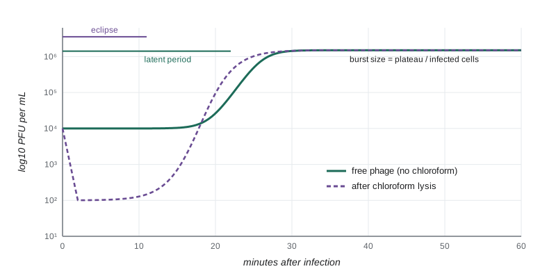
The curve has three parts. During the latent period, the plaque count stays constant, because each infected cell still makes just one plaque, whether it contains one virion or many. The first part of the latent period is the eclipse, during which chloroform-treated samples show no infectious virus at all: the infecting genomes have been uncoated, and no new virions have been assembled yet. The eclipse is the strongest evidence that viruses do not grow and divide but are assembled from parts. At the end of the latent period, cells lyse and the count rises to a plateau. The average number of virions released per infected cell, the burst size, is the ratio of the final count to the number of infected cells. For phage T4 in rich medium at 37 °C, the latent period is about 25 minutes and the burst size about 100 to 200; for animal viruses, cycles take hours to days and yields range from tens to many thousands of virions per cell.
import numpy as np

rng = np.random.default_rng(330)
t = np.arange(0, 61, 5)                          # minutes after infection
infected_cells = 2.0e5                           # per mL after dilution (from MOI and adsorption)
latent, rise, burst = 22, 8, 120
frac_lysed = 1 / (1 + np.exp(-(t - latent - rise / 2) / (rise / 6)))
pfu = infected_cells * (1 + (burst - 1) * frac_lysed)
pfu_obs = pfu * np.exp(rng.normal(0, 0.05, t.size))
for ti, p in zip(t, pfu_obs):
    print(f"{ti:3d} min  {p:10.3g} PFU/mL")
pre = pfu_obs[t <= 15].mean()
plateau = pfu_obs[t >= 45].mean()
print(f"infective centres before lysis = {pre:.3g} per mL")
print(f"burst size = plateau / infective centres = {plateau/pre:.0f}")
half = t[np.argmax(pfu_obs > pre + 0.5 * (plateau - pre))]
print(f"time to half-maximal rise = {half} min (end of latent period is a little earlier)")0 min 2.14e+05 PFU/mL 5 min 2.09e+05 PFU/mL 10 min 2.09e+05 PFU/mL 15 min 2.1e+05 PFU/mL 20 min 4.81e+05 PFU/mL 25 min 8.08e+06 PFU/mL 30 min 2.21e+07 PFU/mL 35 min 2.46e+07 PFU/mL 40 min 2.49e+07 PFU/mL 45 min 2.47e+07 PFU/mL 50 min 2.36e+07 PFU/mL 55 min 2.4e+07 PFU/mL 60 min 2.51e+07 PFU/mL infective centres before lysis = 2.1e+05 per mL burst size = plateau / infective centres = 116 time to half-maximal rise = 30 min (end of latent period is a little earlier)
The simulation recovers the burst size from the ratio of the plateau to the early count of infective centres. In real experiments, two corrections are common: the early count must be measured after unadsorbed phages have been removed or neutralized, otherwise free phages inflate it, and the number of infected cells can be checked independently from the drop in viable bacterial counts.
Lytic and temperate phages
Virulent phages, such as T4, can only follow the lytic cycle: every infection ends in the production of new phages and death of the cell. Temperate phages, such as lambda, have a choice. After infection they may follow the lytic cycle or enter lysogeny, in which the phage genome integrates into the host chromosome as a prophage (or, for some phages, persists as a plasmid) and is copied along with it, while a repressor protein keeps the lytic genes switched off. The host cell, now a lysogen, is immune to further infection by the same phage because the repressor also blocks incoming genomes. The prophage can stay silent for many generations. If the host’s DNA is damaged, the SOS response (lesson 7) leads to cleavage of the repressor, the prophage excises and the lytic cycle begins: the phage abandons a cell that may be about to die.
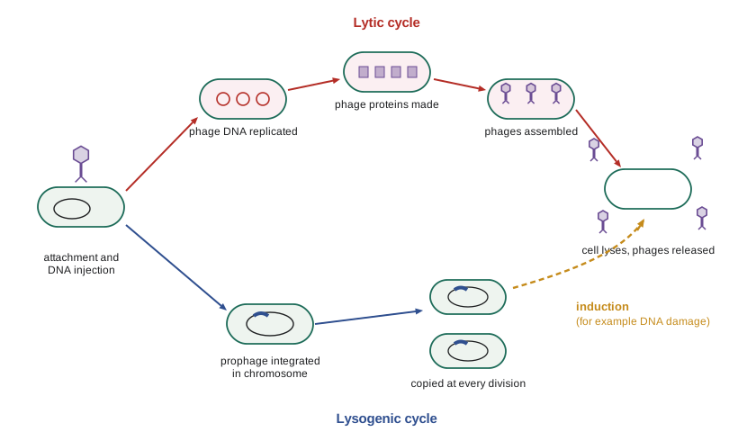
The lambda decision between lysis and lysogeny is one of the best-understood genetic switches in biology, governed by competition between two repressors, CI and Cro, for the same operator sites. Its outcome depends on the physiological state of the cell and on the number of phages infecting it: high multiplicity of infection and starved cells favour lysogeny, a strategy that makes sense when hosts are scarce. Some phages even communicate: the SPbeta family of Bacillus phages release a short peptide signal, called arbitrium, whose accumulation tells later infecting phages that hosts are becoming scarce and tips them towards lysogeny. Prophages are extremely common: most sequenced bacterial genomes contain at least one, and many carry several. Through lysogenic conversion (lesson 7), prophages give their hosts toxins, resistance to other phages and other traits.
Phage therapy
Félix d’Hérelle, who co-discovered bacteriophages in 1917, used them to treat dysentery and cholera in the 1920s and 1930s, and phage therapy was widely practised until antibiotics displaced it in western countries. It continued in the Soviet Union, particularly at the Eliava Institute in Tbilisi, Georgia, and in Poland. The rise of multidrug-resistant bacteria has revived interest. Phages have attractive properties as therapeutics: they are highly specific, sparing the normal microbiota; they replicate at the site of infection; and new phages can be isolated against resistant strains. They also have drawbacks. Their narrow host range means that the infecting strain must be matched with suitable phages; bacteria evolve phage resistance quickly, so cocktails of several phages are used; the immune system can neutralize them; and temperate phages are avoided because they could transfer toxin or resistance genes.
Well-documented compassionate-use cases have shown what phage therapy can do. In 2016, Tom Patterson, a professor at the University of California, San Diego, recovered from a multidrug-resistant Acinetobacter baumannii infection after intravenous phage treatment assembled by several laboratories, and in 2019 a British teenager with cystic fibrosis received genetically engineered phages against a drug-resistant Mycobacterium abscessus infection. Controlled clinical trials have produced more mixed results, partly because of the difficulty of standardizing personalized products. Regulators treat phages as biological drugs: in Canada, phage therapy is available only through clinical trials or Health Canada’s Special Access Program, and in the United States through FDA-authorized trials or expanded-access (emergency) requests. Phage-derived enzymes such as endolysins, and phages used to control Listeria and Salmonella on foods, approved in both countries as processing aids, are other applications.
Animal virus strategies
Three families of animal viruses illustrate how replication strategy shapes biology. Influenza viruses (Baltimore group V) have eight separate negative-sense RNA segments and replicate in the nucleus. Their error-prone polymerase produces a steady stream of mutations in the surface proteins hemagglutinin and neuraminidase, so antibodies made against last year’s strains bind this year’s less well, a process called antigenic drift that requires seasonal vaccines to be updated each year. When two different influenza viruses infect the same cell, their segments can be mixed in the progeny, a process called reassortment; if a human virus acquires a hemagglutinin segment from an animal virus to which people have no immunity, the result is an antigenic shift, which caused the pandemics of 1957, 1968 and 2009.
Retroviruses (group VI), including HIV, carry two copies of a positive-sense RNA genome and a reverse transcriptase that copies it into double-stranded DNA after entry. The viral integrase inserts that DNA into a host chromosome as a provirus, which is then transcribed by host RNA polymerase II. Integration makes infection permanent: latently infected cells carrying silent proviruses are the reason antiretroviral therapy controls HIV but rarely cures it. Reverse transcriptase is highly error-prone and also switches between the two genome copies, recombining them. Combination antiretroviral therapy attacks several steps at once, reverse transcription, integration, protease processing of viral proteins and entry, using the same logic as combination therapy for tuberculosis (lesson 7).
Coronaviruses (group IV) have the largest known RNA genomes among animal viruses, about 30,000 nucleotides. The genome is translated into large polyproteins that viral proteases cut into the replication machinery, which then makes a nested set of subgenomic mRNAs for the structural proteins. Their proofreading exonuclease keeps the mutation rate lower than that of most RNA viruses, but recombination between related coronaviruses in co-infected hosts is common and has shaped their evolution, including the emergence of SARS-CoV, MERS-CoV and SARS-CoV-2.
Viral evolution
RNA viruses mutate at roughly 10⁻⁶ to 10⁻⁴ substitutions per nucleotide per replication cycle, thousands of times faster than DNA-based organisms per nucleotide. Combined with huge population sizes and short generation times, this means that an infected person may carry a cloud of related variants, sometimes called a quasispecies or mutant swarm, rather than a single sequence. High mutation rates let RNA viruses adapt quickly to new hosts, immune responses and drugs, but they also come close to a limit: many mutations are harmful, and drugs that increase the mutation rate further, such as ribavirin and molnupiravir, can push viruses into lethal mutagenesis.
import numpy as np

viruses = {   # mutation rate (per nucleotide per replication), genome length (nucleotides)
    "poliovirus (RNA, +)": (9e-5, 7_500),
    "influenza A (RNA, -)": (2e-5, 13_600),
    "HIV-1 (retrovirus)": (3e-5, 9_700),
    "SARS-CoV-2 (RNA, proofreading)": (1e-6, 29_900),
    "herpes simplex (dsDNA)": (2e-8, 152_000),
    "E. coli (bacterial genome)": (2e-10, 4_640_000),
}
print(f"{'genome':34s} {'mutations per genome copy':>26s} {'P(no mutation)':>15s}")
for name, (mu, L) in viruses.items():
    U = mu * L
print(f"{name:34s} {U:26.4f} {np.exp(-U):15.3f}")genome mutations per genome copy P(no mutation) poliovirus (RNA, +) 0.6750 0.509 influenza A (RNA, -) 0.2720 0.762 HIV-1 (retrovirus) 0.2910 0.748 SARS-CoV-2 (RNA, proofreading) 0.0299 0.971 herpes simplex (dsDNA) 0.0030 0.997 E. coli (bacterial genome) 0.0009 0.999
The printed genomic rates show the contrast. A poliovirus or influenza genome picks up a new mutation in roughly one copy in two to five, whereas a herpesvirus or bacterial genome is copied without error almost every time. The rates are approximate and vary between studies and cell types, but the orders of magnitude are robust. Genomic sequencing of viruses during outbreaks uses these rates as a molecular clock: by counting differences between viral genomes sampled at known dates, phylogenetic methods estimate when an outbreak began, how fast it is growing and how it has spread between places, an approach that became routine during the COVID-19 pandemic.
Emerging viruses
Most new human viral diseases are zoonoses, infections that spill over from animals. Spillover requires a chain of events: the virus must be abundant in an animal reservoir, people must be exposed to it, it must be able to infect human cells and, to cause an epidemic, it must transmit efficiently between people. Bats, rodents and birds are important reservoirs. Recent examples include HIV from chimpanzee simian immunodeficiency virus in the early twentieth century, SARS in 2002 and 2003, MERS from camels since 2012, Ebola outbreaks in Africa, Zika’s spread through the Americas in 2015 and 2016, and SARS-CoV-2 from 2019. Mpox, endemic in parts of central and west Africa, spread globally among people in 2022 and a new clade caused a public health emergency of international concern declared by the WHO in 2024.
Highly pathogenic avian influenza H5N1 of clade 2.3.4.4b illustrates emergence in progress. Since 2020 it has spread through wild birds on every continent except Australia, devastated poultry flocks and infected many wild mammals. In March 2024 it was found in dairy cattle in the United States, where it went on to affect hundreds of herds in many states and to infect about 70 people by early 2026, mostly farm workers with mild illness, with one death reported in Louisiana in January 2025. In Canada, a British Columbia teenager was critically ill with H5N1 in November 2024, the first locally acquired human case in the country, and repeated testing of milk has so far not detected the virus in Canadian dairy cattle (status reviewed October 2026). The Public Health Agency of Canada, the Canadian Food Inspection Agency and provincial authorities lead surveillance. The key question is whether the virus acquires mutations that allow efficient human-to-human transmission; HSE-245 covers protective measures for workers exposed to infected animals.
Surveillance for emerging viruses increasingly combines clinical testing with genomic sequencing and with wastewater monitoring, in which viral RNA shed in feces is measured in sewage to track infection trends in whole communities without testing individuals. Canada’s National Microbiology Laboratory and the US CDC’s National Wastewater Surveillance System both expanded wastewater programmes during the COVID-19 pandemic and now monitor other targets, such as influenza, RSV, mpox and, in some places, H5 influenza. Lesson 12 returns to how public health laboratories use these tools in outbreak investigation.
Worked problem
Reading a one-step growth experiment
Bacteria at 10⁸ cells per mL are infected with phage at an MOI of 0.1. After adsorption and dilution, plaque counts are 1.0 × 10⁴ PFU per mL from 0 to 20 minutes and 1.5 × 10⁶ PFU per mL from 40 minutes onward. Chloroform-treated samples show no PFU at 10 minutes and 8 × 10⁵ at 25 minutes. Find the burst size and comment on the eclipse and latent periods.
Show solution
- Before lysis, each infected cell is one infective centre, so there are 1.0 × 10⁴ infected cells per mL in the diluted culture.
- Burst size = 1.5 × 10⁶/1.0 × 10⁴ = 150 phages per cell.
- Chloroform samples show no infectious phage at 10 minutes, so the eclipse lasts at least 10 minutes; mature phages are already present inside cells at 25 minutes.
- Free-phage counts are flat until after 20 minutes, so lysis begins sometime between 20 and 40 minutes; more frequent sampling would pin down the latent period.
Answer: A burst size of about 150, an eclipse of at least 10 minutes and a latent period of a little over 20 minutes, typical of T-even phages.
Common mistakes
MistakeThinking virus numbers rise during the latent period because the virus is growing.
InsteadEach infected cell counts as one plaque until it lyses; in the eclipse no infectious virus exists inside cells at all.
MistakeAssuming lysogenic phages are harmless.
InsteadProphages can be induced to lyse their hosts and can carry toxin genes that make bacteria pathogenic.
MistakeConfusing antigenic drift with antigenic shift.
InsteadDrift is gradual change by mutation; shift is the sudden acquisition of a new surface protein by reassortment.
MistakeExpecting a single phage to cure every infection by a bacterial species.
InsteadPhages are strain-specific and resistance evolves quickly, so matched cocktails are needed.
MistakeAssuming emerging viruses usually arise from new mutations in human viruses.
InsteadMost come from animal reservoirs through spillover.
MistakeTreating wastewater signals as case counts.
InsteadWastewater measures shed viral RNA in a community, which tracks trends but depends on shedding, flow and dilution.
Summary and key terms
- Viral replication proceeds through attachment, penetration and uncoating, genome replication and expression, assembly and release; receptors determine host range and tropism.
- The one-step growth curve reveals the eclipse period, the latent period and the burst size of a single infection cycle.
- Virulent phages are always lytic; temperate phages can integrate as prophages, conferring immunity and new traits, and are induced by DNA damage.
- Phage therapy, revived for multidrug-resistant infections, is limited by host specificity, resistance and regulation.
- Influenza evolves by drift and reassortment-driven shift; retroviruses integrate as proviruses; coronaviruses have large proofread RNA genomes.
- RNA viruses mutate at 10⁻⁶ to 10⁻⁴ per nucleotide per replication, enabling rapid adaptation and molecular-clock tracking.
- Most emerging viruses are zoonoses; H5N1 in dairy cattle shows emergence in progress, and wastewater and genomic surveillance track spread.
- tropism
- The range of cells, tissues or hosts that a virus can infect.
- eclipse period
- The early part of infection during which no infectious virions exist inside the cell.
- latent period
- The time from infection to release of new virions.
- burst size
- The average number of virions released per infected cell.
- lysogeny
- A state in which a temperate phage genome persists in the host as a prophage without killing it.
- prophage
- A phage genome integrated into, or replicating with, the host genome.
- induction
- Switching a prophage to the lytic cycle, often triggered by DNA damage.
- antigenic drift
- Gradual change in viral surface proteins through accumulated mutations.
- antigenic shift
- Abrupt change in influenza surface proteins through reassortment of genome segments.
- zoonosis
- An infection transmitted from animals to people.
Practice problems
Work each problem on paper first, then reveal the answer to check.
What decides whether a virus can infect a particular cell type?
Show answer
Mainly the presence of the receptor and co-receptors its attachment proteins bind, plus intracellular factors.
Why is the eclipse period evidence that viruses do not divide?
Show answer
Infectivity disappears entirely and reappears only when new virions are assembled from parts.
In a one-step experiment, infective centres are 5 × 10³ per mL and the plateau is 6 × 10⁵ PFU per mL. What is the burst size?
Show answer
120.
Why are infected cultures diluted after adsorption in a one-step experiment?
Show answer
So released phages cannot infect new cells and start a second cycle during the experiment.
What makes a lysogen immune to superinfection by the same phage?
Show answer
The prophage repressor blocks expression of incoming phage genomes.
Why are temperate phages avoided in phage therapy?
Show answer
They may form lysogens rather than kill, and can transfer toxin or resistance genes.
What causes influenza antigenic shift?
Show answer
Reassortment of genome segments between different influenza viruses in one cell, introducing a new surface protein.
Why is HIV infection rarely cured by antiretroviral therapy?
Show answer
Latent proviruses integrated in long-lived cells are not affected by the drugs.
A genome of 10,000 nucleotides mutates at 5 × 10⁻⁵ per nucleotide per replication. What fraction of copies are error-free?
Show answer
e^{−0.5} ≈ 0.61.
Name two kinds of animal reservoir important for emerging viruses.
Show answer
Bats, rodents, wild birds and livestock are all important.
Lesson quiz · 5 questions
Score 60% or more to complete this lesson.
Create a free account to save your progress, take quizzes and earn a certificate.