
BIOL-330 · Lesson 2 of 12 · 32 min
Microbial cell structure: bacteria and archaea
A prokaryotic cell has no nucleus and, in most species, no membrane-bound organelles, but it is far from a simple bag of enzymes. Its envelope is a layered structure that holds the cell together against osmotic pressure, controls what enters and leaves, anchors motility machines and presents the surface that hosts, viruses and antibiotics encounter. Its cytoplasm is organized by a cytoskeleton, its chromosome is compacted into a nucleoid, and many species build specialized inclusions, compartments and resting forms. BIOL-107 compared prokaryotic and eukaryotic cells in outline. This lesson looks at the structures that matter most to microbiologists and explains why they matter for staining, antibiotic action, pathogenesis and survival.
Shape, size and arrangement
Most bacteria and archaea are cocci (spheres), bacilli (rods) or spirals, with variations such as vibrios (curved rods), spirilla (rigid spirals) and spirochetes (flexible spirals that move with internal flagella). A few are star-shaped, square or branched, and some archaea from salt ponds are flat squares. Shape is maintained by the cell wall, which is laid down under the direction of cytoskeletal proteins: MreB, an actin homologue, guides wall synthesis along the sides of rods, and FtsZ, a tubulin homologue, forms the ring that constricts the cell at division. Cells that divide and stay attached form characteristic arrangements. Streptococci divide in one plane and form chains; staphylococci divide in random planes and form grape-like clusters; Sarcina forms cubes of eight.
Why are most prokaryotes small? A cell exchanges nutrients and wastes across its surface but needs them throughout its volume. For a sphere of radius r, the surface area is 4πr² and the volume is (4/3)πr³, so the ratio of surface area to volume is 3/r. Halving the radius doubles the surface available for each unit of volume. Diffusion also limits size: the average time for a small molecule to diffuse a distance x is roughly x²/(2D), where D is the diffusion coefficient. In water, glucose has D of about 6.7 × 10⁻¹⁰ m² per second, so crossing 1 µm takes under a millisecond, but crossing 100 µm takes several seconds, far too slow to supply a rapidly metabolizing cell from its edge.
import numpy as np

D_glucose = 6.7e-10          # m^2/s in water at 25 C
for name, r_um in [("Mycoplasma", 0.15), ("E. coli (sphere-equivalent)", 0.6),
                   ("yeast", 3.0), ("Epulopiscium (sphere-equivalent)", 40.0)]:
    r = r_um * 1e-6
    sav = 3 / r_um                                   # per micrometre, sphere
    t_cross = (r ** 2) / (2 * D_glucose)             # time to diffuse one radius
    print(f"{name:32s} r = {r_um:6.2f} um  SA/V = {sav:6.2f} per um  diffusion time = {t_cross*1e3:9.3f} ms")

# a rod: cylinder of radius a with hemispherical caps, total length L
def rod_sav(a, L):
    cyl = L - 2 * a
    area = 2 * np.pi * a * cyl + 4 * np.pi * a ** 2
    vol = np.pi * a ** 2 * cyl + (4 / 3) * np.pi * a ** 3
    return area / vol
print(f"E. coli rod 0.5 um wide x 2 um long: SA/V = {rod_sav(0.25, 2.0):.2f} per um")Mycoplasma r = 0.15 um SA/V = 20.00 per um diffusion time = 0.017 ms E. coli (sphere-equivalent) r = 0.60 um SA/V = 5.00 per um diffusion time = 0.269 ms yeast r = 3.00 um SA/V = 1.00 per um diffusion time = 6.716 ms Epulopiscium (sphere-equivalent) r = 40.00 um SA/V = 0.07 per um diffusion time = 1194.030 ms E. coli rod 0.5 um wide x 2 um long: SA/V = 8.73 per um
The output shows the size trap clearly. A Mycoplasma cell has twenty times the surface area per unit volume of a yeast cell and diffusion across it is almost instantaneous. A cell 40 µm in radius would wait seconds for glucose to diffuse to its centre. The giant bacteria solve the problem in different ways. Thiomargarita and its relatives are mostly a large central vacuole filled with nitrate, so the living cytoplasm is a thin layer just under the surface. Epulopiscium, which lives in the guts of surgeonfish, has tens of thousands of copies of its genome distributed around the cell periphery, so that protein synthesis happens close to where it is needed. Thiomargarita magnifica goes further and packages its DNA and ribosomes in membrane-bound compartments, blurring the textbook line between prokaryotes and eukaryotes.
The cytoplasmic membrane
The cytoplasmic membrane of bacteria is a phospholipid bilayer studded with proteins, much like the eukaryotic membranes described in BIOL-107, but it does more work. Because most prokaryotes have no mitochondria or chloroplasts, their respiratory and photosynthetic electron transport chains and their ATP synthases are in this membrane, which therefore maintains the proton motive force that powers ATP synthesis, transport and flagellar rotation. Bacterial membranes lack sterols in most species but contain hopanoids, sterol-like molecules that stabilize the bilayer. Transport proteins use the proton motive force, ATP or the phosphotransferase system, which phosphorylates sugars as they enter, trapping them inside the cell.
Archaeal membranes are chemically different, and the difference is one of the deepest divisions in biology. Bacterial and eukaryotic phospholipids have fatty acids joined to glycerol-3-phosphate by ester bonds. Archaeal lipids have branched isoprenoid chains (phytanyl groups) joined to glycerol-1-phosphate by ether bonds, the opposite stereochemistry. Many hyperthermophilic archaea have tetraether lipids whose long chains span the whole membrane, forming a monolayer instead of a bilayer. Ether bonds resist hydrolysis and the monolayer cannot peel apart at high temperature, which helps explain how archaea such as Pyrolobus and Methanopyrus grow above 100 °C. The lipid divide also has practical uses: archaeal tetraether lipids preserved in sediments are used to reconstruct past ocean temperatures.
Peptidoglycan: the bacterial wall
The cytoplasm of a bacterium is concentrated, so water tends to flow in by osmosis, generating an internal turgor pressure of a few atmospheres in Gram-negative cells and up to about 20 atmospheres in some Gram-positive cells. The cell wall resists that pressure. In almost all bacteria it is made of peptidoglycan (also called murein), a single giant molecule that surrounds the cell like a mesh bag. Its backbone consists of chains of two alternating amino sugars, N-acetylglucosamine (NAG) and N-acetylmuramic acid (NAM), joined by β-1,4 bonds. A short peptide hangs from each NAM, and peptides on neighbouring chains are cross-linked to one another, either directly or through a bridge of amino acids such as the five-glycine bridge of Staphylococcus aureus. The peptides contain d-amino acids and, in many Gram-negative bacteria, diaminopimelic acid, which are found nowhere else in nature.
Peptidoglycan is synthesized in the cytoplasm as a NAM–NAG–pentapeptide unit attached to a lipid carrier called bactoprenol, flipped across the membrane and then joined into the growing wall by two activities: transglycosylation, which extends the sugar chains, and transpeptidation, which forms the peptide cross-links. The transpeptidases are the penicillin-binding proteins, the targets of the β-lactam antibiotics. Because the wall must grow without ever being weakened, synthesis is coupled to the activity of autolysins that cut existing bonds to make room for new material. Antibiotics that block wall synthesis while autolysins keep cutting cause growing cells to burst, which is why β-lactams kill growing bacteria but not dormant ones. Lysozyme, an enzyme in tears, saliva and egg white, breaks the β-1,4 bond between NAM and NAG and is one of the body’s first defences.
| Feature | Gram-positive | Gram-negative |
|---|---|---|
| Peptidoglycan thickness | 20–80 nm, many layers | About 2–7 nm, one or a few layers |
| Outer membrane | Absent | Present, with lipopolysaccharide in the outer leaflet |
| Periplasm | Narrow or absent | Distinct compartment between the membranes |
| Teichoic acids | Present (wall and lipoteichoic acids) | Absent |
| Porins | Absent | Present in the outer membrane |
| Endotoxin (lipid A) | Absent | Present |
| Sensitivity to lysozyme and penicillin | Generally high | Generally lower, because the outer membrane limits access |
| Examples | Staphylococcus, Streptococcus, Bacillus, Clostridium, Listeria | E. coli, Salmonella, Pseudomonas, Neisseria, Vibrio |
Gram-positive and Gram-negative envelopes
In Gram-positive bacteria, peptidoglycan forms a thick wall of many layers directly outside the cytoplasmic membrane. Threaded through it are teichoic acids, polymers of glycerol phosphate or ribitol phosphate that carry a strong negative charge, bind metal ions and help regulate autolysins; lipoteichoic acids are anchored in the membrane. Many surface proteins are covalently attached to the wall by sortase enzymes, including adhesins that help pathogens such as S. aureus attach to host tissues.
The Gram-negative envelope has three layers. Outside the cytoplasmic membrane lies the periplasm, a gel-like compartment containing a thin layer of peptidoglycan and many proteins: binding proteins for transport, enzymes that break down nutrients and, in resistant strains, β-lactamases that destroy penicillins before they reach their targets. The outer membrane is a second lipid bilayer, tied to the peptidoglycan by Braun’s lipoprotein. Its inner leaflet is phospholipid, but its outer leaflet is mostly lipopolysaccharide (LPS). Each LPS molecule has three parts: lipid A, which anchors it in the membrane; a core polysaccharide; and the O-antigen, a long, variable sugar chain that differs between strains and is the basis of serotyping (the O in E. coli O157:H7). Lipid A is the endotoxin of Gram-negative bacteria. Released when cells die or divide, it triggers powerful inflammatory responses, and in the bloodstream it can cause fever, a drop in blood pressure and septic shock. The way the immune system recognizes LPS is a topic for BIOL-380.
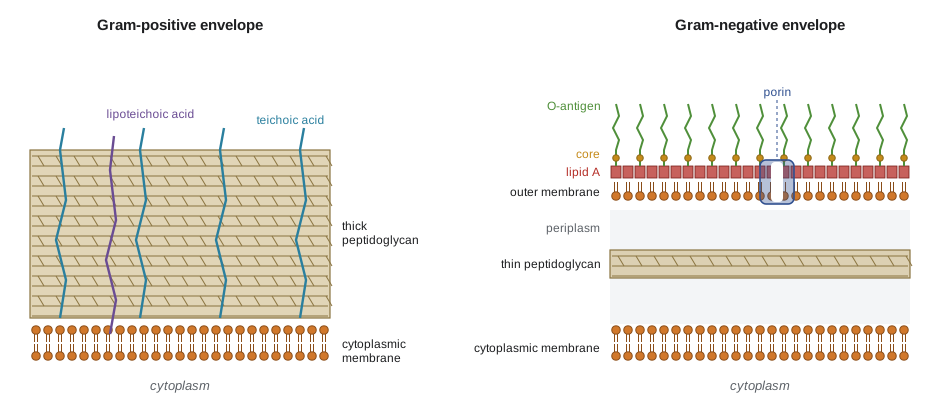
The outer membrane is an effective permeability barrier. Small hydrophilic molecules pass through porins, water-filled channels that exclude molecules larger than about 600 daltons, while the tightly packed LPS layer slows the entry of hydrophobic compounds. Bile salts, detergents, many dyes and large antibiotics such as vancomycin cannot cross it easily. This is why Gram-negative infections are generally harder to treat, why vancomycin is used only against Gram-positive organisms, and why MacConkey agar can use bile salts and crystal violet to suppress Gram-positive bacteria. It is also why reduced porin expression and increased efflux are important resistance mechanisms in Gram-negative pathogens (lesson 5).
Other envelopes
Mycobacteria, including the agents of tuberculosis and leprosy, have a distinctive envelope. Their peptidoglycan is linked to arabinogalactan, a branched polysaccharide, which in turn carries mycolic acids, very long fatty acids with 60 to 90 carbons. Together with other lipids these form an outer layer, sometimes called the mycomembrane, that is waxy, very impermeable and responsible for the acid-fast staining reaction, the slow growth of mycobacteria and their resistance to many disinfectants and antibiotics. Tuberculosis treatment therefore uses drugs aimed at this envelope, such as isoniazid, which blocks mycolic acid synthesis, and ethambutol, which blocks arabinogalactan synthesis, combined for months.
Mycoplasmas have no cell wall at all. They live in osmotically protected environments inside animal and plant hosts, strengthen their membranes with sterols taken from the host, and are naturally resistant to every antibiotic that acts on the wall. Archaea never have peptidoglycan. Most have an S-layer, a two-dimensional crystalline lattice of protein or glycoprotein that forms the only wall in many species. Some methanogens have pseudomurein, a polymer that looks like peptidoglycan but contains N-acetyltalosaminuronic acid in place of NAM, β-1,3 rather than β-1,4 links, and only l-amino acids, so lysozyme and penicillin do not affect it. S-layers are also common in bacteria, where they sit on top of the wall or outer membrane.
Capsules, slime and biofilms
Many bacteria surround themselves with a layer of polysaccharide or, less often, protein. A capsule is organized and firmly attached; a slime layer is loose and easily washed off. Capsules protect pathogens from being engulfed by phagocytes, which is why encapsulated strains of Streptococcus pneumoniae are virulent and unencapsulated ones are not, an observation at the heart of Griffith’s transformation experiment (BIOL-207). Several important vaccines, including those against pneumococcus, Haemophilus influenzae type b and meningococcus, are made from capsular polysaccharides linked to carrier proteins. Extracellular polysaccharides also form much of the matrix of biofilms, the surface-attached communities discussed in lesson 10, which protect their inhabitants from drying, predators, disinfectants and antibiotics.
Appendages: flagella, pili and fimbriae
The bacterial flagellum is a rotary motor, one of the most remarkable machines in biology. A helical filament made of thousands of copies of the protein flagellin is joined by a flexible hook to a basal body embedded in the envelope. Rings of proteins in the basal body act as a rotor, and stator proteins surrounding it let protons (or sodium ions in some marine bacteria) flow down their electrochemical gradient, turning the rotor at speeds of up to about 300 revolutions per second in E. coli and over 1,000 in some Vibrio species. Flagella may be arranged singly at one pole (monotrichous), in tufts (lophotrichous), at both poles (amphitrichous) or all over the cell (peritrichous); the arrangement helps identify species. Spirochetes keep their flagella in the periplasm, where their rotation makes the whole cell corkscrew through viscous fluids such as mucus. The flagellin and the H-antigens used in serotyping are the same molecule: the H7 in E. coli O157:H7 names the flagellar type.
Archaea swim with a structurally unrelated motor, the archaellum, which is built like a bacterial type IV pilus and is driven by ATP hydrolysis rather than a proton gradient. Its existence is another reminder that similar functions have evolved more than once.
Bacteria use flagella to move towards favourable conditions in a process called chemotaxis. A swimming E. coli cell alternates between runs, in which its flagella rotate counterclockwise and bundle together to push the cell forward, and tumbles, in which one or more flagella reverse, the bundle flies apart and the cell reorients at random. Receptors at the cell pole sense attractants and repellents and, through a phosphorylation cascade, change the probability of tumbling. When conditions are improving, tumbles are suppressed and runs lengthen; when they are getting worse, the cell tumbles sooner. The result is a biased random walk that moves cells up gradients, and because the receptors adapt through methylation, the cell responds to changes in concentration rather than absolute levels, over a range of five orders of magnitude.
Pili and fimbriae are thinner, straighter protein filaments. Fimbriae, often hundreds per cell, attach bacteria to surfaces and host cells; the P fimbriae of uropathogenic E. coli bind to sugars on the lining of the urinary tract. Type IV pili extend and retract, pulling cells across surfaces in a jerky motion called twitching motility, and they also take up DNA during transformation. The conjugative or sex pilus links donor and recipient cells during plasmid transfer, the subject of lesson 7.
Inside the cell
The bacterial chromosome is usually a single circular DNA molecule, although some species have linear or multiple chromosomes. It is compacted into the nucleoid by supercoiling and nucleoid-associated proteins and occupies a defined region of the cytoplasm without a surrounding membrane. Plasmids, smaller DNA molecules that replicate independently, carry optional genes such as those for antibiotic resistance, toxin production or the breakdown of unusual compounds. Ribosomes are of the 70S type, made of 30S and 50S subunits, and their differences from the 80S ribosomes of eukaryotic cytoplasm are what allow drugs such as streptomycin and tetracycline to inhibit bacterial protein synthesis selectively.
Many prokaryotes store reserves in inclusions: granules of poly-β-hydroxybutyrate (a carbon store and a source of biodegradable plastics), glycogen, polyphosphate and sulfur. Some have specialized compartments. Carboxysomes are protein-shelled polyhedral bodies that concentrate the CO₂-fixing enzyme RuBisCO in cyanobacteria; gas vesicles are hollow protein cylinders that let aquatic cyanobacteria and archaea float to the right depth; and magnetosomes are membrane-bound crystals of magnetite that align magnetotactic bacteria with Earth’s magnetic field so they can swim along field lines to the sediments they prefer.
Endospores
Some Gram-positive bacteria, notably Bacillus and Clostridium and their relatives, form endospores when nutrients run out. Sporulation is an elaborate developmental programme lasting several hours. The cell divides asymmetrically, the larger mother cell engulfs the smaller forespore, and the mother cell then builds around it a cortex of modified peptidoglycan and a protein coat before lysing to release the mature spore. The spore core is partly dehydrated, rich in calcium dipicolinate, and its DNA is protected by small acid-soluble proteins. Together these features make endospores resistant to heat, drying, radiation and many chemicals. They can survive boiling for hours, and viable spores have been recovered from sediments thousands of years old. When conditions improve, spores germinate within minutes and resume growth.
Endospores matter in practice for three reasons. They cause disease: Clostridium botulinum spores in improperly canned food, Clostridioides difficile spores in hospitals and Bacillus anthracis spores in contaminated soil and animal products. They set the standard for sterilization: an autoclave cycle or a canning process is designed to kill spores, because anything that kills them kills nearly everything else (lesson 4). And they spread easily: spores are not killed by alcohol-based hand sanitizers, which is why soap and water and sporicidal disinfectants are used where C. difficile is present (HSE-245).
Worked problem
Predicting an organism’s sensitivity from its envelope
Three organisms are tested against lysozyme and penicillin G: (a) Staphylococcus aureus, (b) E. coli, (c) Mycoplasma pneumoniae. Predict which are sensitive, and explain.
Show solution
- S. aureus is Gram-positive with an exposed, thick peptidoglycan wall: lysozyme can reach and cut the NAM–NAG bonds, and penicillin can reach the penicillin-binding proteins. Both are expected to act unless the strain makes β-lactamase or an altered target.
- E. coli is Gram-negative: the outer membrane shields its thin peptidoglycan from lysozyme, and penicillin G crosses poorly. Both are much less effective, although agents that disrupt the outer membrane, such as EDTA, make the cells sensitive to lysozyme.
- Mycoplasma has no wall at all, so neither agent has a target.
Answer: Expect sensitivity in S. aureus (subject to acquired resistance), low sensitivity in E. coli and no sensitivity in Mycoplasma. The prediction follows directly from envelope structure.
Common mistakes
MistakeDescribing the Gram-negative wall as thicker because it has more layers.
InsteadGram-negative envelopes have more layers, but the peptidoglycan itself is much thinner than in Gram-positive cells.
MistakeAssuming archaea have peptidoglycan walls.
InsteadArchaea never have peptidoglycan; most have S-layers, and some have pseudomurein, which lysozyme and penicillin do not affect.
MistakeCalling endotoxin a protein toxin secreted by bacteria.
InsteadEndotoxin is lipid A of the lipopolysaccharide in the Gram-negative outer membrane; secreted protein toxins are exotoxins.
MistakeThinking flagella whip back and forth like eukaryotic flagella.
InsteadBacterial flagella are rigid helices turned by a rotary motor at the base, powered by ion flow.
MistakeTreating endospores as a form of reproduction.
InsteadOne cell makes one spore, and one spore makes one cell; sporulation is a survival strategy, not multiplication.
MistakeExpecting alcohol hand rubs to kill all microbes on hands.
InsteadThey do not kill bacterial endospores, so soap and water are used where spore-formers such as C. difficile are a concern.
Summary and key terms
- Small cell size gives prokaryotes a high surface area to volume ratio (3/r for a sphere) and short diffusion times; giant bacteria use vacuoles, polyploidy or compartments to overcome the limit.
- Bacterial membranes carry out respiration and photosynthesis; archaeal membranes use ether-linked isoprenoid lipids and may form heat-stable monolayers.
- Peptidoglycan is a cross-linked NAM–NAG mesh; its transpeptidases are the targets of β-lactams, and lysozyme cuts its sugar backbone.
- Gram-positive cells have thick peptidoglycan with teichoic acids; Gram-negative cells have thin peptidoglycan in a periplasm beneath an outer membrane containing LPS and porins.
- Mycobacteria have a waxy mycolic acid envelope; mycoplasmas have no wall; archaea have S-layers or pseudomurein.
- Capsules resist phagocytosis, flagella are rotary motors driving chemotaxis, and pili mediate attachment, twitching and DNA transfer.
- Endospores are dormant, highly resistant structures that set the standard for sterilization.
- peptidoglycan
- The mesh-like polymer of NAM, NAG and cross-linked peptides that forms the bacterial cell wall.
- penicillin-binding protein
- A transpeptidase that forms peptidoglycan cross-links and is inhibited by β-lactam antibiotics.
- outer membrane
- The second lipid bilayer of Gram-negative bacteria, with lipopolysaccharide in its outer leaflet.
- lipopolysaccharide
- The molecule of the Gram-negative outer leaflet, made of lipid A, core polysaccharide and O-antigen.
- endotoxin
- Lipid A of lipopolysaccharide, which triggers strong inflammatory responses.
- periplasm
- The compartment between the cytoplasmic and outer membranes of Gram-negative bacteria.
- S-layer
- A crystalline surface layer of protein or glycoprotein found in many archaea and bacteria.
- pseudomurein
- A peptidoglycan-like wall polymer of some methanogenic archaea.
- chemotaxis
- Movement directed by chemical gradients, achieved in bacteria by changing the frequency of tumbles.
- endospore
- A dormant, highly resistant structure formed inside some Gram-positive bacteria.
Practice problems
Work each problem on paper first, then reveal the answer to check.
What is the surface area to volume ratio of a spherical cell with a radius of 0.5 µm?
Show answer
3/r = 6 per µm.
About how long does glucose (D = 6.7 × 10⁻¹⁰ m²/s) take to diffuse 10 µm?
Show answer
x²/(2D) = 10⁻¹⁰/(1.34 × 10⁻⁹) ≈ 0.075 s.
Which bond in peptidoglycan does lysozyme cut?
Show answer
The β-1,4 glycosidic bond between NAM and NAG.
Why do β-lactams kill growing cells but not dormant ones?
Show answer
They block cross-linking while autolysins continue cutting the wall during growth, so the weakened wall bursts; dormant cells are not remodelling their walls.
Name the three parts of lipopolysaccharide.
Show answer
Lipid A, core polysaccharide and O-antigen.
Why is vancomycin ineffective against most Gram-negative bacteria?
Show answer
It is too large to pass through porins in the outer membrane.
What two features distinguish archaeal membrane lipids from bacterial lipids?
Show answer
Ether rather than ester linkages, and branched isoprenoid chains on glycerol-1-phosphate.
Why is Mycoplasma resistant to penicillin?
Show answer
It has no cell wall, so there is no peptidoglycan synthesis to inhibit.
How does E. coli move up an attractant gradient?
Show answer
It suppresses tumbles while conditions improve, lengthening runs in the favourable direction.
Give two features of the endospore core that contribute to heat resistance.
Show answer
Dehydration and calcium dipicolinate (plus DNA-protecting small acid-soluble proteins).
Lesson quiz · 5 questions
Score 60% or more to complete this lesson.
Create a free account to save your progress, take quizzes and earn a certificate.