
BIOL-330 · Lesson 8 of 12 · 29 min
Viruses: structure, classification and counting
Viruses are the most abundant biological entities on Earth. A litre of sea water holds around ten billion virus particles, and estimates of the global total run to about 10³¹, roughly ten for every prokaryotic cell. They infect every form of life, from bacteria and archaea to fungi, plants and animals, and they kill an estimated 20 to 40 per cent of marine bacteria every day, releasing nutrients that feed the next generation of plankton. They have shaped the evolution of their hosts, contributed genes to them (about 8 per cent of the human genome consists of the remains of retroviruses) and caused some of the most devastating diseases in history. BIOL-108 introduced viruses as non-cellular parasites. This lesson describes how virus particles are built, how viruses are classified by their genomes and how they are counted, and lesson 9 follows their replication.
What a virus is
A virus is an obligate intracellular parasite consisting of a nucleic acid genome, DNA or RNA but almost never both, enclosed in a protein coat and sometimes a lipid envelope. Outside a cell, the virus particle, or virion, is metabolically inert: it has no ribosomes, makes no ATP and cannot grow or divide. It is a delivery package for a genome. Inside a host cell, the genome takes over the cell’s machinery to make new virions. This life cycle, alternating between an inert extracellular particle and an intracellular replicating phase, sets viruses apart from even the smallest cellular parasites, which, like Rickettsia and Chlamydia, have ribosomes, divide by fission and are inhibited by antibiotics.
Whether viruses are alive is partly a matter of definition. They evolve by natural selection, carry genetic information and reproduce, but only by using the machinery of a living cell. Most microbiologists treat the infected cell, sometimes called the virocell, as the living form of the virus and the virion as its dispersal stage, much as a seed is to a plant.
Virion architecture
The protein shell that surrounds the genome is the capsid, built from many copies of one or a few kinds of protein subunits. Using many identical subunits is economical: a small genome can encode a large shell, and the subunits assemble spontaneously, following the rules of symmetry. The capsid together with the genome is the nucleocapsid. Two kinds of symmetry dominate.
Helical capsids are rods or filaments in which subunits spiral around the nucleic acid. Tobacco mosaic virus, the first virus discovered, is a rigid rod 300 nm long built from 2,130 identical subunits wound around its RNA. The nucleocapsids of influenza, measles and rabies viruses are flexible helices inside an envelope. The length of a helical capsid is set by the length of the genome it packages.
Icosahedral capsids are roughly spherical shells with the symmetry of an icosahedron, a solid with 20 triangular faces, 12 vertices and 30 edges. It is the most efficient way to enclose a volume with identical subunits arranged symmetrically. The simplest icosahedral capsid has 60 identical subunits, three per face. Larger capsids use more subunits in slightly different, quasi-equivalent environments, as described by the theory of Donald Caspar and Aaron Klug (1962). The number of subunits is 60T, where T, the triangulation number, takes values 1, 3, 4, 7, 9, 12, 13 and so on, given by T = h² + hk + k² for non-negative integers h and k. Subunits group into pentamers at the 12 vertices and hexamers elsewhere.
# Caspar-Klug triangulation numbers T = h^2 + h*k + k^2 and capsid sizes
seen = {}
for h in range(0, 5):
    for k in range(0, h + 1):
        T = h * h + h * k + k * k
        if T > 0 and T not in seen:
            seen[T] = (h, k)
for T in sorted(seen)[:9]:
    h, k = seen[T]
    subunits = 60 * T
    pentamers, hexamers = 12, 10 * (T - 1)
    print(f"T = {T:2d} (h={h}, k={k}): {subunits:4d} subunits = {pentamers} pentamers + {hexamers:3d} hexamers")
examples = {1: "satellite tobacco necrosis virus", 3: "poliovirus (pseudo T = 3), many plant viruses",
            4: "hepatitis B virus core (mostly)", 7: "phage HK97, phage lambda", 16: "herpes simplex virus",
            25: "adenovirus (pseudo T = 25)"}
for T, ex in examples.items():
print(f" example T = {T}: {ex}")T = 1 (h=1, k=0): 60 subunits = 12 pentamers + 0 hexamers T = 3 (h=1, k=1): 180 subunits = 12 pentamers + 20 hexamers T = 4 (h=2, k=0): 240 subunits = 12 pentamers + 30 hexamers T = 7 (h=2, k=1): 420 subunits = 12 pentamers + 60 hexamers T = 9 (h=3, k=0): 540 subunits = 12 pentamers + 80 hexamers T = 12 (h=2, k=2): 720 subunits = 12 pentamers + 110 hexamers T = 13 (h=3, k=1): 780 subunits = 12 pentamers + 120 hexamers T = 16 (h=4, k=0): 960 subunits = 12 pentamers + 150 hexamers T = 19 (h=3, k=2): 1140 subunits = 12 pentamers + 180 hexamers example T = 1: satellite tobacco necrosis virus example T = 3: poliovirus (pseudo T = 3), many plant viruses example T = 4: hepatitis B virus core (mostly) example T = 7: phage HK97, phage lambda example T = 16: herpes simplex virus example T = 25: adenovirus (pseudo T = 25)
Many viruses of animals have an envelope, a lipid bilayer taken from a host membrane as the virus buds out of the cell, studded with viral glycoproteins that bind receptors and fuse the envelope with host membranes. Influenza’s hemagglutinin and neuraminidase, HIV’s gp120–gp41 and the spike protein of SARS-CoV-2 are envelope glycoproteins and the main targets of protective antibodies and vaccines. The envelope is the virus’s weak point outside the body: lipid solvents, detergents, soap and alcohol disrupt it, which is why enveloped viruses are the easiest to inactivate (lesson 4). Non-enveloped viruses, such as norovirus, rotavirus, poliovirus and adenovirus, survive longer on surfaces and resist alcohol and drying.
Some viruses have complex architectures. The tailed bacteriophages, such as T4, have an icosahedral head attached to a helical tail with a contractile sheath, baseplate and tail fibres, which together act as a molecular syringe to inject DNA through the bacterial envelope. Poxviruses are brick-shaped, with a complex internal core. Giant viruses such as mimivirus, discovered in 2003 inside amoebae and at first mistaken for a bacterium, have capsids about 500 nm across covered with fibres, and genomes of more than a million base pairs that encode some components of the translation apparatus, such as aminoacyl-tRNA synthetases. Pandoraviruses have genomes of up to about 2.5 million base pairs, larger than those of many bacteria.
Viral genomes
Viral genomes are astonishingly varied. They may be DNA or RNA, single- or double-stranded, linear or circular, in one piece or segmented, and range in size from under 2,000 nucleotides in circoviruses to over 2 million in pandoraviruses. Single-stranded RNA genomes are described by their sense. A positive-sense (plus-strand) RNA can be translated directly by ribosomes, so it behaves as messenger RNA, and the purified RNA of such viruses is infectious on its own. A negative-sense RNA is complementary to mRNA and must first be copied into positive-sense RNA, so these viruses must carry their own RNA-dependent RNA polymerase inside the virion, because host cells do not have one.
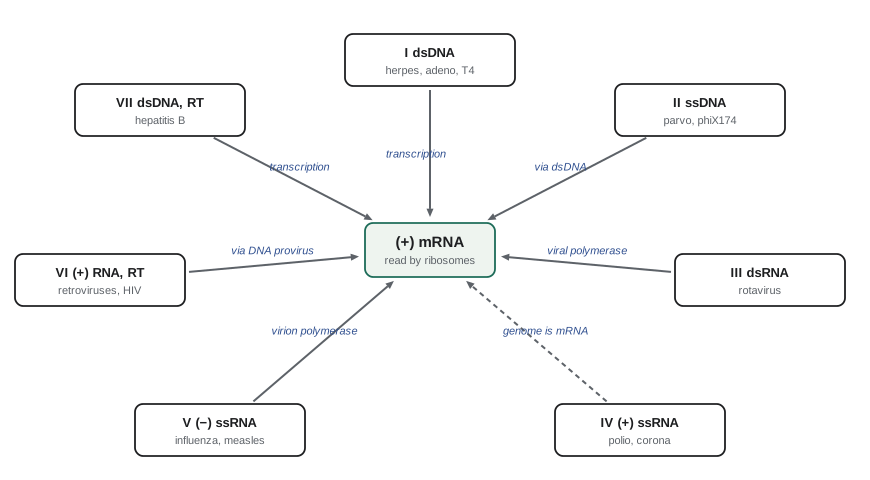
In 1971 David Baltimore proposed classifying viruses by the route their genome takes to produce mRNA. Every virus must make mRNA that host ribosomes can translate, and there are only seven ways to get there from the different genome types. The Baltimore classification remains the most useful way to think about viral replication strategies, because it predicts what enzymes a virus must bring or encode and therefore suggests drug targets.
| Group | Genome | Route to mRNA | Examples |
|---|---|---|---|
| I | Double-stranded DNA | Transcribed by host or viral DNA-dependent RNA polymerase | Herpesviruses, adenoviruses, poxviruses, T4 and lambda phages |
| II | Single-stranded DNA | Converted to dsDNA, then transcribed | Parvoviruses, circoviruses, phage φX174 |
| III | Double-stranded RNA | Viral RNA-dependent RNA polymerase transcribes inside the capsid | Rotaviruses, reoviruses |
| IV | Single-stranded RNA (+) | Genome acts as mRNA; replicated through a negative-strand copy | Poliovirus, hepatitis A, norovirus, coronaviruses, Zika, dengue |
| V | Single-stranded RNA (−) | Virion RNA polymerase transcribes genome into mRNA | Influenza, measles, rabies, Ebola, RSV |
| VI | Single-stranded RNA (+) with DNA intermediate | Reverse transcriptase makes DNA, which integrates and is transcribed | Retroviruses such as HIV |
| VII | Double-stranded DNA with RNA intermediate | Transcribed to RNA, which is reverse transcribed to make new genomes | Hepatitis B virus, cauliflower mosaic virus |
The Baltimore groups also explain differences in mutation rate. RNA-dependent RNA polymerases and reverse transcriptases lack proofreading, so viruses in groups III to VI, and to a lesser extent VII, mutate much faster than DNA viruses that use proofreading DNA polymerases. Coronaviruses are a partial exception: their unusually large RNA genomes, about 30,000 nucleotides, are copied with the help of a proofreading exonuclease (nsp14), which may be what allows such large RNA genomes to exist.
Naming and classifying viruses
Formal virus taxonomy is the responsibility of the International Committee on Taxonomy of Viruses (ICTV). It groups viruses using genome organization, sequence similarity, particle structure and replication strategy into a hierarchy from realm down to species. In 2021 the ICTV began requiring binomial species names in Latinized form, such as Betacoronavirus pandemicum for the species that includes SARS-CoV-2, and by 2023 had converted most existing species names. The species is an abstract category; the virus itself keeps its common name, so SARS-CoV-2 is the virus and Betacoronavirus pandemicum is the species it belongs to. Higher-level groupings based on conserved structural proteins and replication enzymes, such as the realm Riboviria for RNA viruses that use RNA-dependent polymerases, suggest that viruses have several independent origins rather than a single common ancestor.
Viroids, satellites and prions
Several infectious agents are even simpler than viruses. Viroids, discovered by Theodor Diener in 1971, are small circular RNA molecules of about 250 to 400 nucleotides that encode no proteins at all, replicate using host RNA polymerase and cause diseases of crop plants such as potato spindle tuber disease. Satellite viruses and satellite nucleic acids depend on a helper virus for replication; the hepatitis delta virus, which can only infect people already infected with hepatitis B, is the most medically important. Virophages, such as Sputnik, are small viruses that replicate only in cells co-infected with a giant virus and reduce its yield. Prions, the agents of scrapie in sheep, chronic wasting disease in deer, bovine spongiform encephalopathy in cattle and Creutzfeldt–Jakob disease in people, contain no nucleic acid. They are misfolded forms of a normal host protein, PrP, that convert the normal form into the misfolded form, and they resist ordinary sterilization, requiring extended autoclaving or strong alkali for inactivation. Chronic wasting disease is spreading in wild deer, elk and moose populations in western Canada and many US states, and its surveillance is a concern for wildlife and public health agencies in both countries.
Counting viruses
Because viruses cannot be grown on agar, counting them requires either a susceptible host or a molecular method. The most widely used biological method is the plaque assay. A diluted virus sample is mixed with host cells, bacteria for phages or a monolayer of cultured animal cells for animal viruses, and overlaid with soft agar or a semi-solid medium that limits spread to neighbouring cells. Each infectious particle starts a local cycle of infection and lysis that produces a clear zone, or plaque, in the lawn or monolayer. Counting plaques on a plate in the countable range and correcting for dilution gives the titre in plaque-forming units (PFU) per mL, by exactly the same arithmetic as a bacterial plate count (lesson 3).
import numpy as np

rng = np.random.default_rng(330)
true_titre = 1.2e10                       # PFU per mL of a phage lysate
dilutions = [1e-6, 1e-7, 1e-8]
vol = 0.1                                # mL of diluted phage mixed with host and plated
for d in dilutions:
    n = rng.poisson(true_titre * d * vol, 3)
    ok = all(30 <= x <= 300 for x in n)
    note = f"titre = {n.mean() / (vol * d):.3g} PFU/mL" if ok else ("confluent lysis" if n.min() > 300 else "too few")
    print(f"{d:.0e}: plaques {n.tolist()}  {note}")

# Particle-to-PFU ratio: electron microscopy or qPCR counts all genomes or particles
particles_per_mL = 6.0e11                # e.g., from qPCR genome copies (illustrative)
print(f"particle:PFU ratio = {particles_per_mL / true_titre:.0f}:1")1e-06: plaques [1237, 1242, 1163] confluent lysis 1e-07: plaques [98, 73, 117] titre = 9.6e+09 PFU/mL 1e-08: plaques [10, 12, 15] too few particle:PFU ratio = 50:1
Not every virus particle is infectious. Some are defective, some carry damaged genomes, and some simply fail to reach a cell in time. The ratio of total particles, counted by electron microscopy or measured as genome copies by quantitative PCR, to infectious units is called the particle-to-PFU ratio. It is close to 1 for many phages, around 1 to 100 for many animal viruses and can exceed 1,000 for some, such as poliovirus in cell culture. That difference matters in practice: a PCR test that detects viral RNA shows that genomes are present, not necessarily that infectious virus is. Other assays include the TCID₅₀ (tissue culture infectious dose 50 per cent), the dilution at which half of inoculated cultures show cytopathic effects, used for viruses that do not form clear plaques; and hemagglutination assays, which measure viruses such as influenza that clump red blood cells.
Multiplicity of infection and the Poisson distribution
When virus particles are added to a population of cells, they attach at random, so the number infecting each cell follows a Poisson distribution. The average number of infectious particles per cell is the multiplicity of infection (MOI). The fraction of cells receiving exactly k particles is P(k) = e^{−m}m^{k}/k!, where m is the MOI. The fraction left uninfected is e^{−m}, so the fraction infected is 1 − e^{−m}.
import numpy as np
from math import factorial

for m in [0.01, 0.1, 1, 3, 10]:
    p0 = np.exp(-m)
    p1 = m * np.exp(-m)
    multi = 1 - p0 - p1
    print(f"MOI {m:5.2f}: uninfected {p0:6.3f}  single {p1:6.3f}  multiple {multi:6.3f}  infected {1-p0:6.3f}")
# what MOI infects 99% of cells?
print(f"MOI needed for 99% infection = {-np.log(0.01):.2f}")MOI 0.01: uninfected 0.990 single 0.010 multiple 0.000 infected 0.010 MOI 0.10: uninfected 0.905 single 0.090 multiple 0.005 infected 0.095 MOI 1.00: uninfected 0.368 single 0.368 multiple 0.264 infected 0.632 MOI 3.00: uninfected 0.050 single 0.149 multiple 0.801 infected 0.950 MOI 10.00: uninfected 0.000 single 0.000 multiple 1.000 infected 1.000 MOI needed for 99% infection = 4.61
Experimenters choose the MOI according to their purpose. To follow a single synchronous round of infection, every cell must be infected at once, so a high MOI is used; even at an MOI of 3, about 5 per cent of cells escape, and an MOI of about 4.6 is needed to infect 99 per cent. To isolate a clone from one particle or to study rare genetic events, a low MOI such as 0.01 is used, so that almost every infected cell received exactly one particle. Lesson 9 uses a high-MOI one-step growth experiment to measure how many new virions each infected cell releases.
Worked problem
Planning an infection
A flask contains 2 × 10⁶ cultured cells. A virus stock has a titre of 5 × 10⁷ PFU per mL. What volume gives an MOI of 5, and what fraction of cells will remain uninfected?
Show solution
- MOI 5 requires 5 × 2 × 10⁶ = 10⁷ PFU.
- Volume = 10⁷/(5 × 10⁷ per mL) = 0.2 mL.
- The uninfected fraction is e^{−5} ≈ 0.0067, about 0.7 per cent, or roughly 13,000 cells.
Answer: 0.2 mL of stock; about 99.3 per cent of cells are infected. MOI is defined by infectious units, so a stock with a high particle-to-PFU ratio delivers many more particles than this.
Common mistakes
MistakeDescribing viruses as very small bacteria.
InsteadViruses are not cells; they have no ribosomes or metabolism and replicate only inside host cells.
MistakeAssuming a positive PCR result means infectious virus is present.
InsteadPCR detects genomes; the particle-to-PFU ratio shows that many genomes are not infectious.
MistakeExpecting an MOI of 1 to infect every cell.
InsteadBy the Poisson distribution, about 37 per cent of cells remain uninfected at MOI 1.
MistakeConfusing the virus name with the species name.
InsteadSARS-CoV-2 is the virus; Betacoronavirus pandemicum is the ICTV species it belongs to.
MistakeAssuming non-enveloped viruses are easy to inactivate with alcohol.
InsteadNon-enveloped viruses such as norovirus resist alcohol; enveloped viruses are the easy ones.
MistakeCalling prions viruses.
InsteadPrions are misfolded proteins with no nucleic acid.
Summary and key terms
- Viruses are non-cellular obligate parasites: a nucleic acid genome in a protein capsid, sometimes inside a lipid envelope.
- Capsids are helical or icosahedral; icosahedral capsids contain 60T subunits with T = h² + hk + k².
- Envelopes carry the glycoproteins targeted by antibodies but make viruses easy to inactivate; non-enveloped viruses are tougher.
- The Baltimore classification sorts viruses into seven groups by the route from genome to mRNA; RNA viruses and retroviruses lack proofreading and mutate rapidly.
- The ICTV uses binomial species names; viroids, satellites, virophages and prions extend the range of subviral agents.
- Plaque assays give titres in PFU per mL; particle-to-PFU ratios show that many particles are not infectious.
- Infection follows Poisson statistics: the fraction infected at MOI m is 1 − e^{−m}.
- virion
- A complete virus particle outside a host cell.
- capsid
- The protein shell that encloses a viral genome.
- envelope
- A lipid membrane derived from the host that surrounds some virions.
- triangulation number
- T in the Caspar–Klug theory, giving the number of subunits (60T) in an icosahedral capsid.
- positive-sense RNA
- Viral RNA with the same sense as mRNA, translatable directly by ribosomes.
- Baltimore classification
- A grouping of viruses into seven classes by how their genomes produce mRNA.
- plaque-forming unit
- A virus particle or group of particles that produces one plaque.
- particle-to-PFU ratio
- The ratio of total virus particles to infectious units in a preparation.
- multiplicity of infection
- The average number of infectious virus particles per cell.
- viroid
- A small, non-coding, circular infectious RNA that causes plant disease.
Practice problems
Work each problem on paper first, then reveal the answer to check.
How many subunits are in a T = 7 capsid?
Show answer
420.
Why must negative-sense RNA viruses carry a polymerase in the virion?
Show answer
Host cells have no RNA-dependent RNA polymerase to make mRNA from the genome.
Which Baltimore group uses reverse transcriptase to make new genomes from an RNA intermediate?
Show answer
Group VII (hepadnaviruses such as hepatitis B); group VI uses it at the start of infection.
Why is purified poliovirus RNA infectious but purified influenza RNA is not?
Show answer
Poliovirus RNA is positive-sense and acts as mRNA; influenza RNA is negative-sense and needs the viral polymerase.
A 10⁻⁸ dilution plated at 0.1 mL gives 64 plaques. What is the titre?
Show answer
64/(0.1 × 10⁻⁸) = 6.4 × 10¹⁰ PFU per mL.
qPCR finds 2 × 10¹² genome copies per mL in that sample. What is the particle-to-PFU ratio?
Show answer
2 × 10¹²/6.4 × 10¹⁰ ≈ 31:1.
At MOI 2, what fraction of cells is uninfected?
Show answer
e^{−2} ≈ 0.135.
What MOI infects 95 per cent of cells?
Show answer
−ln(0.05) ≈ 3.0.
Why are giant viruses challenging to the definition of a virus?
Show answer
They are larger than some bacteria and encode parts of the translation machinery.
What makes prions so hard to inactivate?
Show answer
They are proteins with highly stable misfolded structures, resistant to standard heat and chemical treatments.
Lesson quiz · 5 questions
Score 60% or more to complete this lesson.
Create a free account to save your progress, take quizzes and earn a certificate.