
BIOL-330 · Lesson 1 of 12 · 34 min
The microbial world and how we study it
Microbiology is the study of organisms too small to see clearly without a microscope, together with the viruses that infect them. That definition sounds like a statement about size, but the real subject is far bigger than the cells themselves. Microorganisms carried out most of the chemistry that made Earth habitable, they still drive the cycles of carbon, nitrogen, sulfur and iron, and they outnumber every other form of life by many orders of magnitude. Current estimates put the number of bacterial and archaeal cells on Earth at around 10³⁰, most of them in deep sediments, soils and the oceans. A single gram of fertile soil can hold a billion cells belonging to thousands of species; a millilitre of sea water holds about a million cells and ten times as many viruses.
BIOL-108 introduced the three domains of life and gave a first tour of bacteria, archaea and viruses, and BIOL-107 explained how light and electron microscopes work and what limits their resolution. This course goes deeper. It asks how microbial cells are built and why their structures matter for medicine and industry, how microbial populations grow and how that growth is measured and controlled, how microbes make a living from almost any chemical energy source, how their genes move between unrelated cells, how viruses are organized and replicate, how microbial communities are studied, and how microbiology is put to work in food, industry and public health. Along the way we use the same quantitative tools a working microbiologist uses: exponential growth, dilution arithmetic, survival kinetics, minimum inhibitory concentrations, Poisson statistics and diversity indices.
What counts as a microorganism
The word microorganism is a practical label rather than a branch of the tree of life. It covers bacteria and archaea (the two prokaryotic domains), microscopic eukaryotes such as yeasts, moulds, algae and protozoa, and, by convention, viruses, even though viruses are not cells and cannot reproduce on their own. Microbiologists also study subviral agents: viroids, which are naked infectious RNA molecules that cause plant diseases, satellite viruses that depend on a helper virus, and prions, which are misfolded proteins rather than any kind of microbe but are often handled by the same laboratories. This course concentrates on bacteria, archaea and viruses. Microbial eukaryotes appear where they matter for food, industry and ecology, and their biology is treated more fully in protist and mycology courses.
Size is the feature everyone notices first. A typical bacterium such as Escherichia coli is a rod about 1 to 2 µm long and 0.5 to 1 µm wide, with a volume of roughly 1 µm³, which is about a femtolitre. Most archaea are similar in size. Yeast cells are 5 to 10 µm across, and many protozoa are tens of micrometres long. Viruses are much smaller: poliovirus is about 30 nm in diameter, influenza about 100 nm and the large poxviruses about 250 to 350 nm. A few organisms break the pattern. Thiomargarita magnifica, described from Caribbean mangroves in 2022, has cells up to about a centimetre long, and giant viruses such as mimivirus are larger than some bacteria. Lesson 2 explains why most prokaryotes stay small and how the giants get around the limits.
| Agent | Typical size | Cell type | Example |
|---|---|---|---|
| Small virus | 20–50 nm | Not a cell | Poliovirus (about 30 nm) |
| Large virus | 100–400 nm | Not a cell | Influenza (about 100 nm); vaccinia (about 300 nm) |
| Giant virus | 400–1,500 nm | Not a cell | Mimivirus (about 750 nm including fibres) |
| Small bacterium | 0.2–0.5 µm | Prokaryotic | Mycoplasma species |
| Typical bacterium or archaeon | 0.5–5 µm | Prokaryotic | E. coli, Bacillus subtilis, Haloferax volcanii |
| Giant bacterium | 100 µm to about 1 cm | Prokaryotic | Epulopiscium, Thiomargarita magnifica |
| Yeast | 5–10 µm | Eukaryotic | Saccharomyces cerevisiae |
| Protozoan | 10–200 µm | Eukaryotic | Giardia, Paramecium |
How the science began
Antonie van Leeuwenhoek, a Dutch cloth merchant, made the first reliable observations of bacteria in the 1670s and 1680s with single-lens microscopes he ground himself, describing the ‘animalcules’ in pond water and in plaque scraped from his teeth. For almost two centuries afterwards, microbes were curiosities. The questions that turned microbiology into a science were practical: why does wine spoil, where does putrefaction come from, and what causes infectious disease?
Louis Pasteur answered the first two in the 1850s and 1860s. He showed that fermentation and spoilage are carried out by living microorganisms, that different microbes produce different products, and, with his swan-necked flask experiments, that broth sterilized by heat stays clear indefinitely if dust-borne microbes cannot reach it. The experiments buried the idea of spontaneous generation and led directly to pasteurization, gentle heating that kills spoilage organisms and pathogens without boiling the product. John Tyndall then showed that some boiled infusions still spoiled because they contained heat-resistant forms, which Ferdinand Cohn identified as bacterial endospores. Joseph Lister applied the germ theory to surgery in the 1860s, using carbolic acid to reduce wound infection, and his antiseptic methods cut post-operative mortality sharply.
Robert Koch supplied the methods and the logic that linked particular microbes to particular diseases. Working on anthrax and then tuberculosis, he grew bacteria in pure culture on solid media, first on slices of potato and then on gelatin and agar, an idea suggested by Fanny Hesse from her kitchen, where agar was used to set jellies. Koch’s colleague Julius Petri designed the shallow covered dish that still carries his name. Koch’s postulates, stated in their mature form in the 1880s, set out the evidence needed to say that a microbe causes a disease.
| Postulate | What it requires | Modern limitation |
|---|---|---|
| 1 | The microbe is found in every case of the disease and not in healthy hosts | Asymptomatic carriers are common, as with Salmonella Typhi and many viruses |
| 2 | The microbe can be isolated and grown in pure culture | Many pathogens, including Treponema pallidum and Mycobacterium leprae, cannot be grown in ordinary media |
| 3 | The cultured microbe causes the disease when introduced into a susceptible host | Ethics forbids human challenge for most diseases; animal models may not reproduce human disease |
| 4 | The same microbe is re-isolated from the experimentally infected host | Depends on postulates 2 and 3 being possible |
Because so many organisms fail one or more postulates, modern practice uses an expanded set of criteria. Stanley Falkow’s molecular Koch’s postulates ask whether a particular gene is found in pathogenic strains, whether disrupting it reduces virulence and whether restoring it restores virulence. Sequence-based criteria, proposed by Fredricks and Relman in 1996, ask whether a microbial nucleic acid sequence is present in diseased tissue, is more abundant where the disease is worse and disappears with recovery. These criteria were essential for identifying agents such as hepatitis C virus and Tropheryma whipplei, which cannot easily be cultured, and they were used in early 2020 to identify SARS-CoV-2 as the cause of COVID-19 within weeks.
Looking at microbes
The light microscope remains the microbiologist’s first tool. BIOL-107 showed that its resolution is limited by the wavelength of light and the numerical aperture of the objective to about 0.2 µm, so a 1 µm bacterium is resolved but its internal structures are not. Microbiology uses several variants of light microscopy. Bright-field microscopy shows stained cells against a bright background. Phase-contrast microscopy converts small differences in refractive index into differences in brightness, so living, unstained cells, their motility and their endospores can be seen. Dark-field microscopy collects only scattered light and makes thin organisms such as spirochetes visible. Fluorescence microscopy detects cells labelled with fluorescent dyes or antibodies, and fluorescence in situ hybridization (FISH) uses labelled DNA probes against ribosomal RNA to identify particular groups of organisms in a mixed sample without culturing them. Electron microscopy reveals envelopes, flagella, ribosomes and virus particles, and cryo-electron tomography can image frozen whole cells at near-molecular detail.
Because most bacteria are almost transparent, staining is central to routine work. A simple stain such as methylene blue or crystal violet colours every cell and shows shape, size and arrangement: cocci or rods, chains or clusters, pairs or tetrads. Differential stains separate cells into groups according to their structure, and the most important of them is the Gram stain, devised by the Danish physician Hans Christian Gram in 1884. It is still the first test performed on many clinical specimens because it takes a few minutes and immediately narrows the possibilities.
The logic of the Gram stain
The Gram stain is a four-step procedure, and its result depends on the structure of the cell envelope, which lesson 2 describes in detail. A heat-fixed smear is first flooded with crystal violet, which stains all cells purple. Iodine is then added as a mordant: it forms large crystal violet–iodine complexes inside the cells. The decolourizing step, a brief wash with alcohol or an acetone–alcohol mixture, is the critical one. In Gram-positive cells, the thick, highly cross-linked peptidoglycan wall dehydrates and shrinks, trapping the large dye complexes inside. In Gram-negative cells, the alcohol dissolves the lipid-rich outer membrane, and the thin peptidoglycan layer cannot hold the complexes, so the cells lose their colour. Finally a counterstain, usually safranin, colours the now-colourless Gram-negative cells pink or red, while Gram-positive cells stay purple.
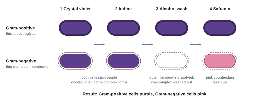
Each step can go wrong, and recognizing the errors is part of reading a slide. Over-decolourizing washes the dye out of Gram-positive cells and makes them look Gram-negative; under-decolourizing leaves Gram-negative cells purple. Old cultures of Gram-positive organisms often stain variably because their walls are being broken down. Thick smears decolourize unevenly. Laboratories therefore include control organisms of known Gram reaction on every slide. Some organisms do not fit the scheme at all. Mycobacteria, which have a waxy envelope rich in mycolic acids, resist the Gram stain and are detected instead by the acid-fast (Ziehl–Neelsen) stain, in which carbol fuchsin driven in by heat is retained even after an acid–alcohol wash. Mycoplasmas have no cell wall and cannot be Gram stained in a meaningful way. Archaea stain in either colour depending on their envelope, so a Gram result says nothing about whether a cell is a bacterium or an archaeon.
Other special stains highlight particular structures. Endospore stains use malachite green driven into the resistant spore coat with heat, then a safranin counterstain for the vegetative cells. Negative stains such as India ink or nigrosin colour the background and leave capsules as bright haloes around cells. Flagella are too thin to see in an ordinary light microscope, but special stains thicken them with a precipitate so that their number and arrangement can be seen.
Worked problem
Reading a Gram stain result
A Gram stain of a cerebrospinal fluid specimen shows pink, kidney-shaped cocci in pairs, many inside white blood cells. The control slide shows purple Staphylococcus cells and pink E. coli cells. What can be concluded, and what cannot?
Show solution
- The controls stained correctly, so the decolourizing step worked and the pink colour can be trusted.
- Pink means Gram-negative; kidney-shaped cocci in pairs are called diplococci.
- Gram-negative diplococci inside neutrophils in spinal fluid are the classic appearance of Neisseria meningitidis.
- The stain alone cannot confirm the species or its antibiotic susceptibility: culture, molecular tests and susceptibility testing must follow.
Answer: The appearance strongly suggests meningococcal meningitis and justifies urgent treatment and public health notification, but confirmation and susceptibility results come from further testing. Clinical decision-making of this kind is treated in health and medicine courses; here the point is the logic of the stain.
Growing microbes
A pure culture contains a single kind of organism derived from one cell. Pure cultures are the foundation of classical microbiology because only then can growth, biochemistry and genetics be attributed to one organism. They are obtained by spreading a sample thinly over the surface of a solid medium so that individual cells are separated. Each cell grows into a visible colony of millions of identical descendants. The streak plate method dilutes cells mechanically by drawing a loop over successive sectors of a plate; the spread plate and pour plate methods dilute the sample first in liquid. Colonies differ in size, shape, edge, elevation, colour and texture, and those differences are a first clue to identity.
Culture media are designed for particular purposes. A defined (synthetic) medium has a known chemical composition, which is essential for studying nutrition and metabolism. A complex medium contains ingredients such as peptones, yeast extract or blood whose exact composition is unknown but which supports a wide range of organisms. Selective media favour some organisms over others, for example by including bile salts that inhibit most Gram-positive bacteria, high salt that only staphylococci and a few others tolerate, or an antibiotic. Differential media let different organisms grow but make them look different: on MacConkey agar, lactose fermenters produce acid and form pink colonies, while non-fermenters stay colourless; on blood agar, organisms that completely lyse red cells produce clear zones (beta-hemolysis). Enrichment cultures use selective conditions in liquid to increase the proportion of a rare organism before plating, a strategy pioneered by Martinus Beijerinck and Sergei Winogradsky for isolating soil bacteria with unusual metabolisms.
| Medium type | Purpose | Example |
|---|---|---|
| Defined | Known composition for nutrition and physiology studies | Glucose–mineral salts medium for E. coli |
| Complex | Supports many organisms; composition not exactly known | Tryptic soy agar, nutrient broth |
| Selective | Suppresses unwanted organisms | Mannitol salt agar (7.5 per cent NaCl) for staphylococci |
| Differential | Distinguishes organisms by a visible reaction | MacConkey agar (lactose fermentation); blood agar (hemolysis) |
| Enrichment | Raises the proportion of a rare organism before isolation | Selenite broth for Salmonella from stool or food |
Aseptic technique, the set of practices that keep cultures pure and keep the worker safe, is learned in teaching laboratories with organisms of the lowest risk. Its principles are simple: work near a flame or in a biological safety cabinet, sterilize tools before and after use, keep containers covered, and treat every culture as potentially hazardous. Lesson 12 returns to how biosafety levels and laboratory licensing protect workers and the public; HSE-245 covers infection prevention in workplaces more generally.
The great plate count anomaly
For a century microbiology depended on culture, and culture has a large blind spot. When the number of cells seen in a sample under the microscope is compared with the number of colonies that grow on plates, the plate count is usually lower by a factor of a hundred to ten thousand. Staley and Konopka named this the great plate count anomaly in 1985. Some of the missing cells are dead, but most are alive and simply do not grow under laboratory conditions. They may need partners that supply essential metabolites, very low nutrient concentrations, unusual temperatures or gases, or more time than an experimenter is willing to wait.
Molecular methods closed much of the gap. In the 1970s Carl Woese compared the sequences of small-subunit ribosomal RNA, which every cell has and which changes slowly enough to record deep evolutionary relationships, and discovered that the prokaryotes formed two fundamentally different groups: Bacteria and Archaea. In the 1980s Norman Pace and colleagues showed that ribosomal RNA genes could be amplified and sequenced directly from environmental DNA, revealing whole phyla that had never been cultured. Lesson 10 shows how 16S rRNA sequencing and metagenomics are used to describe microbial communities today. Culture remains important, because only cultured organisms can be studied fully, and new methods such as diffusion chambers incubated in the natural environment and high-throughput dilution culture are steadily bringing more of the uncultured majority into the laboratory. One success was teixobactin, an antibiotic reported in 2015 from a soil bacterium grown in an iChip diffusion device.
Identifying what has grown
Classical identification relied on shape, Gram reaction and a panel of biochemical tests: whether an organism ferments particular sugars, produces catalase or oxidase, reduces nitrate, hydrolyses urea or uses citrate. Commercial test strips combine twenty or so reactions into a numerical profile that is matched against a database. Clinical and food laboratories now rely increasingly on two faster methods. MALDI-TOF mass spectrometry ionizes proteins, mostly ribosomal proteins, from a smear of a colony and compares the resulting spectrum with reference spectra; it identifies most common bacteria and yeasts in minutes for a few dollars per sample. Sequencing of the 16S rRNA gene, or of the whole genome, identifies organisms that biochemical tests and mass spectrometry cannot, and whole-genome sequencing also reveals resistance genes, virulence genes and the relatedness of isolates, which is why it has become the standard tool for outbreak investigation (lesson 12).
Counting cells under the microscope
Microscopy also gives total cell counts. A counting chamber, such as the Petroff–Hausser chamber used for bacteria, is a slide with a grid of known area and a coverslip held a known distance above it, so each grid square holds a known volume. Counting the cells in several squares and dividing by the volume gives the concentration. Direct counts include dead cells, and very dilute samples give too few cells to count, but the method is quick and needs no incubation. Fluorescent DNA stains such as DAPI or SYBR Green make direct counting of environmental samples much easier, and paired live/dead stains distinguish cells with intact and damaged membranes.
import numpy as np

rng = np.random.default_rng(330)
# Petroff-Hausser chamber: each small square is 0.05 mm x 0.05 mm, depth 0.02 mm
side_mm, depth_mm = 0.05, 0.02
vol_mL = side_mm * side_mm * depth_mm / 1000      # 1 mL = 1000 mm^3
true_conc = 2.4e8                                  # cells per mL in the diluted sample
expected_per_square = true_conc * vol_mL
counts = rng.poisson(expected_per_square, size=16) # 16 squares counted
mean = counts.mean()
conc = mean / vol_mL
se = np.sqrt(counts.sum()) / len(counts) / vol_mL  # Poisson standard error
print(f"volume per square = {vol_mL:.1e} mL")
print("counts:", counts.tolist())
print(f"mean per square = {mean:.2f}")
print(f"estimated concentration = {conc:.3g} cells/mL  (95% CI about +/- {1.96*se:.2g})")
dilution = 10
print(f"original suspension (sample was diluted 1:{dilution}) = {conc*dilution:.3g} cells/mL")volume per square = 5.0e-08 mL counts: [16, 16, 8, 5, 11, 10, 12, 15, 10, 18, 14, 14, 14, 13, 15, 14] mean per square = 12.81 estimated concentration = 2.56e+08 cells/mL (95% CI about +/- 3.5e+07) original suspension (sample was diluted 1:10) = 2.56e+09 cells/mL
Each small square of this chamber holds 5 × 10⁻⁸ mL, so a concentration of about 2 × 10⁸ cells per mL gives a dozen cells per square, a comfortable number to count. Cells fall into squares at random, so the counts follow a Poisson distribution whose variance equals its mean. Counting about 200 cells in total gives a standard error of about 7 per cent (the square root of 200 divided by 200), which is why protocols specify a minimum total count rather than a fixed number of squares. If the suspension was diluted before loading, the result is multiplied back by the dilution factor. The same Poisson reasoning reappears in lesson 3 for colony counts and in lesson 8 for viruses.
Microbes and people
Most microbes are harmless or beneficial. They make bread, cheese, yogurt, wine and soy sauce; they produce antibiotics, enzymes, vitamins and vaccines; they treat sewage and degrade pollutants; and the communities living on and in the human body help digest food and resist infection. A small minority cause disease, and infectious disease remains one of the leading causes of death worldwide, especially in low-income countries and in young children. Antimicrobial resistance, the subject of lesson 5, is steadily eroding the drugs that made bacterial infection treatable. The applied lessons at the end of the course show how one body of knowledge supports food safety, industrial production and public health, and how biosafety rules let that work proceed without putting workers or the public at risk.
Common mistakes
MistakeTreating Gram-negative as a synonym for bacteria that are harmful.
InsteadThe Gram reaction reflects envelope structure only. Both Gram groups include harmless, beneficial and pathogenic species.
MistakeAssuming that a Gram-variable result means a mixed culture.
InsteadOld Gram-positive cultures, thick smears and errors in decolourizing all give variable results; check controls and culture age first.
MistakeConcluding that an organism is absent because nothing grew on a plate.
InsteadMost environmental microbes do not grow on standard media. Absence of growth shows only that the organism did not grow under those conditions.
MistakeExpecting every pathogen to satisfy Koch’s postulates.
InsteadMany cannot be cultured, many infections are asymptomatic, and human challenge is rarely ethical. Molecular and epidemiological criteria fill the gap.
MistakeForgetting to multiply a chamber count by the dilution factor.
InsteadConcentrations always refer to a stated sample. Track every dilution from the original material.
MistakeAssuming archaea will stain Gram-negative because they are not bacteria.
InsteadArchaea stain either way depending on their envelope; the Gram stain does not identify domains.
Summary and key terms
- Microorganisms are defined by size and practice, not by lineage: they include bacteria, archaea, microbial eukaryotes and, by convention, viruses.
- Pasteur, Koch and their contemporaries turned microbiology into a science by linking specific microbes to fermentation, spoilage and disease and by developing pure culture on solid media.
- Koch’s postulates are applied flexibly; molecular and sequence-based criteria identify agents that cannot be cultured.
- The Gram stain separates cells by envelope structure: thick peptidoglycan retains crystal violet–iodine complexes, while thin peptidoglycan with an outer membrane does not.
- Selective, differential and enrichment media and aseptic technique allow particular organisms to be isolated in pure culture.
- Most environmental microbes have never been cultured; ribosomal RNA sequencing revealed the archaea and the uncultured majority.
- Direct counts in a chamber follow Poisson statistics, so precision depends on the total number of cells counted.
- microorganism
- An organism too small to be seen clearly without a microscope; by convention the term also covers viruses.
- pure culture
- A population of cells all descended from a single cell.
- Koch’s postulates
- Criteria for establishing that a particular microbe causes a particular disease.
- Gram stain
- A differential stain that separates bacteria into Gram-positive (purple) and Gram-negative (pink) groups according to envelope structure.
- mordant
- A substance, such as iodine in the Gram stain, that fixes a dye in the cell.
- acid-fast stain
- A differential stain that detects organisms, such as mycobacteria, whose waxy envelope retains carbol fuchsin after an acid–alcohol wash.
- selective medium
- A medium that suppresses the growth of some organisms while allowing others to grow.
- differential medium
- A medium on which different organisms produce visibly different colonies.
- great plate count anomaly
- The observation that far fewer cells grow on plates than are seen under the microscope.
- MALDI-TOF
- Matrix-assisted laser desorption ionization time-of-flight mass spectrometry, used to identify microbes from protein spectra.
Practice problems
Work each problem on paper first, then reveal the answer to check.
Name the four reagents of the Gram stain in order.
Show answer
Crystal violet, iodine, alcohol (or acetone–alcohol) decolourizer and safranin counterstain.
Which step of the Gram stain is the critical, differential one?
Show answer
Decolourizing, because that is when Gram-negative cells lose the dye complex.
A smear of Gram-positive cocci appears pink. Give two possible explanations.
Show answer
Over-decolourizing, or an old culture whose walls are breaking down.
Why are mycobacteria identified with an acid-fast stain rather than a Gram stain?
Show answer
Their waxy mycolic acid envelope resists the Gram reagents but retains carbol fuchsin after acid–alcohol.
Give one reason why a pathogen might fail Koch’s first postulate.
Show answer
Asymptomatic carriers harbour the organism without disease.
What makes MacConkey agar both selective and differential?
Show answer
Bile salts and crystal violet inhibit Gram-positive bacteria (selective), and lactose with a pH indicator shows fermenters as pink colonies (differential).
A chamber square holds 5 × 10⁻⁸ mL and averages 8 cells. What is the concentration?
Show answer
8/(5 × 10⁻⁸) = 1.6 × 10⁸ cells per mL.
About how many cells must be counted for a 5 per cent standard error?
Show answer
Since SE/count = 1/√N, N = 400.
What did Carl Woese discover by comparing small-subunit rRNA sequences?
Show answer
That prokaryotes comprise two distinct domains, Bacteria and Archaea.
Why is culture still important despite sequencing?
Show answer
Only cultured organisms can be studied fully for physiology, genetics and susceptibility, and culture yields products such as new antibiotics.
Lesson quiz · 5 questions
Score 60% or more to complete this lesson.
Create a free account to save your progress, take quizzes and earn a certificate.