
BIOL-350 · Lesson 8 of 12 · 30 min
Circulation
Diffusion is fast over short distances and hopelessly slow over long ones. The time needed for a molecule to diffuse a distance x rises with x squared: oxygen crosses 10 µm of tissue in about a twentieth of a second, but would take about three hours to cross 1 cm. Small, thin or slow animals, such as flatworms, cnidarians and sponges, manage without a circulatory system because every cell is close to the surface or to a gut cavity. Larger, active animals need bulk flow, the movement of fluid driven by pressure, to carry oxygen, nutrients, hormones, heat and wastes over long distances. This lesson compares the circulatory systems of animals, explains the physics of flow through vessels and the pumping action of the heart, and shows how fluid moves between blood and tissues across capillary walls. Clinical cardiovascular physiology is treated in MED-111.
Open and closed circulations
In an open circulatory system, found in most molluscs and in arthropods, a heart pumps a fluid called hemolymph into vessels that empty into body spaces, collectively called the hemocoel, where it bathes the tissues directly before returning to the heart through openings called ostia. Pressures are low and flow is slow, but open systems work well for many animals. In insects, the circulatory system plays only a minor role in gas transport, because oxygen is delivered directly to tissues by the tracheal system (lesson 9); the hemolymph carries nutrients, hormones and immune cells. In a closed circulatory system, found in vertebrates, annelids and cephalopod molluscs, blood stays within vessels, and exchange with tissues takes place across the thin walls of capillaries. Closed systems allow higher pressures, faster flow and more precise distribution of blood to particular organs.
Cephalopods show how activity drives circulatory design. Octopuses and squid have closed circulations with three hearts: two branchial hearts that pump blood through the gills and a systemic heart that pumps oxygenated blood to the body. Their blood carries oxygen bound to hemocyanin, a copper-containing protein dissolved in the plasma, which has a lower oxygen-carrying capacity than vertebrate blood. Squid, which jet continuously at high speed, operate close to the limits of their oxygen supply, using most of the oxygen in their blood even at rest.
Evolution of the vertebrate heart
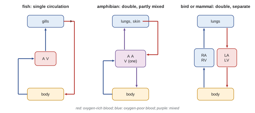
Fish have a single circulation. A two-chambered heart, with one atrium and one ventricle in series, pumps deoxygenated blood to the gills, where it is oxygenated, and the blood then flows on to the body without returning to the heart. Because blood loses much of its pressure in the narrow gill capillaries, systemic pressure in fish is low. The transition to air breathing changed this. Lungfish and amphibians have partially divided hearts and a double circulation, in which blood returns to the heart after passing through the lungs (or, in amphibians, also the skin) before being pumped to the body. The amphibian ventricle is undivided, but its structure and the spiral valve of the outflow tract keep oxygenated and deoxygenated streams largely separate.
Non-crocodilian reptiles have a ventricle incompletely divided by a muscular ridge, and crocodilians have a fully divided ventricle with a unique connection, the foramen of Panizza, between the two aortas. In these animals, blood can be shunted away from the lungs during diving or breath-holding, which may save energy or aid digestion and is clearly adaptive in animals that breathe intermittently. Birds and mammals independently evolved fully divided four-chambered hearts with complete separation of the pulmonary and systemic circuits. This lets the right ventricle pump blood through the delicate lungs at low pressure while the left ventricle drives blood around the body at much higher pressure, an arrangement essential for high metabolic rates.
The cardiac cycle and the pacemaker
Vertebrate hearts are myogenic: the heartbeat originates in the heart muscle itself, in specialized pacemaker cells. In mammals and birds, the pacemaker is the sinoatrial node in the wall of the right atrium. Pacemaker cells have no stable resting potential. Their membrane slowly depolarizes between beats, largely through hyperpolarization-activated cation channels that carry the funny current, until it reaches the threshold for calcium-dependent action potentials. The impulse spreads through the atria, is delayed at the atrioventricular node, which lets the atria finish emptying into the ventricles, and then spreads rapidly through the ventricles via the bundle of His and Purkinje fibers. Many crustacean hearts, by contrast, are neurogenic: their beat is driven by a cardiac ganglion of a few neurons, a valuable model for studying how small neural networks generate rhythms.
Each cardiac cycle consists of systole, when the ventricles contract and eject blood, and diastole, when they relax and fill. Valves between atria and ventricles and at the exits of the ventricles keep blood flowing in one direction. The volume ejected by one ventricle per beat is the stroke volume, and the product of stroke volume and heart rate is cardiac output. The autonomic nervous system adjusts both: sympathetic stimulation and adrenaline speed the pacemaker and increase the force of contraction, while parasympathetic stimulation through the vagus nerve slows the heart.
The Frank–Starling mechanism
The heart also adjusts its output intrinsically. In the 1890s Otto Frank and in the 1910s Ernest Starling found that the more a ventricle is filled during diastole, the more forcefully it contracts and the more blood it ejects. This Frank–Starling mechanism ensures that, over a series of beats, each ventricle pumps out whatever volume it receives, so the outputs of the left and right ventricles stay matched without any nervous control. Its basis lies partly in the length–tension relationship described in lesson 6: cardiac muscle normally works on the ascending limb of the curve, so stretch increases filament overlap, and stretch also increases the sensitivity of the myofilaments to calcium. In fish, which lack strong sympathetic control of the heart in many species, the Frank–Starling mechanism is particularly important; a trout's heart can more than double its stroke volume when filling pressure rises slightly.
Pressure, flow and resistance
Blood flows from regions of higher pressure to regions of lower pressure. For steady flow through a tube, flow equals the pressure difference divided by the resistance, an analog of Ohm's law. For smooth, laminar flow of a fluid through a cylindrical tube, Jean Poiseuille showed in the 1840s that resistance depends on the fluid's viscosity, the length of the tube and, most dramatically, on the fourth power of its radius.
The fourth-power dependence means that small changes in radius have large effects on flow. Halving the radius of an arteriole increases its resistance sixteenfold. This is how animals redistribute blood: the smooth muscle in the walls of arterioles constricts or relaxes in response to nerves, hormones and local signals such as low oxygen, carbon dioxide and nitric oxide, adjusting flow to each organ. During exercise, blood flow to working muscles can rise more than twentyfold while flow to the gut falls. During a dive, seals constrict the arteries to most organs and keep flow mainly to the heart and brain (lesson 10).
import math

# Poiseuille's law: flow through a vessel with fixed pressure difference
eta = 3.5e-3      # blood viscosity, Pa s (about 3.5 times water)
L = 1.0e-3        # vessel length, m (1 mm arteriole segment)
dP = 400.0        # pressure drop along the segment, Pa (about 3 mmHg)

def flow(r):
    R = 8 * eta * L / (math.pi * r ** 4)
    return dP / R          # m^3 per s

r0 = 25e-6        # arteriole radius 25 um
q0 = flow(r0)
print(f"baseline flow: {q0 * 1e9 * 60:.2f} uL per min")
for factor in (1.25, 1.10, 0.90, 0.80, 0.50):
    q = flow(r0 * factor)
    print(f"radius x {factor:4.2f}: flow x {q / q0:6.2f}")

# mean velocity in the arteriole versus in a capillary bed with the same total flow
area_art = math.pi * r0 ** 2
cap_r, n_caps = 3e-6, 300
area_caps = n_caps * math.pi * cap_r ** 2
print(f"mean velocity in arteriole: {q0 / area_art * 1000:.2f} mm/s; "
f"in {n_caps} capillaries it supplies: {q0 / area_caps * 1000:.2f} mm/s")baseline flow: 1.05 uL per min radius x 1.25: flow x 2.44 radius x 1.10: flow x 1.46 radius x 0.90: flow x 0.66 radius x 0.80: flow x 0.41 radius x 0.50: flow x 0.06 mean velocity in arteriole: 8.93 mm/s; in 300 capillaries it supplies: 2.07 mm/s
Widening the arteriole by a quarter more than doubles its flow; narrowing it by half cuts flow to about one sixteenth. The final lines illustrate another principle: blood slows down as it enters the capillary beds, because although each capillary is narrow, their combined cross-sectional area is larger than that of the vessel that feeds them. Slow flow in capillaries gives time for exchange. Real blood is not a simple fluid. It contains red cells that deform and line up in narrow vessels, so its apparent viscosity falls in vessels below about 0.3 mm in diameter, the Fåhræus–Lindqvist effect, and pulsatile, branching flow departs from Poiseuille's assumptions. The law is still the best starting point for understanding how vessel radius controls flow.
Arteries, capillaries and veins
Arteries have thick walls rich in elastic fibers and smooth muscle. Large elastic arteries near the heart expand during systole and recoil during diastole, smoothing the pulsatile output of the heart into a more continuous flow, an effect known as the Windkessel, after the air chamber of old fire engines. Arterioles, with their muscular walls, are the main site of resistance and control. Capillaries are tubes of a single layer of endothelial cells, about 5 to 10 µm across, just wide enough for red cells to squeeze through in single file. Veins are thin-walled, compliant vessels that hold most of the blood volume; valves and the squeezing action of skeletal muscles help return blood to the heart against gravity.
Blood pressure in systemic arteries varies widely among vertebrates. Mean arterial pressure is about 20 to 40 mmHg in many fish, about 90 to 100 mmHg in most mammals regardless of size, and higher in birds. The giraffe, whose brain may sit two meters above its heart, has a mean arterial pressure at heart level of about 200 mmHg, roughly twice that of other mammals, so that pressure at the head is normal. Its legs, where pressure is very high, are protected from swelling by tight skin and thick-walled vessels, an example of how hydrostatic pressure shapes the circulation of tall animals. Snakes show a similar pattern: tree-climbing species have hearts closer to the head and higher blood pressures than aquatic species, in which the surrounding water supports the blood column.
Capillary exchange and Starling forces
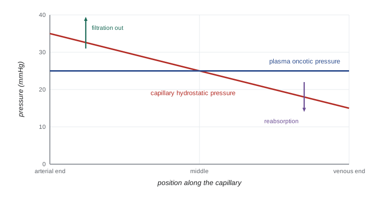
Oxygen, carbon dioxide and other small lipid-soluble molecules diffuse directly across capillary walls. Water and small solutes move through gaps between endothelial cells, driven by two opposing forces described by Ernest Starling in 1896. The hydrostatic pressure of blood in the capillary pushes fluid out into the interstitial space. The colloid osmotic pressure, or oncotic pressure, of plasma proteins, which cannot easily cross the wall, draws fluid back in. In a typical mammalian capillary, hydrostatic pressure falls from about 35 mmHg at the arterial end to about 15 mmHg at the venous end, while plasma oncotic pressure stays at about 25 mmHg. Fluid therefore tends to leave at the arterial end and return at the venous end, although modern measurements suggest that in many tissues net filtration occurs along most of the capillary.
# Net filtration along a capillary (classical Starling model, mmHg)
Pa, Pv = 35.0, 15.0     # capillary hydrostatic pressure at arterial and venous ends
Pi = 0.0                # interstitial hydrostatic pressure
pi_c, pi_i = 25.0, 3.0  # plasma and interstitial oncotic pressures
sigma = 0.9             # reflection coefficient for plasma proteins

def net(x, Pa=Pa, pi_c=pi_c):
    Pc = Pa + (Pv - Pa) * x            # linear fall along the capillary, x from 0 to 1
    return (Pc - Pi) - sigma * (pi_c - pi_i)

def summary(name, **kw):
    pts = [net(i / 10, **kw) for i in range(11)]
    filt = sum(p for p in pts if p > 0) / 10
    reab = -sum(p for p in pts if p < 0) / 10
    print(f"{name:34s} arterial {pts[0]:6.1f}  venous {pts[-1]:6.1f}  "
          f"filtered {filt:5.2f}  reabsorbed {reab:5.2f}  net out {filt - reab:5.2f}")

summary("normal")
summary("arteriolar dilation (Pa 45)", Pa=45.0)
summary("low plasma protein (pi_c 15)", pi_c=15.0)normal arterial 15.2 venous -4.8 filtered 6.56 reabsorbed 0.84 net out 5.72 arteriolar dilation (Pa 45) arterial 25.2 venous -4.8 filtered 11.88 reabsorbed 0.66 net out 11.22 low plasma protein (pi_c 15) arterial 24.2 venous 4.2 filtered 15.62 reabsorbed 0.00 net out 15.62
Under normal conditions, the small net outward filtration in this model is returned to the blood by the lymphatic system, a network of thin vessels that collects excess interstitial fluid and proteins and empties them into large veins. When capillary pressure rises, as when arterioles dilate or venous pressure increases in heart failure, or when plasma protein concentration falls, as in starvation or liver disease, filtration exceeds lymphatic capacity and fluid accumulates in tissues as edema. The swollen bellies of children with severe protein malnutrition, kwashiorkor, are a tragic illustration of Starling's principle.
Scaling the heart
Heart mass in mammals is about 0.6 percent of body mass across a wide range of sizes, and stroke volume scales roughly in proportion to body mass. Heart rate scales with mass to about the −0.25 power (lesson 2), so cardiac output scales with mass to about the 0.75 power, matching metabolic rate. A resting shrew's heart may beat 800 or more times per minute, and hummingbirds exceed 1,000 beats per minute in flight; a resting blue whale's heart beats only a few times per minute. In 2019 Jeremy Goldbogen and colleagues used a suction-cup tag with electrodes to record the heart rate of a free-diving blue whale off California: it fell to as low as two beats per minute at depth and rose to over 30 at the surface.
Worked problem
Blood flow during exercise
At rest, a dog's cardiac output is 2.4 L per minute, with 15 percent going to skeletal muscle. During hard running, cardiac output rises to 12 L per minute and 85 percent goes to muscle, while mean arterial pressure rises only slightly, from 100 to 110 mmHg. By what factor has muscle blood flow increased, and what has happened to the resistance of the muscle vascular bed?
Show solution
- Resting muscle flow: 0.15 × 2.4 = 0.36 L per minute.
- Exercising muscle flow: 0.85 × 12 = 10.2 L per minute, an increase of about 28 times.
- Resistance = pressure / flow. At rest: 100 / 0.36 ≈ 278 units; in exercise: 110 / 10.2 ≈ 10.8 units.
- Muscle vascular resistance has fallen by a factor of about 26.
- By Poiseuille's law, a 26-fold fall in resistance requires the effective radius of the resistance vessels to increase by about 26^(1/4) ≈ 2.3 times, assuming no change in length or viscosity; recruitment of extra capillaries also contributes.
Answer: Muscle blood flow rises about 28-fold, achieved mostly by a roughly 26-fold fall in the resistance of the muscle vessels, while vessels to the gut and other organs constrict so that arterial pressure is maintained.
Common mistakes
MistakeThinking an open circulatory system means an animal is less advanced or less active.
InsteadInsects are highly active with open circulations because their tracheal system, not the blood, delivers oxygen.
MistakeSaying the heartbeat of all animals is started by nerves.
InsteadVertebrate hearts are myogenic; some crustacean hearts are neurogenic.
MistakeAssuming flow is proportional to vessel radius.
InsteadBy Poiseuille's law, resistance varies with the fourth power of radius, so flow is extremely sensitive to radius.
MistakeThinking blood flows fastest in capillaries because they are narrow.
InsteadTotal cross-sectional area is greatest in capillary beds, so flow velocity is lowest there.
MistakeIgnoring the lymphatic system in fluid balance.
InsteadLymphatics return the excess filtered fluid and protein; their failure causes edema even with normal Starling forces.
MistakeTreating the crocodilian heart's shunt as a defect.
InsteadControlled shunting away from the lungs is adaptive during diving and breath-holding.
Summary and key terms
- Diffusion time rises with distance squared, so large active animals need bulk flow.
- Open circulations bathe tissues in hemolymph at low pressure; closed circulations keep blood in vessels and allow high pressure and control.
- Vertebrate hearts evolved from the two-chambered fish heart to the four-chambered, fully divided hearts of birds and mammals.
- The vertebrate heart is myogenic; cardiac output equals heart rate times stroke volume, adjusted by autonomic nerves and the Frank–Starling mechanism.
- Flow equals pressure difference divided by resistance; resistance varies with the fourth power of vessel radius.
- Capillary exchange of fluid is governed by hydrostatic and oncotic Starling forces, with lymphatics returning excess fluid.
- Cardiac output scales with metabolic rate; heart rate falls with size, from shrews to whales.
- open circulatory system
- A circulation in which hemolymph leaves vessels and bathes tissues directly.
- closed circulatory system
- A circulation in which blood remains within vessels and exchanges through capillary walls.
- double circulation
- A circulation in which blood passes through the heart twice per circuit, once for lungs and once for body.
- myogenic heart
- A heart whose beat originates in specialized muscle cells.
- sinoatrial node
- The pacemaker region of the bird and mammal heart.
- stroke volume
- The volume of blood ejected by a ventricle in one beat.
- cardiac output
- The volume of blood pumped per minute by one ventricle.
- Frank–Starling mechanism
- The increase in stroke volume when the ventricle is filled more during diastole.
- Poiseuille's law
- The relationship giving resistance to laminar flow as proportional to viscosity and length and inversely proportional to radius to the fourth power.
- oncotic pressure
- The osmotic pressure exerted by plasma proteins that draws fluid into capillaries.
- Starling forces
- The hydrostatic and oncotic pressures that determine fluid movement across capillary walls.
- edema
- Accumulation of excess fluid in the interstitial spaces.
Practice problems
Work each problem on paper first, then reveal the answer to check.
How long would oxygen take to diffuse 1 mm if D = 2 × 10⁻⁹ m² per s?
Show answer
t ≈ x²/2D = (10⁻³)²/(4 × 10⁻⁹) ≈ 250 s, about 4 minutes.
Why do insects manage with an open circulation despite high activity?
Show answer
Oxygen is delivered by the tracheal system, not the blood.
What is the advantage of a fully divided four-chambered heart?
Show answer
It separates pulmonary and systemic circuits, allowing low pressure in the lungs and high pressure in the body.
A heart beats 70 times per minute with a stroke volume of 75 mL. What is cardiac output?
Show answer
70 × 75 = 5,250 mL, about 5.3 L per minute.
By how much does resistance change if a vessel's radius is reduced by 20 percent?
Show answer
(1/0.8)⁴ ≈ 2.44 times higher.
Explain the Frank–Starling mechanism and why it matters.
Show answer
Greater diastolic filling stretches the muscle and increases stroke volume, so the ventricles pump out what they receive and stay matched.
Why is blood velocity lowest in capillaries?
Show answer
Their total cross-sectional area is the largest of any vessel class.
Give two causes of edema in terms of Starling forces.
Show answer
Raised capillary hydrostatic pressure (for example heart failure) and lowered plasma oncotic pressure (for example protein malnutrition).
Why is giraffe arterial pressure so high?
Show answer
To push blood about two meters up to the brain against gravity.
Where is the pacemaker in mammals, and what current drives its slow depolarization?
Show answer
The sinoatrial node; the funny current through hyperpolarization-activated cation channels (with calcium currents).
Lesson quiz · 5 questions
Score 60% or more to complete this lesson.
Create a free account to save your progress, take quizzes and earn a certificate.