
BIOL-350 · Lesson 5 of 12 · 30 min
Synapses, integration and sensory transduction
An action potential carries a signal along one cell, but behavior depends on what happens when signals pass between cells and when many signals are combined. This lesson follows information across the synapse, explains how a neuron integrates thousands of inputs into a decision to fire or not, and shows how sensory receptor cells convert physical stimuli, such as light, pressure and chemicals, into electrical signals. The aim is the physiological level needed for the rest of the course: how the nervous system controls muscles, glands and organs, and how animals sense their internal and external environment. Detailed treatment of synaptic plasticity, sensory systems, motor circuits, and learning and memory belongs to BIOL-420 Neurobiology.
Electrical and chemical synapses
Synapses come in two kinds. At an electrical synapse, the membranes of two cells are joined by gap junctions, clusters of channels made of connexins in vertebrates and innexins in invertebrates, through which ions flow directly from one cell to the next. Electrical synapses are fast, with almost no delay, and usually bidirectional. They synchronize groups of cells, as in the escape circuits of crayfish, the electric organs of some fish and the inferior olive of the mammalian brain, and gap junctions link heart muscle cells and smooth muscle cells, as lessons 6 and 8 show. At a chemical synapse, the presynaptic cell releases a neurotransmitter into a narrow cleft about 20 to 40 nm wide; the transmitter diffuses across and binds receptors on the postsynaptic cell. Chemical synapses are slower, introducing a delay of about 0.5 to 1 millisecond, but they can amplify signals, make them excitatory or inhibitory, and change their strength with experience.
Transmitter release
When an action potential reaches a presynaptic terminal, it opens voltage-gated calcium channels. Calcium enters and binds synaptotagmin, a sensor protein on synaptic vesicles, triggering the fusion of vesicles with the plasma membrane through the SNARE protein complex and releasing their contents by exocytosis. The vertebrate neuromuscular junction, where a motor neuron contacts a skeletal muscle fiber, has been the model synapse since the 1950s, when Bernard Katz and colleagues showed that transmitter is released in packets. Even with no nerve stimulation, they recorded small, spontaneous depolarizations of the muscle membrane of about 0.5 mV, which they called miniature end-plate potentials. Each corresponds to the release of the contents of one vesicle, a quantum of several thousand acetylcholine molecules. A nerve impulse releases many quanta at once.
In low-calcium solutions, release is reduced, and a nerve impulse sometimes releases no quanta at all, a failure. Del Castillo and Katz showed in 1954 that the numbers of quanta released followed a Poisson distribution. If the mean number released per impulse, the quantal content m, is small, the probability of zero quanta is e to the minus m, so the proportion of failures gives an estimate of m that can be checked against the ratio of mean response size to the size of a single quantum. The agreement between the two estimates was strong evidence for the quantal hypothesis.
import math, random

random.seed(350)
q = 0.40           # mean amplitude of one quantum, mV (miniature end-plate potential)
q_sd = 0.08        # quantum-to-quantum variability, mV
m_true = 1.3       # true mean quantal content in low-calcium saline
trials = 400

def poisson(lam):
    L, k, p = math.exp(-lam), 0, 1.0
    while True:
        p *= random.random()
        if p < L: return k
        k += 1

responses = []
for _ in range(trials):
    k = poisson(m_true)
    responses.append(sum(random.gauss(q, q_sd) for _ in range(k)))

failures = sum(1 for r in responses if r == 0)
m_fail = math.log(trials / failures)
m_mean = (sum(responses) / trials) / q
print(f"failures: {failures} of {trials}")
print(f"m from failures   = ln({trials}/{failures}) = {m_fail:.2f}")
print(f"m from mean / q   = {sum(responses)/trials:.3f} / {q} = {m_mean:.2f}")
counts = {}
for r in responses:
    k = max(1, round(r / q)) if r > 0 else 0
    counts[k] = counts.get(k, 0) + 1
for k in range(6):
    exp = trials * m_fail**k * math.exp(-m_fail) / math.factorial(k)
print(f" {k} quanta: observed {counts.get(k, 0):4d} Poisson expected {exp:6.1f}")failures: 122 of 400 m from failures = ln(400/122) = 1.19 m from mean / q = 0.485 / 0.4 = 1.21 0 quanta: observed 122 Poisson expected 122.0 1 quanta: observed 143 Poisson expected 144.9 2 quanta: observed 85 Poisson expected 86.0 3 quanta: observed 33 Poisson expected 34.0 4 quanta: observed 11 Poisson expected 10.1 5 quanta: observed 6 Poisson expected 2.4
The two estimates of quantal content agree closely, and the distribution of responses sorted into multiples of the quantal size matches the Poisson prediction. Real experiments gave the same kind of agreement and established three ideas that hold at synapses throughout the animal kingdom: transmitter is packaged in vesicles, release is probabilistic, and calcium entry controls the probability of release. Synaptic strength can therefore be changed by altering the number of release sites, the probability of release at each site or the size of the postsynaptic response to each quantum.
Postsynaptic receptors
Neurotransmitters act on two broad classes of receptor. Ionotropic receptors are ligand-gated ion channels: binding of the transmitter opens the channel within a millisecond. The nicotinic acetylcholine receptor at the neuromuscular junction admits sodium and potassium and depolarizes the muscle. Glutamate receptors of the AMPA and NMDA types mediate most fast excitation in the vertebrate brain; GABA-A and glycine receptors open chloride channels and mediate fast inhibition. Metabotropic receptors are G protein-coupled receptors that act through second messengers, as described in BIOL-214; they produce slower, longer-lasting and more widespread effects. Muscarinic acetylcholine receptors in the heart, adrenergic receptors for noradrenaline and adrenaline, and most receptors for neuropeptides are metabotropic.
| Transmitter | Main receptor types | Typical effect | Examples of roles |
|---|---|---|---|
| Acetylcholine | Nicotinic (ionotropic), muscarinic (metabotropic) | Excitatory at muscle; inhibitory on heart (muscarinic) | Neuromuscular junction, autonomic ganglia, parasympathetic targets |
| Glutamate | AMPA, NMDA, kainate; metabotropic mGluRs | Fast excitation | Most excitatory synapses in vertebrate brain; insect neuromuscular junction |
| GABA | GABA-A (Cl⁻ channel), GABA-B (metabotropic) | Inhibition | Vertebrate brain; inhibitory motor neurons of crustaceans and insects |
| Glycine | Glycine receptor (Cl⁻ channel) | Inhibition | Vertebrate spinal cord and brainstem |
| Noradrenaline and adrenaline | α and β adrenergic (metabotropic) | Varies by receptor | Sympathetic targets; arousal |
| Serotonin, dopamine | Mostly metabotropic | Modulation | Mood, reward, motor control; serotonin modulates aggression in crustaceans |
| Neuropeptides | Metabotropic | Slow modulation | Pain, feeding, stress, reproduction |
Transmitter action must be terminated quickly for synapses to transmit rapid signals. At the neuromuscular junction, acetylcholinesterase in the cleft hydrolyzes acetylcholine within a millisecond. Organophosphate insecticides and nerve agents inhibit this enzyme, so acetylcholine accumulates and causes uncontrolled muscle activity followed by paralysis; this is why organophosphate poisoning is an occupational hazard in agriculture. Other transmitters, including glutamate, GABA, noradrenaline and serotonin, are removed by transporters that carry them back into neurons or glial cells. Many drugs act on these transporters, including antidepressants that block serotonin reuptake.
Integration: summation and the decision to fire
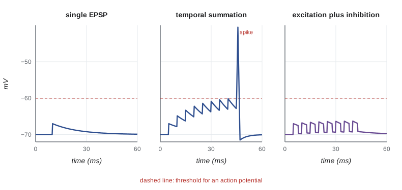
A single excitatory postsynaptic potential (EPSP) at a central synapse is usually small, often less than 1 mV, far below the 10 to 20 mV of depolarization needed to reach threshold. A neuron fires only when many EPSPs add together. In temporal summation, EPSPs arriving in rapid succession at the same synapse build on one another before each decays; in spatial summation, EPSPs from different synapses arriving at about the same time add together. Inhibitory postsynaptic potentials (IPSPs) subtract from the total. Inhibition through chloride channels can also work without changing the membrane potential much, by shunting: opening chloride channels lowers membrane resistance so that excitatory currents produce smaller voltage changes. The summed potential spreads passively, with decay governed by the cable properties described in lesson 4, to the axon initial segment, which has a high density of sodium channels and the lowest threshold in the neuron. There the decision to fire is made.
import math

# Temporal summation of EPSPs at the axon initial segment (teaching model).
# Each EPSP: amplitude A, rises instantly, decays with membrane time constant tau.
A, tau, threshold = 3.0, 15.0, 10.0     # mV, ms, mV of depolarization needed

def peak_depolarization(interval_ms, n_inputs, tau=tau):
    # n EPSPs arriving every interval_ms; return the peak summed depolarization
    v = 0.0
    peak = 0.0
    for i in range(n_inputs):
        v = v * math.exp(-interval_ms / tau) + A if i else A
        peak = max(peak, v)
    return peak

print("interval   10 inputs   20 inputs   (threshold 10 mV)")
for interval in (2, 5, 10, 20, 40):
    p10, p20 = peak_depolarization(interval, 10), peak_depolarization(interval, 20)
    print(f"{interval:5d} ms   {p10:6.1f} mV   {p20:6.1f} mV   "
          f"{'fires' if max(p10, p20) >= threshold else 'silent'}")
# a longer time constant promotes temporal summation
print(f"tau = 40 ms, 10 inputs every 10 ms: {peak_depolarization(10, 10, 40.0):.1f} mV")interval 10 inputs 20 inputs (threshold 10 mV)
2 ms 17.7 mV 22.4 mV fires
5 ms 10.2 mV 10.6 mV fires
10 ms 6.2 mV 6.2 mV silent
20 ms 4.1 mV 4.1 mV silent
40 ms 3.2 mV 3.2 mV silent
tau = 40 ms, 10 inputs every 10 ms: 12.4 mVInputs every 2 or 5 milliseconds summate past threshold; inputs every 10 milliseconds or more stay below it, and at 20 or 40 milliseconds they barely build at all, because each EPSP has largely decayed before the next arrives. A neuron with a longer membrane time constant integrates over a longer window: with a time constant of 40 ms instead of 15 ms, inputs every 10 ms now reach threshold. Real neurons are more complex, because dendrites contain voltage-gated channels that can amplify or limit inputs, and synapses far out on dendrites are attenuated more than those near the soma. But the principle holds: a neuron is an integrator whose output, a train of action potentials, depends on the timing, location and sign of thousands of inputs.
Neuromodulation and the autonomic nervous system
Beyond fast point-to-point signaling, nervous systems use neuromodulators, such as serotonin, dopamine, octopamine in insects and many neuropeptides, that change the properties of neurons and synapses over seconds to hours. The stomatogastric ganglion of crabs and lobsters, a circuit of about 30 neurons that drives rhythmic movements of the stomach, has shown that the same anatomical circuit can produce different rhythms depending on which modulators are present, a lesson with implications for all nervous systems.
In vertebrates, the autonomic nervous system controls internal organs. Its sympathetic division, with noradrenaline as the main transmitter at targets, prepares the body for action: it raises heart rate and blood pressure, dilates airways, mobilizes glucose and diverts blood to muscles. Its parasympathetic division, using acetylcholine acting on muscarinic receptors, promotes rest and digestion: it slows the heart and stimulates the gut. The two divisions often act on the same organ in opposite directions, giving fine control, a theme that recurs in the control of circulation (lesson 8), breathing (lesson 9) and digestion (lesson 12). The enteric nervous system, a network of neurons in the gut wall, can run digestive reflexes on its own. Adrenaline released from the adrenal medulla extends sympathetic effects through the blood, linking the nervous and endocrine systems (lesson 7).
Sensory transduction
Sensory receptors convert stimulus energy into electrical signals, a process called transduction. Receptors are classified by the stimulus they detect: mechanoreceptors for touch, stretch, sound and gravity; thermoreceptors for warmth and cold; chemoreceptors for taste, smell and internal chemistry such as blood oxygen and carbon dioxide; photoreceptors for light; nociceptors for damaging stimuli; and, in some animals, electroreceptors and magnetoreceptors. In every case the first step is a change in membrane conductance that produces a graded receptor potential, whose size reflects the stimulus intensity. In receptors that are neurons themselves, such as touch receptors and the stretch receptors of muscle spindles, the receptor potential triggers action potentials in the same cell; in others, such as vertebrate hair cells and taste cells, the receptor cell releases transmitter onto a sensory neuron.
Mechanotransduction often uses channels that open when the membrane or attached structures are stretched. In the hair cells of the vertebrate inner ear and lateral line, deflection of the hair bundle pulls on fine tip links that open mechanically gated channels within microseconds, fast enough to encode sound at thousands of cycles per second. Piezo channels, identified by Ardem Patapoutian's group in 2010, mediate touch and proprioception in mammals, and TRP channels, studied by David Julius and others, detect temperature and chemical irritants: TRPV1 responds to heat and to capsaicin from chili peppers, TRPM8 to cold and menthol. Julius and Patapoutian shared the 2021 Nobel Prize in Physiology or Medicine for these discoveries. Vertebrate rod photoreceptors work in reverse of most receptors: in darkness they are depolarized by an inward current through cyclic GMP-gated channels, and light, acting through rhodopsin and a G protein cascade, closes these channels and hyperpolarizes the cell. A single photon can produce a detectable response in a rod.
Coding intensity and change
Because action potentials are all-or-none, stimulus intensity is coded mainly by firing frequency and by the number of receptors recruited. Over a wide range, firing rate increases roughly with the logarithm of stimulus intensity, or as a power function of intensity, which compresses an enormous range of stimulus intensities into the limited range of firing rates, from a few to a few hundred impulses per second. Receptors also differ in how they respond to a maintained stimulus. Tonic receptors, such as muscle spindles and many joint receptors, keep firing as long as the stimulus lasts and signal the stimulus magnitude. Phasic receptors, such as Pacinian corpuscles in the skin, respond mainly to changes and adapt quickly, signaling vibration and the onset or end of a stimulus. Adaptation lets the nervous system ignore constant background stimuli, which is why you stop feeling your clothes, and emphasize change, which is usually what matters for survival.
Some animals detect stimuli that people cannot. Pit vipers sense infrared radiation through pit organs using TRPA1 channels that respond to slight warming. Sharks, rays and platypuses detect weak electric fields produced by the muscles of their prey; the ampullae of Lorenzini of sharks can detect field gradients of a few nanovolts per centimeter. Many migratory birds, sea turtles and salmon sense Earth's magnetic field, though the mechanism, possibly involving light-dependent cryptochrome molecules in birds' eyes or magnetite particles, is still debated. These senses are discussed in BIOL-420; here they serve as a reminder that sensory physiology is shaped by each animal's ecology.
Worked problem
A reflex arc in numbers
When a doctor taps a person's patellar tendon, the knee jerk occurs about 25 to 30 ms later. The pathway from the muscle spindle to the spinal cord and back to the quadriceps is about 0.9 m each way, carried by large myelinated axons conducting at about 70 m/s, with one synapse in the spinal cord. Is the timing consistent with a monosynaptic reflex?
Show solution
- Conduction time: 1.8 m / 70 m/s ≈ 26 ms.
- One central synaptic delay ≈ 0.5–1 ms.
- Neuromuscular transmission and the delay before muscle force develops add a few milliseconds.
- Predicted total ≈ 28–31 ms, close to the observed latency.
- A pathway with several interneurons would add one synaptic delay for each additional synapse, plus slower conduction in thinner interneuron axons.
Answer: Yes. Most of the latency is conduction time in the long peripheral axons, and only one central synapse fits within the measured delay. The stretch reflex is indeed monosynaptic, as shown by Charles Sherrington and later by intracellular recordings.
Common mistakes
MistakeThinking every chemical synapse is excitatory.
InsteadWhether a synapse excites or inhibits depends on the receptor and the ions it passes, not on the transmitter alone.
MistakeTreating a single EPSP as sufficient to fire a central neuron.
InsteadMost central EPSPs are below 1 mV; many must summate to reach threshold at the axon initial segment.
MistakeSaying transmitter release is triggered by sodium entry.
InsteadCalcium entry through voltage-gated calcium channels triggers vesicle fusion.
MistakeAssuming receptor potentials are all-or-none.
InsteadReceptor potentials are graded with stimulus intensity; only the action potentials they trigger are all-or-none.
MistakeDescribing rods as depolarized by light.
InsteadVertebrate rods hyperpolarize in light because light closes cyclic GMP-gated channels.
MistakeAssuming adaptation means a receptor is damaged.
InsteadAdaptation is a normal property that emphasizes change and suppresses constant stimuli.
Summary and key terms
- Electrical synapses use gap junctions for fast, often bidirectional transmission; chemical synapses use neurotransmitters and receptors.
- Calcium entry triggers vesicle fusion; transmitter is released in quanta, and release is probabilistic (Poisson statistics at low m).
- Ionotropic receptors give fast responses; metabotropic receptors give slower, modulatory ones; transmitter action is ended by enzymes or uptake.
- Neurons integrate EPSPs and IPSPs by temporal and spatial summation; the decision to fire is made at the axon initial segment.
- The autonomic nervous system's sympathetic and parasympathetic divisions control internal organs, often in opposition.
- Sensory receptors transduce stimuli into graded receptor potentials using channels such as hair-cell transduction channels, Piezo and TRP channels.
- Intensity is coded by firing rate and recruitment; tonic receptors signal magnitude and phasic receptors signal change.
- electrical synapse
- A synapse at which current flows directly between cells through gap junctions.
- chemical synapse
- A synapse at which a neurotransmitter released by one cell acts on receptors of another.
- quantum
- The packet of transmitter released by fusion of one synaptic vesicle.
- quantal content
- The mean number of quanta released by one presynaptic impulse.
- ionotropic receptor
- A receptor that is itself a ligand-gated ion channel.
- metabotropic receptor
- A receptor that acts through G proteins and second messengers.
- EPSP
- An excitatory postsynaptic potential, a depolarization that brings the cell closer to threshold.
- IPSP
- An inhibitory postsynaptic potential, which moves the cell away from threshold or shunts excitation.
- temporal summation
- Addition of postsynaptic potentials arriving in quick succession at the same synapse.
- spatial summation
- Addition of postsynaptic potentials arriving at about the same time from different synapses.
- transduction
- The conversion of stimulus energy into an electrical signal by a receptor.
- adaptation
- A decline in a receptor's response during a maintained stimulus.
Practice problems
Work each problem on paper first, then reveal the answer to check.
Give two advantages of chemical synapses over electrical synapses.
Show answer
They can amplify signals, be excitatory or inhibitory, and change their strength (plasticity).
In a low-calcium experiment, 120 of 300 impulses produce failures. Estimate the quantal content.
Show answer
m = ln(300/120) = ln 2.5 ≈ 0.92.
What triggers vesicle fusion, and which protein senses it?
Show answer
Calcium entry; synaptotagmin.
How do organophosphates disrupt neuromuscular transmission?
Show answer
They inhibit acetylcholinesterase, so acetylcholine accumulates in the cleft.
Explain shunting inhibition.
Show answer
Opening chloride channels lowers membrane resistance, so excitatory currents produce smaller depolarizations even if the membrane potential changes little.
Why does a longer membrane time constant favor temporal summation?
Show answer
Each EPSP decays more slowly, so successive EPSPs overlap and add.
Name two effects of sympathetic activation and two of parasympathetic activation.
Show answer
Sympathetic: faster heart, airway dilation. Parasympathetic: slower heart, stimulation of digestion.
What channels detect capsaicin and menthol?
Show answer
TRPV1 (capsaicin) and TRPM8 (menthol).
Distinguish tonic and phasic receptors and give an example of each.
Show answer
Tonic receptors fire throughout a stimulus (muscle spindle); phasic receptors respond mainly to change (Pacinian corpuscle).
Why does light hyperpolarize a vertebrate rod?
Show answer
Light activates rhodopsin and a cascade that lowers cyclic GMP, closing the channels that carry the dark current.
Lesson quiz · 5 questions
Score 60% or more to complete this lesson.
Create a free account to save your progress, take quizzes and earn a certificate.