
BIOL-350 · Lesson 7 of 12 · 30 min
Endocrine control
Nervous signals are fast and targeted: an action potential travels down a particular axon to a particular muscle. Many physiological processes, though, need control that is slower, longer lasting and broadcast to many tissues at once. Growth, molting, metamorphosis, reproduction, the response to stress, the regulation of blood glucose, salt and water, and seasonal changes in physiology are all coordinated by hormones, chemical messengers released into the blood or other body fluids that act on distant target cells. This lesson describes the main classes of hormones and how they act, explains the vertebrate hypothalamic–pituitary axes and the feedback that controls them, and compares endocrine control in insects and amphibians, where hormones orchestrate some of the most dramatic transformations in the animal kingdom. Detailed human endocrinology is covered in MED-202.
What hormones are
A hormone, in the classical definition, is a substance secreted by an endocrine gland into the blood and carried to target cells elsewhere in the body. The definition has broadened. Neurohormones are secreted by neurons into the blood, such as oxytocin and vasopressin released from the posterior pituitary. Paracrine signals act on nearby cells, and autocrine signals act on the secreting cell itself. Many organs not traditionally called glands secrete hormones: the heart secretes atrial natriuretic peptide, adipose tissue secretes leptin, the stomach secretes ghrelin, and bone secretes fibroblast growth factor 23. The distinction between nervous and endocrine control is also blurred: the same molecule, such as noradrenaline, can act as a neurotransmitter at a synapse and as a hormone when released by the adrenal medulla.
Hormones fall into three chemical classes, and their chemistry largely determines how they travel and act. Peptide and protein hormones, the largest class, are made as larger precursors, stored in vesicles and released by exocytosis. They are water-soluble, travel freely in plasma, have short half-lives of minutes, and act on receptors at the cell surface. Steroid hormones, made from cholesterol by the gonads, adrenal cortex and placenta, and the ecdysteroids of arthropods, are lipid-soluble. They are not stored but synthesized on demand, travel bound to carrier proteins, have longer half-lives, and cross membranes to bind intracellular receptors that act as transcription factors. Amine hormones, derived from amino acids, include the catecholamines, which behave like peptides, and the thyroid hormones, which behave like steroids.
| Class | Examples | Solubility and transport | Receptor location | Speed and duration of effect |
|---|---|---|---|---|
| Peptides and proteins | Insulin, glucagon, growth hormone, vasopressin, prolactin, insect prothoracicotropic hormone | Water-soluble; free in plasma | Cell surface (GPCRs, receptor tyrosine kinases, cytokine receptors) | Fast onset; minutes to hours |
| Steroids | Cortisol, aldosterone, testosterone, estradiol, ecdysone | Lipid-soluble; bound to carrier proteins | Intracellular (nuclear receptors) | Slower onset; hours to days |
| Catecholamines | Adrenaline, noradrenaline, dopamine | Water-soluble | Cell surface (adrenergic GPCRs) | Seconds to minutes |
| Thyroid hormones | Thyroxine (T4), triiodothyronine (T3) | Lipid-soluble; bound to carrier proteins | Intracellular (nuclear receptors) | Slow onset; days |
| Lipid-derived (insects) | Juvenile hormone (a sesquiterpenoid) | Lipid-soluble; bound to carrier proteins | Intracellular (Met receptor) | Hours to days |
Hormone concentration, binding and clearance
Hormones circulate at extraordinarily low concentrations, typically 10⁻¹² to 10⁻⁹ M, so their receptors must bind them with high affinity. The response of a target cell depends on the fraction of receptors occupied, which for simple binding follows a hyperbolic relationship governed by the dissociation constant Kd, the hormone concentration at which half the receptors are occupied. Target cells regulate their sensitivity by changing receptor numbers: prolonged high hormone levels often cause down-regulation, as in insulin resistance, while some hormones up-regulate their own or other receptors, as estradiol increases oxytocin receptors in the uterus before birth. Many responses are amplified by intracellular cascades, so a maximal response can occur when only a small fraction of receptors is occupied, a phenomenon called spare receptors.
The concentration of a hormone in plasma reflects the balance between its secretion and its removal. Most hormones are cleared by the liver and kidneys in a first-order process, so their concentration decays exponentially after secretion stops, with a characteristic half-life. A short half-life allows rapid changes in signaling. Many hormones are also secreted in pulses rather than continuously: gonadotropin-releasing hormone is released every 30 to 120 minutes in mammals, and target cells respond to the pulsatile pattern but become desensitized when exposed to constant levels, a fact exploited clinically when long-acting analogues are used to suppress the reproductive axis.
import math

# Plasma concentration of a hormone with first-order clearance and pulsatile secretion
# dC/dt = S(t)/V - k C ;  k = ln2 / half-life
def simulate(half_life_min, pulse_every_min, pulse_amount, V=3.0, hours=6, dt=0.1):
    k = math.log(2) / half_life_min
    C, t, trace = 0.0, 0.0, []
    while t <= hours * 60:
        secreting = (t % pulse_every_min) < 5.0          # each pulse lasts 5 min
        S = pulse_amount / 5.0 if secreting else 0.0     # amount per minute
        C += (S / V - k * C) * dt
        if t >= 3 * 60: trace.append(C)                  # record after 3 h (steady pattern)
        t += dt
    return min(trace), max(trace), sum(trace) / len(trace)

print("hormone (illustrative)        half-life  trough   peak    mean   peak/trough")
for name, hl in (("short-lived peptide", 4.0), ("intermediate", 20.0), ("protein-bound steroid", 90.0)):
    lo, hi, mean = simulate(hl, 60.0, 30.0)
    ratio = hi / lo if lo > 0.01 else float("inf")
    shown = f"{ratio:7.1f}" if ratio < 1000 else "  >1000"
print(f"{name:28s} {hl:5.0f} min {lo:6.2f} {hi:6.2f} {mean:6.2f} {shown}")hormone (illustrative) half-life trough peak mean peak/trough short-lived peptide 4 min 0.00 6.73 0.96 >1000 intermediate 20 min 1.53 10.50 4.78 6.9 protein-bound steroid 90 min 13.26 24.69 19.27 1.9
With hourly pulses, a hormone with a four-minute half-life swings from near zero to a sharp peak each hour, so target cells receive a clearly pulsed signal; a hormone with a 90-minute half-life fluctuates less than twofold, and the pulses are largely smoothed away. Because the mean concentration depends on both secretion rate and half-life, a change in clearance, for example in liver or kidney disease, can alter hormone levels even when secretion is unchanged. Binding to carrier proteins prolongs the half-life of steroids and thyroid hormones by protecting them from clearance, and only the small free fraction is biologically active.
The hypothalamus and the pituitary
In vertebrates, the hypothalamus at the base of the brain links the nervous and endocrine systems through the pituitary gland, which hangs beneath it. The posterior pituitary is an extension of the brain. Neurosecretory cells in the hypothalamus send axons down into it and release two peptide neurohormones directly into the blood: vasopressin, also called antidiuretic hormone, which acts on the kidney to conserve water (lesson 11), and oxytocin, which triggers milk ejection and uterine contraction. Their non-mammalian equivalents, such as arginine vasotocin and isotocin, regulate water balance and reproduction in fish, amphibians, reptiles and birds.
The anterior pituitary is a true gland. Neurosecretory cells in the hypothalamus release releasing and inhibiting hormones into a portal system of blood vessels that carries them directly to the anterior pituitary, where they control the secretion of six major hormones. Four of these act on other endocrine glands, forming axes: thyroid-stimulating hormone (TSH) acts on the thyroid, adrenocorticotropic hormone (ACTH) on the adrenal cortex, and the gonadotropins, follicle-stimulating hormone (FSH) and luteinizing hormone (LH), on the gonads. Growth hormone and prolactin act more directly on many tissues. Roger Guillemin and Andrew Schally shared part of the 1977 Nobel Prize for isolating the first hypothalamic releasing hormones, using hundreds of thousands of sheep and pig hypothalami obtained from slaughterhouses.
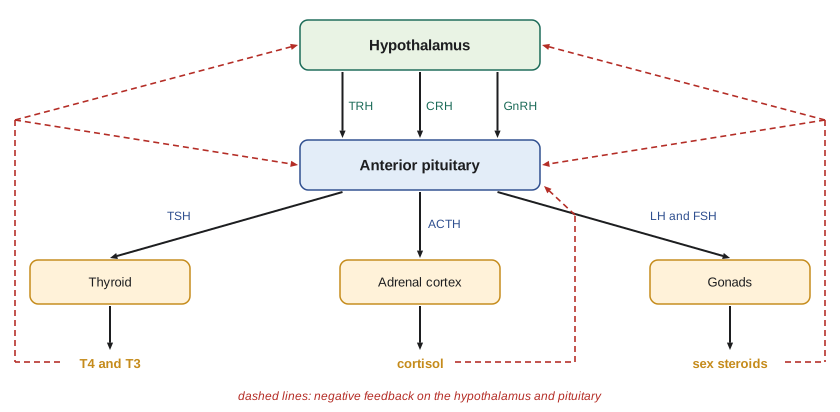
Each axis is controlled by negative feedback. Thyroid hormones inhibit the secretion of both thyrotropin-releasing hormone (TRH) by the hypothalamus and TSH by the pituitary; cortisol inhibits corticotropin-releasing hormone (CRH) and ACTH. This long-loop feedback stabilizes hormone levels, and its disruption is the basis of many clinical tests and disorders: in primary hypothyroidism, where the thyroid fails, TSH rises because the feedback signal is missing. In some cases feedback switches sign. As estradiol rises in the late follicular phase of the mammalian ovarian cycle, it switches from inhibiting to stimulating LH release, producing the positive-feedback LH surge that triggers ovulation.
The stress response
When an animal faces a threat, two systems respond. Within seconds, the sympathetic nervous system and the adrenal medulla release noradrenaline and adrenaline, which raise heart rate and blood pressure, mobilize glucose and fatty acids and divert blood to muscles, the fight-or-flight response described by Walter Cannon. Over minutes to hours, the hypothalamic–pituitary–adrenal (HPA) axis raises glucocorticoids, cortisol in most mammals and fish, corticosterone in rodents, birds, reptiles and amphibians. Glucocorticoids mobilize energy by promoting gluconeogenesis and the breakdown of protein and fat, suppress processes that can wait, such as reproduction, growth and some immune responses, and act on the brain to shape behavior and memory.
Acute stress responses are adaptive; chronic activation is costly. Wildlife physiologists measure glucocorticoids in blood, feces, hair and feathers to assess the condition of populations. Snowshoe hares in the Yukon show raised glucocorticoid levels when predators are abundant during the decline phase of their roughly ten-year population cycle, and Rudy Boonstra and colleagues have argued that this predator-induced stress reduces reproduction and may contribute to the delayed recovery of hare populations. Interpreting such measurements requires care, because glucocorticoid levels also vary with season, time of day, reproductive state and the handling needed to obtain a sample, and blood levels rise within a few minutes of capture.
Hormones and energy balance
Blood glucose is regulated by the opposing actions of insulin and glucagon from the islets of the pancreas. When glucose rises after a meal, beta cells release insulin, which promotes glucose uptake into muscle and fat and glycogen synthesis in the liver; when glucose falls, alpha cells release glucagon, which promotes glycogen breakdown and glucose production. Insulin was discovered in Toronto in 1921–1922 by Frederick Banting, Charles Best, James Collip and John Macleod, a discovery that transformed the treatment of type 1 diabetes. Comparative endocrinology adds perspective: birds have blood glucose levels roughly twice those of mammals of similar size and are relatively insensitive to insulin, while many carnivorous fish tolerate large swings in blood glucose.
Longer-term energy balance is signaled by leptin, secreted by fat cells in proportion to fat mass, which acts on the hypothalamus to reduce food intake and permit energy-expensive processes such as reproduction. Ghrelin from the stomach signals hunger. Thyroid hormones set the overall rate of metabolism in endotherms; they increase mitochondrial biogenesis and the activity of the sodium pump, and their levels change seasonally in many northern mammals and birds. In fish, amphibians and reptiles, thyroid hormones have less effect on resting metabolism but are essential for development.
Metamorphosis: insects
Arthropods grow by molting, shedding the old exoskeleton and expanding a new one before it hardens. In insects, molting is controlled by the steroid hormone 20-hydroxyecdysone, made from ecdysone secreted by the prothoracic glands under the control of prothoracicotropic hormone from the brain. Whether a molt produces another larva, a pupa or an adult depends on juvenile hormone, secreted by the corpora allata. When juvenile hormone is high, a molt produces another larval stage; when it falls in the final larval stage, the next molt produces a pupa (in insects with complete metamorphosis) and then an adult. Vincent Wigglesworth showed this in the 1930s with the blood-sucking bug Rhodnius, joining insects at different stages so that they shared blood, and Carroll Williams later extracted juvenile hormone from silk moths.
The ecdysis behavior itself, the sequence of movements that splits and sheds the old cuticle, is triggered by a cascade of peptide hormones including ecdysis-triggering hormone and eclosion hormone, and the new cuticle is hardened by bursicon. Because juvenile hormone and ecdysteroids are unique to arthropods, synthetic mimics and blockers have been developed as insecticides. Methoprene, a juvenile hormone mimic, is used against mosquito larvae and fleas: treated larvae cannot complete metamorphosis into reproductive adults.
Metamorphosis: amphibians
In amphibians, metamorphosis is driven by thyroid hormones. As a tadpole approaches metamorphosis, the hypothalamus releases corticotropin-releasing hormone, which in amphibians stimulates TSH as well as ACTH. Thyroxine and triiodothyronine rise and act on nuclear receptors in tissues throughout the body, with tissue-specific results: the tail and gills are resorbed by programmed cell death, legs grow, the gut is remodeled from a long herbivorous tube into a short carnivorous one, the skin changes, and the liver begins to make urea rather than ammonia. J. F. Gudernatsch showed in 1912 that feeding tadpoles thyroid tissue made them metamorphose early. Glucocorticoids act together with thyroid hormones and accelerate metamorphosis, so tadpoles in drying ponds or under predation risk can metamorphose earlier, at a smaller size.
Because amphibian metamorphosis depends on thyroid signaling, tadpoles are sensitive indicators of chemicals that disrupt it, and tests using Xenopus tadpoles are part of international testing guidelines for endocrine disruptors. Endocrine-disrupting chemicals, including some pesticides, industrial compounds and pharmaceuticals, can mimic or block hormones. Feminization of male fish downstream of wastewater outfalls exposed to synthetic estrogens has been documented in Canada and the United States, and a whole-lake experiment at the IISD Experimental Lakes Area in northwestern Ontario, in which very low concentrations of the contraceptive estrogen ethinylestradiol were added to a lake, led to the collapse of the fathead minnow population within a few years, as reported by Karen Kidd and colleagues in 2007.
# A simple negative feedback model of a hypothalamic-pituitary-gland axis
# (teaching model; arbitrary units).  R = releasing hormone, P = pituitary hormone,
# G = gland hormone.  G inhibits both R and P.
def run(gland_capacity=1.0, extra_G=0.0, days=10.0, dt=0.001):
    R, P, G = 1.0, 1.0, 1.0
    t = 0.0
    while t < days:
        R += (1.0 / (1.0 + G ** 2) * 2.0 - R) * 2.0 * dt
        P += (R / (1.0 + G ** 2) * 2.0 - P) * 3.0 * dt
        G += (gland_capacity * P - G) * 0.5 * dt + extra_G * dt * 0.5
        t += dt
    return R, P, G

cases = (("normal axis", 1.0, 0.0),
         ("gland damaged (capacity x 0.3)", 0.3, 0.0),
         ("hormone given as a drug", 1.0, 1.5))
print("case                              R      P      G")
for name, cap, extra in cases:
    R, P, G = run(cap, extra)
print(f"{name:32s} {R:5.2f} {P:5.2f} {G:5.2f}")case R P G normal axis 1.00 1.00 1.00 gland damaged (capacity x 0.3) 1.44 2.08 0.62 hormone given as a drug 0.49 0.24 1.74
The model reproduces two patterns that endocrinologists use every day. When the gland is damaged, its hormone falls, feedback weakens, and the pituitary hormone roughly doubles, the pattern of primary hypothyroidism with high TSH. When the end hormone is supplied from outside, as with long-term glucocorticoid treatment or anabolic steroid use, the releasing and pituitary hormones are suppressed, and the gland itself can atrophy from lack of stimulation. The model is far simpler than any real axis, which includes pulsatile release, circadian rhythms and binding proteins, but feedback logic explains the direction of change.
Worked problem
Interpreting a hormone profile
A captive bird shows lethargy and poor molt. Plasma thyroxine is low. In bird A, TSH is high; in bird B, TSH is low. A test injection of TRH raises TSH sharply in bird B. Where is the defect in each bird?
Show solution
- Low thyroxine with high TSH means the pituitary is responding to weak feedback; the thyroid itself is failing (primary hypothyroidism) in bird A.
- Low thyroxine with low TSH means the pituitary is not driving the thyroid; the defect lies in the pituitary or hypothalamus in bird B.
- In bird B, TRH raises TSH, so the pituitary can respond; the problem is insufficient TRH from the hypothalamus.
- Possible causes in bird A include iodine deficiency or thyroid damage; in bird B, hypothalamic disease.
Answer: Bird A has a thyroid defect; bird B has a hypothalamic defect (tertiary hypothyroidism). Reading the levels of hormones at successive steps of the axis, and testing each step, localizes the defect.
Common mistakes
MistakeAssuming all hormones act on cell-surface receptors.
InsteadSteroid and thyroid hormones cross membranes and act on intracellular nuclear receptors.
MistakeConfusing total hormone concentration with active hormone.
InsteadFor bound hormones such as steroids and thyroid hormones, only the free fraction is biologically active.
MistakeThinking the posterior pituitary makes its own hormones.
InsteadVasopressin and oxytocin are made by hypothalamic neurons and released from their axon terminals in the posterior pituitary.
MistakeExpecting feedback always to be negative.
InsteadSome axes switch to positive feedback, as estradiol does before the LH surge.
MistakeAssuming juvenile hormone triggers molting.
InsteadEcdysteroids trigger molting; juvenile hormone determines whether the molt produces a larva, pupa or adult.
MistakeTreating a single glucocorticoid measurement as a reliable index of chronic stress.
InsteadLevels vary with time of day, season, reproductive state and handling; sampling must be standardized.
Summary and key terms
- Hormones are chemical messengers carried in body fluids; neurohormones, paracrine and autocrine signals broaden the classical definition.
- Peptides act on surface receptors and act quickly; steroids and thyroid hormones act on nuclear receptors and act slowly.
- Hormone effect depends on receptor occupancy (Kd), receptor number, half-life and pulsatility of secretion.
- The hypothalamus controls the posterior pituitary by neurosecretion and the anterior pituitary by releasing hormones in a portal system.
- Hypothalamic–pituitary axes are controlled by negative feedback, which explains common clinical patterns.
- Catecholamines and glucocorticoids coordinate the acute stress response; chronic activation is costly.
- Insect metamorphosis is controlled by ecdysteroids and juvenile hormone; amphibian metamorphosis by thyroid hormones, making both targets for endocrine disruption.
- hormone
- A chemical messenger secreted into body fluids that acts on distant target cells.
- neurohormone
- A hormone secreted by a neuron into the blood.
- dissociation constant
- The hormone concentration at which half the receptors are occupied.
- down-regulation
- A reduction in receptor number in response to prolonged high hormone levels.
- half-life
- The time for a hormone's plasma concentration to fall by half after secretion stops.
- portal system
- Blood vessels that carry hypothalamic releasing hormones directly to the anterior pituitary.
- tropic hormone
- A hormone that controls the secretion of another endocrine gland.
- HPA axis
- The hypothalamic–pituitary–adrenal axis that controls glucocorticoid secretion.
- glucocorticoid
- A steroid hormone of the adrenal cortex, such as cortisol or corticosterone, that mobilizes energy.
- ecdysteroid
- A steroid hormone of arthropods that triggers molting.
- juvenile hormone
- An insect hormone that maintains larval characters at each molt.
- endocrine disruptor
- A chemical that interferes with hormone synthesis, transport or action.
Practice problems
Work each problem on paper first, then reveal the answer to check.
Give one example each of a peptide, a steroid and an amine hormone.
Show answer
Insulin (peptide), cortisol (steroid), adrenaline or thyroxine (amine).
A hormone has a Kd of 1 nM. What fraction of receptors is occupied at 3 nM?
Show answer
3/(3 + 1) = 0.75.
Why do steroid hormones typically have longer half-lives than peptides?
Show answer
They circulate bound to carrier proteins, which protect them from clearance.
Explain why TSH is high in primary hypothyroidism.
Show answer
Low thyroid hormone removes negative feedback, so the hypothalamus and pituitary increase TRH and TSH secretion.
Which hormones are released from the posterior pituitary, and where are they made?
Show answer
Vasopressin and oxytocin, made in hypothalamic neurosecretory cells.
What causes the LH surge before ovulation?
Show answer
Rising estradiol switches from negative to positive feedback on GnRH and LH secretion.
Contrast the fast and slow components of the stress response.
Show answer
Fast: sympathetic and adrenal medullary catecholamines; slow: HPA axis glucocorticoids.
What happens if juvenile hormone remains high during the last larval molt?
Show answer
The insect molts into another, often supernumerary, larval stage instead of a pupa.
Which hormone drives amphibian metamorphosis, and why are tadpoles used to test chemicals?
Show answer
Thyroid hormones; tadpoles respond sensitively to chemicals that disrupt thyroid signaling.
What did the Experimental Lakes Area ethinylestradiol experiment show?
Show answer
Very low concentrations of a synthetic estrogen caused the collapse of a fathead minnow population.
Lesson quiz · 5 questions
Score 60% or more to complete this lesson.
Create a free account to save your progress, take quizzes and earn a certificate.