
BIOL-350 · Lesson 6 of 12 · 30 min
Muscle and movement
Muscle turns chemical energy into force and movement. It powers walking, flying, swimming, breathing, the heartbeat, the movement of food through the gut and the constriction of blood vessels. Muscle tissue makes up 40 percent or more of the body mass of many vertebrates, and its properties set limits on how fast animals can run, how high they can jump and how long they can keep going. BIOL-214 described actin filaments, myosin motors and the sliding filament mechanism at the molecular level. This lesson builds up from the sarcomere to whole muscles, compares the main types of muscle, and examines some of the extreme muscles that evolution has produced, from the asynchronous flight muscles of flies to the superfast sound-producing muscles of rattlesnakes.
Structure of skeletal muscle
A vertebrate skeletal muscle is made of long, multinucleate cells called muscle fibers, formed by the fusion of many precursor cells. Each fiber is packed with myofibrils, and each myofibril is divided along its length into repeating units called sarcomeres, about 2 to 3 µm long, which give skeletal and cardiac muscle their striped, or striated, appearance. A sarcomere runs from one Z disc to the next. Thin filaments, made mainly of actin together with the regulatory proteins tropomyosin and troponin, are anchored at the Z discs and extend toward the center. Thick filaments, made of myosin, occupy the center of the sarcomere, forming the dark A band, and are held in place by the giant elastic protein titin, which runs from the Z disc to the middle of the sarcomere. The light I band contains only thin filaments.
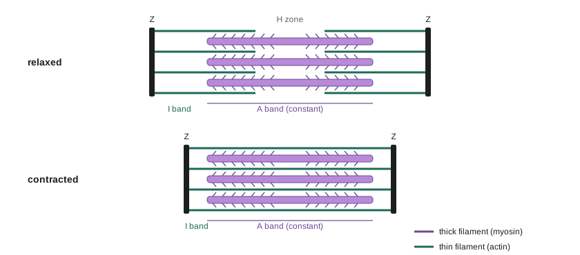
When a muscle contracts, the A band stays the same width while the I bands and the central H zone narrow, as Andrew Huxley and Rolf Niedergerke, and independently Hugh Huxley and Jean Hanson, showed in 1954. Neither filament shortens; they slide past one another. The sliding is driven by cross-bridges, the heads of myosin molecules, which bind to actin, pivot in a power stroke that pulls the thin filament toward the center of the sarcomere, release when they bind a new ATP molecule, and re-cock as ATP is hydrolyzed. Each cycle moves the filaments about 5 to 10 nm and uses one ATP. Without ATP, cross-bridges remain locked to actin, which is why muscles stiffen after death in rigor mortis.
Excitation–contraction coupling
A skeletal muscle fiber contracts when its motor neuron fires. Acetylcholine released at the neuromuscular junction opens nicotinic receptors and triggers an action potential in the muscle membrane, the sarcolemma, which spreads along the fiber and down into it through transverse tubules, or T tubules, invaginations of the membrane that reach every myofibril. Where T tubules meet the sarcoplasmic reticulum, the muscle's internal calcium store, voltage-sensing dihydropyridine receptors in the T-tubule membrane are mechanically linked to ryanodine receptors, the calcium release channels of the sarcoplasmic reticulum. Depolarization changes the shape of the dihydropyridine receptors, which pull open the ryanodine receptors, and calcium floods into the cytoplasm, raising its concentration about a hundredfold, from about 0.1 µM to 10 µM.
Calcium binds troponin C, which shifts tropomyosin away from the myosin-binding sites on actin, allowing cross-bridges to form. When the action potentials stop, calcium pumps (SERCA) return calcium to the sarcoplasmic reticulum, tropomyosin covers the binding sites again, and the muscle relaxes. Relaxation therefore costs energy too: the calcium pumps may use a quarter to a half of the ATP consumed during contraction. In the inherited condition malignant hyperthermia, found in humans and pigs, mutant ryanodine receptors leak calcium when exposed to certain anesthetic gases, and the resulting uncontrolled contraction and calcium pumping produce dangerous heat. In vertebrate cardiac muscle, coupling works differently: calcium entering through the dihydropyridine receptors themselves triggers calcium release from the sarcoplasmic reticulum, calcium-induced calcium release.
The length–tension relationship
The force a muscle fiber can produce depends on its length, because force depends on how many cross-bridges can form, which depends on the overlap between thick and thin filaments. In 1966 A. M. Gordon, Andrew Huxley and F. J. Julian measured the maximum isometric force of single frog muscle fibers held at different sarcomere lengths. Force was greatest over a plateau of sarcomere lengths from about 2.0 to 2.2 µm, where every myosin head in the overlapping region could reach actin. At longer lengths force fell linearly to zero at about 3.6 µm, where thick and thin filaments no longer overlapped. At shorter lengths force also fell, as thin filaments from opposite ends collided and thick filaments butted against the Z discs. The result was a striking confirmation of the sliding filament theory.
# Length-tension relationship from filament overlap (frog muscle, Gordon, Huxley and Julian 1966)
thick = 1.60      # thick filament length, um
bare = 0.15       # bare zone without heads at the middle of the thick filament, um
thin = 1.00       # each thin filament (including half Z disc), um
plateau_hi = thin * 2 + bare          # 2.15: all heads overlap thin filaments
zero_hi = thick + 2 * thin            # 3.60: no overlap left

def force(sl):
    # relative isometric force (1.0 on the plateau); simplified ascending limb
    if sl >= zero_hi: return 0.0
    if sl >= plateau_hi: return (zero_hi - sl) / (zero_hi - plateau_hi)
    if sl >= 2.0: return 1.0
    if sl >= 1.65: return 1.0 - 0.6 * (2.0 - sl) / (2.0 - 1.65)      # thin filaments overlap
    return max(0.0, 0.4 - 1.6 * (1.65 - sl))                          # thick filaments hit Z discs

print(" sarcomere length (um)   relative force")
for sl in (1.3, 1.6, 1.9, 2.0, 2.15, 2.6, 3.0, 3.4, 3.6):
print(f" {sl:4.2f} {force(sl):5.2f} " + "#" * int(round(force(sl) * 30))) sarcomere length (um) relative force
1.30 0.00
1.60 0.32 ##########
1.90 0.83 #########################
2.00 1.00 ##############################
2.15 1.00 ##############################
2.60 0.69 #####################
3.00 0.41 ############
3.40 0.14 ####
3.60 0.00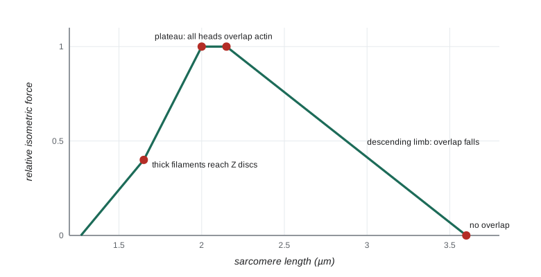
In the body, most muscles operate near the top of the curve. The skeleton constrains muscle length, and the lengths of muscles and the positions of their attachments are matched so that sarcomeres work near their optimum over the normal range of joint movement. Passive tension, mainly from titin, rises steeply at long lengths and protects sarcomeres from being pulled apart.
Force, velocity and power
A muscle shortening against a light load moves quickly; against a heavy load it moves slowly, and against a load equal to its maximum isometric force it does not shorten at all. A. V. Hill described this relationship in 1938 with a hyperbolic equation derived from measurements of frog muscle. The force–velocity curve follows because cross-bridges attached for less time during fast shortening, and some are dragged into negative positions where they resist motion, so fewer contribute to force. Power, the product of force and velocity, is zero at both ends of the curve and reaches a maximum at about one third of maximum force and one third of maximum velocity. Animals tend to use their muscles near this point when power matters, for example in jumping, sprinting and flight.
# Hill's force-velocity equation and power output
# (F + a)(v + b) = (F0 + a) b, with a/F0 = b/vmax = 0.25 (typical of vertebrate muscle)
F0 = 1.0          # maximum isometric force (relative)
vmax = 1.0        # maximum shortening velocity (relative)
k = 0.25
a, b = k * F0, k * vmax

def velocity(F):
    return (F0 + a) * b / (F + a) - b

print("  F/F0    v/vmax   power (F v)")
best = (0, 0, 0)
for i in range(0, 11):
    F = i / 10
    v = velocity(F)
    p = F * v
    best = max(best, (p, F, v))
    print(f"  {F:4.1f}   {v:6.3f}    {p:6.3f}")
fine = max((F * velocity(F), F) for F in [i / 1000 for i in range(1001)])
print(f"maximum power {fine[0]:.3f} at F/F0 = {fine[1]:.2f}, v/vmax = {velocity(fine[1]):.2f}")F/F0 v/vmax power (F v) 0.0 1.000 0.000 0.1 0.643 0.064 0.2 0.444 0.089 0.3 0.318 0.095 0.4 0.231 0.092 0.5 0.167 0.083 0.6 0.118 0.071 0.7 0.079 0.055 0.8 0.048 0.038 0.9 0.022 0.020 1.0 0.000 0.000 maximum power 0.095 at F/F0 = 0.31, v/vmax = 0.31
Peak power occurs at about 0.31 of maximum force and 0.31 of maximum velocity, delivering only about a tenth of the product of maximum force and maximum velocity. Because the curvature constant differs between muscles, the exact optimum varies, but the principle is general and explains why vertebrates evolved gears in their skeletons and why bicycles have gears too: by changing the leverage, an animal or a cyclist can keep muscles shortening near the velocity that gives maximum power.
From twitch to tetanus: grading force
A single action potential in a muscle fiber produces a twitch, a brief contraction lasting tens to hundreds of milliseconds. Because the twitch lasts much longer than the action potential, a second action potential arriving before the fiber has relaxed produces additional force, summation. At high stimulation frequencies, the twitches fuse into a smooth, sustained contraction, a fused tetanus, with a force several times that of a single twitch, because calcium stays high and the elastic elements in series with the cross-bridges stay stretched.
Whole muscles grade their force in two ways. First, by rate coding: changing the firing frequency of motor neurons. Second, by recruitment: changing the number of motor units active. A motor unit is one motor neuron and all the muscle fibers it innervates, from a few fibers in muscles controlling the eye to more than a thousand in large leg muscles. According to Henneman's size principle, motor units are recruited in order of size: small motor neurons, which innervate a few slow, fatigue-resistant fibers, are recruited first, and large motor neurons, which innervate many fast, powerful but fatigable fibers, are recruited only for strong contractions. Fine control at low forces and powerful contractions when needed both follow from this simple rule.
Fiber types
Skeletal muscle fibers differ in the isoform of myosin they express, which determines how fast they contract, and in their metabolism. Slow oxidative fibers (type I) contain slow myosin, many mitochondria, much myoglobin and a dense capillary supply; they are red, resist fatigue and suit posture and endurance. Fast glycolytic fibers (type IIb or IIx) contain fast myosin, few mitochondria and large glycogen stores; they are pale, powerful and fatigue quickly. Fast oxidative-glycolytic fibers (type IIa) are intermediate. The proportions vary with species, muscle and training: the breast muscles of migratory birds such as geese are dark and oxidative; those of chickens and grouse, which fly only in short bursts, are pale. In fish, the slow red muscle runs in a thin band along the side of the body and powers sustained cruising, while the bulk of the body is white muscle used for bursts.
| Property | Slow oxidative (I) | Fast oxidative-glycolytic (IIa) | Fast glycolytic (IIx/IIb) |
|---|---|---|---|
| Myosin ATPase rate | Slow | Fast | Fastest |
| Mitochondria and capillaries | Many | Many to moderate | Few |
| Myoglobin (color) | High (red) | High (red–pink) | Low (white) |
| Main ATP source | Oxidative phosphorylation | Both | Glycolysis |
| Fatigue resistance | High | Intermediate | Low |
| Typical use | Posture, endurance, cruising fish | Sustained running, flight in some birds | Sprinting, bursts, escape |
Cardiac and smooth muscle
Cardiac muscle is striated like skeletal muscle but its cells are short, branched and usually have a single nucleus. They are joined end to end by intercalated discs containing gap junctions, so action potentials spread from cell to cell and the heart contracts as a unit. The cardiac action potential has a long plateau, lasting hundreds of milliseconds, produced by L-type calcium channels, which keeps the muscle refractory until it has relaxed; the heart therefore cannot be tetanized, which would be fatal. The heart's rhythm and the regulation of its force are covered in lesson 8.
Smooth muscle lacks sarcomeres, so it is not striated. Actin and myosin filaments are arranged obliquely and anchored to dense bodies in the cytoplasm and membrane. Smooth muscle lines blood vessels, airways, the gut, the bladder and the uterus. It contracts slowly and can sustain force for long periods at very low energy cost. It is regulated not by troponin but mainly by calcium–calmodulin activation of myosin light-chain kinase, which phosphorylates myosin. In the latch state, dephosphorylated cross-bridges remain attached and hold tension economically, an extreme version of which is the catch muscle of bivalve molluscs: the adductor muscle of a mussel or scallop can hold its shell shut for hours or days using almost no ATP, a state controlled by the protein twitchin.
Extreme muscles
Insects that beat their wings faster than about 100 times a second, including flies, bees, wasps and beetles, use asynchronous flight muscle. Instead of contracting once for every nerve impulse, these muscles are activated by stretch: when the thorax is deformed by one set of muscles, the stretch triggers contraction of the antagonist set, which deforms the thorax back. Calcium stays high throughout flight, and nerve impulses arrive only every few wingbeats to keep the muscles active. The thorax and wings form a resonant system, and wingbeat frequency is set by its mechanical properties rather than by the nervous system. Some midges beat their wings about 1,000 times per second. By avoiding calcium cycling on every beat, asynchronous muscle saves the cost of pumping calcium and can devote space to mitochondria and myofibrils.
Synchronous superfast muscles take the other route. The shaker muscles in the tails of rattlesnakes, the sound-producing muscles of toadfish swim bladders and the syrinx muscles of songbirds contract and relax up to 100 to 250 times a second, each contraction triggered by its own action potential. Lawrence Rome and colleagues showed that these muscles achieve their speed with an abundant sarcoplasmic reticulum that pumps calcium extremely quickly, fast cross-bridge detachment and very short sarcomeres, at the cost of producing little force. The trade-off between speed and force in muscle is one of the clearest examples of how a single molecular machine has been tuned for very different jobs.
Worked problem
Why can a flea jump so high?
A flea about 1.5 mm long can jump over 15 cm high, accelerating in less than a millisecond. Vertebrate muscle produces a maximum power output of roughly 200 to 400 W per kilogram of muscle. Estimate the power needed per kilogram of muscle if leg muscles make up about 10 percent of the flea's 0.5 mg body mass and the take-off lasts 1 ms. Can muscle alone explain the jump?
Show solution
- Energy for a 15 cm jump: m g h = 0.5 × 10⁻⁶ kg × 9.8 × 0.15 ≈ 7.4 × 10⁻⁷ J.
- Delivered in 1 ms, power = 7.4 × 10⁻⁷ / 10⁻³ ≈ 7.4 × 10⁻⁴ W.
- Muscle mass ≈ 0.05 mg = 5 × 10⁻⁸ kg, so power per kilogram ≈ 7.4 × 10⁻⁴ / 5 × 10⁻⁸ ≈ 15,000 W/kg.
- That is dozens of times higher than any muscle can deliver.
- Fleas, like locusts and froghoppers, use a catapult: muscles contract slowly to store energy in an elastic pad of resilin, which is released almost instantly.
Answer: Muscle alone cannot explain the jump; the power must be amplified by elastic energy storage. Power amplification with springs and latches is common in small, fast animals, from the strikes of mantis shrimp to the tongues of chameleons.
Common mistakes
MistakeSaying the filaments shorten when a muscle contracts.
InsteadThick and thin filaments keep their length; they slide past one another, narrowing the I band and H zone.
MistakeThinking ATP is needed only for cross-bridge power strokes.
InsteadATP is also needed to detach cross-bridges and to pump calcium back into the sarcoplasmic reticulum during relaxation.
MistakeAssuming maximum force and maximum power occur together.
InsteadMaximum power occurs at about a third of maximum force and velocity; maximum force occurs at zero velocity.
MistakeTreating all muscle as regulated by troponin.
InsteadSmooth muscle is regulated mainly through calcium–calmodulin and myosin light-chain kinase.
MistakeDescribing asynchronous flight muscle as contracting once per nerve impulse.
InsteadIt is activated by stretch and contracts many times per impulse; the wingbeat frequency is set by thoracic mechanics.
MistakeSaying cardiac muscle can sum to tetanus like skeletal muscle.
InsteadIts long refractory period prevents tetanus, allowing the heart to relax and fill between beats.
Summary and key terms
- Skeletal muscle fibers contain myofibrils made of sarcomeres; contraction occurs as thin and thick filaments slide, driven by cross-bridge cycles that use ATP.
- Excitation–contraction coupling links the action potential to calcium release through dihydropyridine and ryanodine receptors; calcium binding to troponin allows cross-bridges to form.
- Isometric force depends on filament overlap, giving the length–tension relationship.
- Force and velocity trade off along Hill's hyperbola; power peaks near a third of maximum force and velocity.
- Force is graded by summation, tetanus, rate coding and size-ordered recruitment of motor units.
- Fiber types differ in myosin isoforms and metabolism; cardiac muscle cannot be tetanized; smooth muscle holds force economically.
- Asynchronous flight muscles and superfast synchronous muscles show how muscle has been tuned for extreme frequencies.
- sarcomere
- The repeating contractile unit of striated muscle, from one Z disc to the next.
- cross-bridge
- A myosin head that binds actin and generates force during its power stroke.
- excitation–contraction coupling
- The sequence linking a muscle action potential to calcium release and contraction.
- T tubule
- An invagination of the muscle membrane that carries action potentials into the fiber.
- sarcoplasmic reticulum
- The internal membrane store from which calcium is released and to which it is pumped back.
- ryanodine receptor
- The calcium release channel of the sarcoplasmic reticulum.
- length–tension relationship
- The dependence of isometric force on sarcomere length, reflecting filament overlap.
- force–velocity relationship
- The inverse, hyperbolic relationship between load and shortening velocity described by Hill.
- tetanus
- A sustained contraction produced by fused twitches at high stimulation frequency.
- motor unit
- A motor neuron and all the muscle fibers it innervates.
- size principle
- Motor units are recruited in order from smallest to largest.
- asynchronous muscle
- Muscle that contracts many times per nerve impulse, activated by stretch, as in insect flight muscle.
Practice problems
Work each problem on paper first, then reveal the answer to check.
Which bands of the sarcomere narrow during contraction and which stays constant?
Show answer
The I band and H zone narrow; the A band stays constant.
List the steps of excitation–contraction coupling in skeletal muscle.
Show answer
Neuromuscular transmission, muscle action potential, spread into T tubules, dihydropyridine receptors pull open ryanodine receptors, calcium release, calcium binds troponin, cross-bridges form.
Why does rigor mortis occur?
Show answer
Without ATP, myosin heads cannot detach from actin, so muscles stay locked.
At what sarcomere length does a frog muscle fiber produce zero isometric force on the descending limb, and why?
Show answer
About 3.6 µm, where thick and thin filaments no longer overlap.
Using Hill's equation with a/F₀ = 0.25, at what fraction of maximum force does power peak?
Show answer
About 0.31 of maximum force.
Distinguish rate coding from recruitment.
Show answer
Rate coding changes motor neuron firing frequency; recruitment changes the number of active motor units.
Why are migratory geese breast muscles dark and chicken breast muscles pale?
Show answer
Geese use oxidative fibers with myoglobin for sustained flight; chickens use glycolytic fibers for brief bursts.
Why can't the heart be tetanized?
Show answer
The cardiac action potential has a long plateau and refractory period that outlast most of the contraction.
How do bivalve catch muscles hold the shell closed cheaply?
Show answer
Cross-bridges remain attached in a latch-like state controlled by twitchin, using very little ATP.
How do asynchronous flight muscles achieve very high wingbeat frequencies?
Show answer
Stretch activation and resonance of the thorax produce many contractions per nerve impulse without cycling calcium each beat.
Lesson quiz · 5 questions
Score 60% or more to complete this lesson.
Create a free account to save your progress, take quizzes and earn a certificate.