
BIOL-107 · Lesson 5 of 12 · 26 min
Cell structure: prokaryotic and eukaryotic cells
Lesson 1 drew the big distinction between prokaryotic and eukaryotic cells. This lesson opens them up. We tour a bacterial cell, then an animal cell and a plant cell, organelle by organelle, asking for each structure what it is made of and what job it does. Think of the eukaryotic cell as a city with districts: a control center, factories, a shipping department, power stations, waste recycling and a transport network. The analogy is imperfect, but it helps to remember that every compartment exists to solve a problem.
This is an overview. BIOL-214 (Cell Biology) returns to each organelle in depth, including how proteins are sorted to the right compartment and how the cytoskeleton is built. Here the aim is a clear map.
Inside a bacterial cell
A typical bacterium such as Escherichia coli is a rod about 1 µm wide and 2 µm long. Despite its size, it is highly organized.
| Structure | Description | Function |
|---|---|---|
| Plasma membrane | Phospholipid bilayer with proteins | Controls what enters and leaves; site of respiration and, in some bacteria, photosynthesis |
| Cell wall | Peptidoglycan (sugar chains cross-linked by short peptides) | Gives shape and stops the cell bursting from water uptake |
| Outer membrane (Gram-negative bacteria) | A second membrane outside a thin wall | Extra barrier; contains lipopolysaccharide |
| Capsule | Sticky layer of polysaccharide (not in all bacteria) | Attachment to surfaces; protection from the immune system |
| Nucleoid | Region containing the circular chromosome | Stores genetic information |
| Plasmids | Small extra circles of DNA | Carry optional genes, such as antibiotic resistance |
| Ribosomes (70S) | RNA and protein particles | Make proteins |
| Flagella | Long helical protein filaments driven by a rotary motor | Swimming |
| Pili (fimbriae) | Short hair-like protein appendages | Attachment; some transfer DNA between cells |
The cell wall matters medically. Bacteria with a thick peptidoglycan layer retain a purple dye in the Gram stain (Gram-positive); those with a thin wall and an outer membrane do not (Gram-negative). Penicillin and related antibiotics block the enzymes that build peptidoglycan, which human cells do not have, so they harm bacteria without harming us. That is a general principle: many antibiotics target structures, such as 70S ribosomes or peptidoglycan, that differ between bacteria and eukaryotes. Microbiology (BIOL-330) develops this further.
The eukaryotic cell: an overview
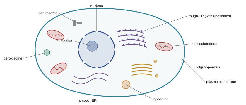
Eukaryotic cells are typically 10 to 100 µm across, and their key innovation is internal membranes that divide the cell into compartments. Each compartment can maintain its own chemical conditions, such as the acidic interior of a lysosome, and concentrate particular enzymes and substrates together. The membranes of the nucleus, endoplasmic reticulum, Golgi apparatus, lysosomes and vesicles form a connected network called the endomembrane system, exchanging material through small membrane bubbles called vesicles.
The nucleus: the information center
The nucleus holds most of the cell's DNA. It is enclosed by the nuclear envelope, a double membrane perforated by nuclear pores, large protein complexes that control traffic in and out: RNA molecules and ribosome parts leave; proteins needed for DNA work enter. Inside, DNA is wrapped around proteins called histones to form chromatin. When the cell prepares to divide, chromatin condenses into visible chromosomes (lesson 12). A dense region called the nucleolus assembles ribosome subunits from RNA and protein.
The nucleus is where genes are read. The flow of genetic information, which Francis Crick called the central dogma, runs from DNA to RNA to protein. In transcription, a gene's DNA sequence is copied into messenger RNA (mRNA) in the nucleus. The mRNA leaves through a nuclear pore, and in translation, ribosomes in the cytoplasm read it three bases at a time. Each three-base codon specifies one amino acid or a stop signal. BIOL-207 covers the machinery; the short program below shows the logic of reading a message.
# Translating a short mRNA with part of the standard genetic code
code = {"AUG": "Met", "UUU": "Phe", "UUC": "Phe", "GGC": "Gly", "GGA": "Gly",
        "AAA": "Lys", "GAA": "Glu", "UGG": "Trp", "CUG": "Leu", "GCU": "Ala",
        "UAA": "STOP", "UAG": "STOP", "UGA": "STOP"}

def translate(mrna):
    start = mrna.find("AUG")              # ribosomes begin at the first AUG
    protein = []
    for i in range(start, len(mrna) - 2, 3):
        aa = code[mrna[i:i + 3]]
        if aa == "STOP":
            break
        protein.append(aa)
    return "-".join(protein)

print(translate("GCAUGUUUGGCAAAGAAUGGCUGUAAGC"))
print(translate("CCAUGGCUGCUUUCUAGAAA"))Met-Phe-Gly-Lys-Glu-Trp-Leu Met-Ala-Ala-Phe
Ribosomes and the endoplasmic reticulum: making proteins
Ribosomes are not surrounded by membrane; they are particles of RNA and protein, about 25 nm across, that build proteins. Free ribosomes in the cytosol make proteins that will stay in the cytosol or go to the nucleus, mitochondria or peroxisomes. Ribosomes bound to the endoplasmic reticulum make proteins destined for membranes, for lysosomes or for export from the cell.
The endoplasmic reticulum (ER) is a maze of flattened sacs and tubes continuous with the outer nuclear membrane. Its two regions do different jobs. Rough ER is studded with ribosomes that feed new polypeptides into the ER interior (the lumen), where they fold and often receive sugar chains. Smooth ER lacks ribosomes; it makes lipids such as phospholipids and steroids, detoxifies drugs (liver cells have a great deal of it), and stores calcium ions, which muscle cells release to trigger contraction.
The Golgi apparatus: processing and shipping
Vesicles bud from the ER carrying new proteins and travel to the Golgi apparatus, a stack of flattened membrane sacs named after Camillo Golgi, who saw it in 1898. The Golgi modifies proteins further, trimming and adding sugar chains, and sorts them by destination. Vesicles leaving the far side of the stack carry proteins to the plasma membrane, to lysosomes, or out of the cell by exocytosis. A cell in the pancreas that secretes digestive enzymes, or a plasma cell making antibodies, has an extensive ER and Golgi.
Worked problem
Tracing an antibody
A plasma cell makes antibodies and secretes them into the blood. List, in order, the structures an antibody molecule passes through from the gene to the outside of the cell.
Show solution
- The antibody genes are transcribed into mRNA in the nucleus.
- The mRNA leaves through nuclear pores and is translated by ribosomes that attach to the rough ER.
- The new polypeptides enter the ER lumen, fold, pair up and receive sugar chains.
- Transport vesicles carry them to the Golgi apparatus, where sugar chains are modified and the protein is sorted.
- Secretory vesicles carry them to the plasma membrane and release them by exocytosis.
Answer: Nucleus → nuclear pore → ribosome on rough ER → ER lumen → transport vesicle → Golgi apparatus → secretory vesicle → plasma membrane (exocytosis).
Lysosomes, peroxisomes and vacuoles
Lysosomes are membrane sacs of digestive enzymes that break down proteins, fats, carbohydrates and nucleic acids. Their interior is kept at about pH 4.5 to 5 by proton pumps, and their enzymes work best at that pH, a safety feature: if a lysosome leaks, its enzymes are much less active in the neutral cytosol. Lysosomes digest material the cell takes in, such as bacteria engulfed by white blood cells, and recycle worn-out organelles in a process called autophagy (self-eating), whose mechanisms earned Yoshinori Ohsumi the 2016 Nobel Prize in Physiology or Medicine. In lysosomal storage diseases such as Tay–Sachs disease, a missing enzyme lets undigested material build up, damaging nerve cells.
Peroxisomes are small single-membrane organelles that carry out oxidation reactions, including breaking down long fatty acids and detoxifying alcohol in liver cells. These reactions produce hydrogen peroxide, which is toxic, so peroxisomes also contain catalase, which converts it to water and oxygen.
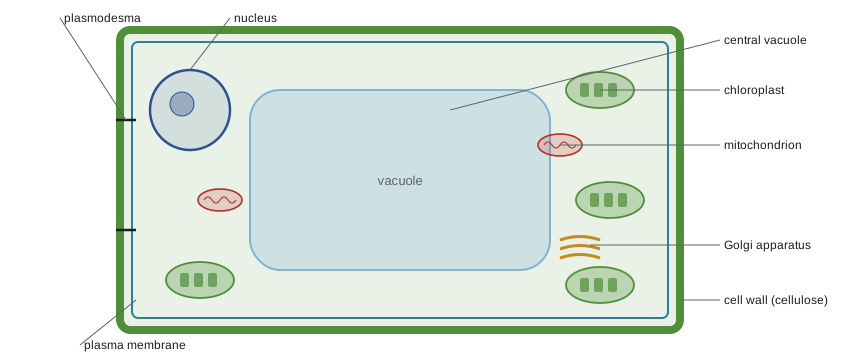
Vacuoles are large membrane-bound sacs. Plant cells usually have a large central vacuole that can fill up to 80 or 90 percent of the cell. It stores water, ions and pigments, holds waste, and pushes the cytoplasm against the wall, keeping the cell firm (turgid). Freshwater protists have contractile vacuoles that pump out excess water.
Mitochondria and chloroplasts: energy converters
Mitochondria release energy from food molecules through cellular respiration (lesson 9), making most of the cell's ATP. Each has a smooth outer membrane and a highly folded inner membrane; the folds, called cristae, increase the surface area for the proteins that make ATP. The space inside the inner membrane, the matrix, contains enzymes, mitochondrial DNA and ribosomes. Cells with high energy demands, such as heart muscle, have thousands.
Chloroplasts, found in plants and algae, capture light energy and use it to build sugars (lesson 10). They have two envelope membranes and a third internal membrane system of flattened sacs called thylakoids, often stacked into grana, which hold the chlorophyll. The fluid around them is the stroma. As lesson 1 explained, both organelles descend from bacteria and keep their own DNA and ribosomes.
The cytoskeleton and the cell surface
The cytoplasm is crisscrossed by a network of protein filaments, the cytoskeleton, which gives the cell shape, anchors organelles, provides tracks for transport and drives movement. It has three main types of filaments: microfilaments of actin, intermediate filaments, and microtubules of tubulin. Cilia and flagella in eukaryotes are built from microtubules and beat with a whip-like motion, quite different from the rotating bacterial flagellum. Lesson 11 describes the cytoskeleton more fully.
Outside the plasma membrane, animal cells secrete an extracellular matrix of proteins such as collagen and sugar-rich molecules, which holds tissues together and passes signals. Plant cells build a rigid cell wall of cellulose fibers embedded in other polysaccharides. Neighboring cells connect through junctions: tight junctions seal gaps between animal cells, desmosomes rivet them together, gap junctions let small molecules pass between them, and in plants channels called plasmodesmata link the cytoplasm of adjacent cells through the walls.
Plant and animal cells compared
| Structure | Animal cells | Plant cells |
|---|---|---|
| Cell wall | Absent; extracellular matrix instead | Present, made of cellulose |
| Chloroplasts | Absent | Present in green tissues |
| Central vacuole | Absent or small vacuoles only | Large central vacuole |
| Mitochondria | Present | Present (plants respire too) |
| Centrioles | Present in the centrosome | Absent in most flowering plants |
| Lysosomes | Present | Lysosome-like functions carried out mainly by the vacuole |
| Shape | Variable, often rounded | Fixed, often box-like, set by the wall |
| Connections | Tight junctions, desmosomes, gap junctions | Plasmodesmata |
A common misconception is that plant cells have chloroplasts instead of mitochondria. In fact plant cells have both: chloroplasts make sugar in the light, and mitochondria break sugar down to make ATP day and night. Root cells, which receive no light, have mitochondria but no chloroplasts.
Structure fits function
Different cell types emphasize different organelles according to their jobs. Asking why a cell has lots of a particular organelle is a good way to check your understanding.
| Cell type | Abundant structure | Why |
|---|---|---|
| Pancreatic acinar cell | Rough ER and Golgi | Secretes large amounts of digestive enzymes |
| Liver cell | Smooth ER and peroxisomes | Detoxifies drugs and alcohol; processes lipids |
| Heart muscle cell | Mitochondria | Needs a continuous supply of ATP to contract |
| Macrophage (white blood cell) | Lysosomes | Digests engulfed bacteria and debris |
| Intestinal lining cell | Microvilli on the surface | Increases surface area for absorbing nutrients |
| Leaf palisade cell | Chloroplasts | Captures light for photosynthesis |
| Sperm cell | Mitochondria and a flagellum | Swims long distances |
Common mistakes
MistakePlant cells have chloroplasts instead of mitochondria.
InsteadPlant cells have mitochondria as well as chloroplasts; they respire continuously and photosynthesize only in light.
MistakeRibosomes are membrane-bound organelles.
InsteadRibosomes are RNA–protein particles with no membrane, found in all cells, including prokaryotes.
MistakeThe cell wall and the plasma membrane are the same thing.
InsteadThe plasma membrane is a phospholipid bilayer that controls transport; the wall is a rigid layer outside it (in plants, fungi and most bacteria).
MistakeBacteria have no internal organization.
InsteadBacteria lack membrane-bound organelles but have a nucleoid, ribosomes, a cytoskeleton and specialized membrane regions.
MistakeLysosome enzymes would digest the whole cell if they leaked.
InsteadLysosomal enzymes work best at pH 4.5 to 5 and are much less active in the neutral cytosol, which limits damage from small leaks.
Summary and key terms
- Bacteria have a plasma membrane, usually a peptidoglycan wall, a nucleoid, plasmids, 70S ribosomes and often flagella and pili; many antibiotics target bacterial-specific structures.
- Eukaryotic cells are divided into membrane-bound compartments; the nucleus, ER, Golgi, lysosomes and vesicles form the endomembrane system.
- The nucleus holds chromatin and controls gene expression; information flows from DNA to RNA to protein.
- Rough ER makes proteins for membranes, lysosomes and export; smooth ER makes lipids, detoxifies and stores calcium.
- The Golgi modifies and sorts proteins and ships them in vesicles; lysosomes digest and recycle; peroxisomes oxidize and detoxify.
- Mitochondria (respiration) and chloroplasts (photosynthesis) convert energy and have their own DNA.
- Plant cells have a cellulose wall, chloroplasts and a large central vacuole; animal cells have centrioles, lysosomes and an extracellular matrix.
- nucleus
- The double-membrane organelle that contains a eukaryotic cell’s chromosomes.
- chromatin
- DNA wrapped around histone proteins, the form of chromosomes in a non-dividing nucleus.
- ribosome
- An RNA–protein particle that synthesizes proteins by reading mRNA.
- endoplasmic reticulum
- A network of membranes; rough ER makes proteins and smooth ER makes lipids.
- Golgi apparatus
- A stack of membrane sacs that modifies, sorts and ships proteins.
- lysosome
- An acidic organelle containing digestive enzymes.
- endomembrane system
- The connected membranes of the nucleus, ER, Golgi, lysosomes, vesicles and plasma membrane.
- peptidoglycan
- The sugar–peptide polymer that forms bacterial cell walls.
- central vacuole
- A large plant organelle that stores water and solutes and keeps the cell turgid.
- codon
- A sequence of three mRNA bases that specifies one amino acid or a stop signal.
Practice problems
Work each problem on paper first, then reveal the answer to check.
Why does penicillin harm bacteria but not human cells?
Show answer
It blocks enzymes that build peptidoglycan cell walls, which human cells do not have.
What is the difference between Gram-positive and Gram-negative bacteria?
Show answer
Gram-positive bacteria have a thick peptidoglycan wall that retains crystal violet; Gram-negative bacteria have a thin wall plus an outer membrane.
Name the parts of the endomembrane system.
Show answer
Nuclear envelope, rough and smooth ER, Golgi apparatus, lysosomes, vesicles and vacuoles, and the plasma membrane.
A cell secretes large amounts of protein hormone. Which organelles would you expect to be well developed?
Show answer
Rough ER and Golgi apparatus, with many secretory vesicles.
What does the nucleolus do?
Show answer
It assembles ribosomal subunits from ribosomal RNA and proteins.
Translate the mRNA AUG-GGC-UUU-UAA using the codon table in this lesson.
Show answer
Met-Gly-Phe, then stop.
Why are lysosomal enzymes kept in an acidic compartment?
Show answer
They work best at pH 4.5 to 5, and keeping them separate protects the rest of the cell; they are much less active in the neutral cytosol.
What is the function of cristae in mitochondria?
Show answer
They increase the surface area of the inner membrane for the proteins of respiration and ATP synthesis.
List three structures found in plant cells but not animal cells.
Show answer
Cellulose cell wall, chloroplasts and a large central vacuole (also plasmodesmata).
Why do heart muscle cells contain many mitochondria?
Show answer
They contract continuously and need a constant large supply of ATP.
Lesson quiz · 5 questions
Score 60% or more to complete this lesson.
Create a free account to save your progress, take quizzes and earn a certificate.