
BIOL-107 · Lesson 3 of 12 · 28 min
The chemistry of life II: macromolecules
Cells build almost everything they need from four classes of large organic molecules: carbohydrates, lipids, proteins and nucleic acids. Three of them are polymers, long chains made by linking many similar small units called monomers. Learning how these molecules are built, and how their structure determines what they do, is the foundation for the rest of the course.
Building and breaking polymers
Cells assemble polymers by dehydration synthesis (also called a condensation reaction): two monomers are joined by a covalent bond, and a molecule of water is released. The reverse reaction, hydrolysis, uses water to break the bond. Digestion is mostly hydrolysis: enzymes in your gut break starch into glucose, proteins into amino acids and fats into fatty acids and glycerol. Building polymers costs energy, which the cell supplies from ATP (lesson 8); breaking them releases a little.
| Class | Monomer | Bond between monomers | Main jobs |
|---|---|---|---|
| Carbohydrates | Monosaccharides (for example glucose) | Glycosidic bond | Energy source and storage; structure (cellulose, chitin); cell recognition |
| Lipids | Not true polymers; built from fatty acids and glycerol and other parts | Ester bonds (in fats and phospholipids) | Energy storage; membranes; hormones; insulation |
| Proteins | Amino acids (20 standard types) | Peptide bond | Enzymes, transport, structure, signaling, defense, movement |
| Nucleic acids | Nucleotides | Phosphodiester bond | Store and express genetic information; some RNAs act as catalysts |
Carbohydrates: sugars and their polymers
Carbohydrates contain carbon, hydrogen and oxygen, usually in the ratio CH2O. The simplest are monosaccharides, single sugar units such as glucose, fructose and galactose (all C6H12O6, differing in arrangement) and the five-carbon sugars ribose and deoxyribose found in nucleic acids. In water, glucose exists mostly as a ring.
Two monosaccharides joined by a glycosidic bond form a disaccharide: sucrose (table sugar) is glucose plus fructose, and lactose (milk sugar) is glucose plus galactose. Many people lose the enzyme lactase after childhood and cannot break down lactose well, which is why lactose intolerance is common; in some populations with a long history of dairy farming, a genetic change keeps lactase active into adulthood.
Long chains of monosaccharides are polysaccharides. Small differences in how the units are linked produce very different materials:
| Polysaccharide | Monomer and linkage | Shape | Role |
|---|---|---|---|
| Starch (amylose and amylopectin) | Glucose, alpha 1–4 links (with 1–6 branches in amylopectin) | Helical coils and branches | Energy storage in plants |
| Glycogen | Glucose, alpha 1–4 with many 1–6 branches | Highly branched | Energy storage in animal liver and muscle |
| Cellulose | Glucose, beta 1–4 links | Straight chains bundled by hydrogen bonds into fibrils | Plant cell walls; the most abundant organic molecule on Earth |
| Chitin | N-acetylglucosamine, beta 1–4 links | Straight, strong sheets | Fungal cell walls and insect exoskeletons |
Starch and cellulose are both made entirely of glucose, yet you can digest starch but not cellulose. The difference is the orientation of the glycosidic bond: alpha links in starch, beta links in cellulose. Human enzymes fit alpha links only. Cows and termites digest cellulose with the help of microbes in their guts that make cellulases. For us, cellulose is dietary fiber, which helps keep the gut working.
Carbohydrates also act as identity tags. Short sugar chains attached to proteins and lipids on the cell surface (glycoproteins and glycolipids) help cells recognize each other. The ABO blood groups, for example, are defined by small differences in sugar chains on red blood cells.
Lipids: fats, phospholipids and steroids
Lipids are a mixed group united by one property: they are mostly hydrophobic and do not dissolve well in water. They are not polymers in the strict sense, because they are not long chains of repeating units, but they are assembled from smaller parts.
Fats (triglycerides) consist of one glycerol molecule joined by ester bonds to three fatty acids. A fatty acid is a long hydrocarbon chain, usually 14 to 22 carbons, with a carboxyl group at one end. In a saturated fatty acid every carbon carries as many hydrogens as it can, with no double bonds, so the chains are straight and pack tightly; such fats, like butter, are solid at room temperature. Unsaturated fatty acids have one or more carbon–carbon double bonds, usually in the cis arrangement, which puts a kink in the chain. They pack loosely and form oils. Industrial partial hydrogenation once produced trans fats, which raise the risk of heart disease; Canada banned partially hydrogenated oils in foods in 2018, and the US Food and Drug Administration removed them from the list of ingredients generally recognized as safe in the same year.
Fats store energy densely: about 37 kJ per gram, compared with about 17 kJ per gram for carbohydrates and proteins. A person carries enough fat for weeks of energy, but only about a day's worth of glycogen.
Phospholipids are the main lipids of membranes. In a phospholipid, glycerol carries two fatty acids and a phosphate group linked to a small polar molecule such as choline. The result is amphipathic: a polar, charged head and two non-polar tails. In water, phospholipids spontaneously form a double layer, the lipid bilayer, with tails inside and heads facing the water. Lesson 6 develops this into the fluid mosaic model of membranes.
Steroids have a skeleton of four fused carbon rings. Cholesterol is a steroid found in animal membranes, where it adjusts fluidity, and it is the starting material for steroid hormones such as testosterone, estrogen and cortisol. Plants use similar molecules called phytosterols.
Proteins: the workers of the cell
Proteins do most of the work in cells. Enzymes speed up reactions; transporters and channels move substances across membranes; structural proteins such as collagen and keratin give strength; receptors and hormones carry signals; antibodies defend; motor proteins such as myosin produce movement. A typical human cell makes thousands of different proteins.
Every protein is a chain of amino acids. Each amino acid has a central carbon bonded to a hydrogen, an amino group, a carboxyl group and a variable side chain, called the R group. There are 20 standard amino acids, and their side chains differ in size, charge and polarity: some are non-polar (leucine, valine), some polar but uncharged (serine, glutamine), some acidic and negatively charged at pH 7 (aspartate, glutamate), and some basic and positively charged (lysine, arginine). The R groups give each protein its chemistry.
Amino acids join by peptide bonds between the carboxyl group of one and the amino group of the next, releasing water. A chain is called a polypeptide; it has an amino end (N-terminus) and a carboxyl end (C-terminus). A protein is one or more polypeptides folded into a specific shape. How the cell reads a gene to choose the order of amino acids is previewed in lesson 5 and developed in BIOL-207.
Four levels of protein structure
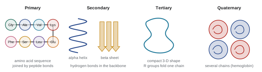
A protein's function depends on its three-dimensional shape, and that shape is usually determined by its amino acid sequence. Biochemists describe four levels of structure:
| Level | What it is | Held together by |
|---|---|---|
| Primary | The sequence of amino acids in the chain | Peptide bonds (covalent) |
| Secondary | Local folding into alpha helices and beta sheets | Hydrogen bonds between backbone atoms |
| Tertiary | The overall three-dimensional shape of one polypeptide | Interactions between R groups: hydrophobic clustering, hydrogen bonds, ionic bonds, disulfide bridges |
| Quaternary | The arrangement of two or more polypeptides in one protein | The same interactions as tertiary structure, between chains |
A single change in primary structure can change everything. In sickle cell disease, one amino acid in the beta chain of hemoglobin, glutamate (charged), is replaced by valine (non-polar). The non-polar patch makes hemoglobin molecules stick together when oxygen is low, deforming red blood cells into sickle shapes that block small blood vessels.
Heat, extreme pH, high salt or certain chemicals can disrupt the weak interactions that hold a protein's shape. The protein unfolds, or denatures, and loses its function. Cooking an egg denatures its proteins irreversibly. Fever is dangerous at very high temperatures partly because some proteins begin to lose their shape. In the cell, helper proteins called chaperones assist folding and refold damaged proteins; misfolded proteins that clump together are involved in diseases such as Alzheimer's and Parkinson's.
Predicting a protein's shape from its sequence was a famous unsolved problem for 50 years. In 2020 an artificial intelligence system, AlphaFold, predicted structures with accuracy close to experiment for many proteins, and its developers shared the 2024 Nobel Prize in Chemistry with David Baker, who designs new proteins. Experimental methods such as cryo-electron microscopy (lesson 4) remain essential for checking predictions and for structures the software gets wrong.
# Molecular mass of a short peptide from average residue masses (daltons)
residue = {"G": 57.05, "A": 71.08, "S": 87.08, "V": 99.13, "L": 113.16,
           "K": 128.17, "E": 129.12, "F": 147.18, "R": 156.19, "W": 186.21}
water = 18.02

def peptide_mass(seq):
    return sum(residue[a] for a in seq) + water   # add one water for the two chain ends

for seq in ["GAVL", "KLVFFAE", "RWEKLS"]:
    print(f"{seq:<8} {len(seq)} residues, {peptide_mass(seq):7.1f} Da")
print("A 300-residue protein is roughly", round(300 * 110 / 1000), "kDa")GAVL 4 residues, 358.4 Da KLVFFAE 7 residues, 853.0 Da RWEKLS 6 residues, 817.9 Da A 300-residue protein is roughly 33 kDa
A handy rule of thumb follows from the program: the average amino acid residue weighs about 110 daltons, so a protein of 300 amino acids weighs about 33 kilodaltons (kDa). Biologists use this constantly when they read the size of a protein band on a gel (lesson 4).
Nucleic acids: information molecules
Nucleic acids store and transmit genetic information. There are two kinds: deoxyribonucleic acid (DNA) and ribonucleic acid (RNA). Both are polymers of nucleotides. Each nucleotide has three parts: a five-carbon sugar (deoxyribose in DNA, ribose in RNA), a phosphate group, and a nitrogen-containing base. DNA uses the bases adenine (A), guanine (G), cytosine (C) and thymine (T); RNA uses uracil (U) in place of thymine.
Nucleotides link through phosphodiester bonds between the sugar of one and the phosphate of the next, forming a sugar–phosphate backbone with the bases sticking out. Each strand has a direction, written from the 5′ end to the 3′ end, named after carbon atoms in the sugar.
DNA is usually a double helix: two strands wound around each other, running in opposite directions, with bases paired in the middle by hydrogen bonds. A always pairs with T (two hydrogen bonds) and G with C (three hydrogen bonds). This complementary base pairing, worked out by James Watson and Francis Crick in 1953 using X-ray images made by Rosalind Franklin and Maurice Wilkins's group, explains how DNA is copied: each strand serves as a template for a new partner. RNA is usually single-stranded and folds on itself; it carries messages from genes to ribosomes and forms parts of the ribosome itself. Details of replication, transcription and translation belong to BIOL-207.
Worked problem
Complementary strands and base composition
One strand of a DNA segment reads 5′-ATGCGTTA-3′. Write the complementary strand, and find the percentage of G plus C in the double-stranded segment.
Show solution
- Pair each base: A with T, T with A, G with C, C with G. The partner of ATGCGTTA is TACGCAAT.
- The partner runs in the opposite direction, so written 5′ to 3′ it is 5′-TAACGCAT-3′.
- Count G and C in both strands: the first strand has G, C, G (3), the second has C, G, C (3). Total 6 out of 16 bases.
- 6/16 = 37.5 percent G plus C.
Answer: Complement 5′-TAACGCAT-3′; the segment is 37.5 percent G plus C. Because G pairs with C, the amount of G always equals the amount of C in double-stranded DNA (Chargaff’s rule).
# Complementary strand and GC content
pair = {"A": "T", "T": "A", "G": "C", "C": "G"}

def reverse_complement(seq):
    return "".join(pair[b] for b in reversed(seq))

def gc_percent(seq):
    return 100 * sum(b in "GC" for b in seq) / len(seq)

s = "ATGCGTTA"
rc = reverse_complement(s)
print("strand:     5'-" + s + "-3'")
print("complement: 5'-" + rc + "-3'")
print("GC content:", gc_percent(s + rc), "%")strand: 5'-ATGCGTTA-3' complement: 5'-TAACGCAT-3' GC content: 37.5 %
Nucleotides have other jobs too. ATP (adenosine triphosphate) is the cell's main short-term energy carrier (lesson 8). NAD+ and FAD carry electrons in metabolism (lessons 9 and 10). Cyclic AMP is a common signaling molecule inside cells (lesson 11).
Putting the four classes together
A useful habit is to ask, for any cell structure, which macromolecules it is made of. A membrane is a phospholipid bilayer with cholesterol, studded with proteins and decorated with carbohydrates. A ribosome is RNA plus protein. A chromosome is DNA wrapped around proteins. A plant cell wall is mostly cellulose. Muscle is largely protein. By the end of the course you should be able to describe any part of a cell in these chemical terms.
Common mistakes
MistakeStarch and cellulose are different because they are made of different sugars.
InsteadBoth are made of glucose; they differ in the type of glycosidic link (alpha in starch, beta in cellulose), which changes their shape and digestibility.
MistakeAll fats are bad for health.
InsteadFats are essential for energy storage, membranes and hormones; health effects depend on the type and amount, with industrial trans fats the clearest harm.
MistakeDenaturing a protein breaks its peptide bonds.
InsteadDenaturation disrupts the weak interactions of secondary, tertiary and quaternary structure; the primary sequence of peptide bonds usually stays intact.
MistakeDNA and RNA are made of the same four bases.
InsteadDNA uses thymine and RNA uses uracil; their sugars also differ (deoxyribose versus ribose).
MistakeLipids are polymers just like proteins.
InsteadLipids are assembled from smaller parts such as glycerol and fatty acids, but they are not long chains of repeating monomers.
Summary and key terms
- Carbohydrates, lipids, proteins and nucleic acids are the four classes of biological macromolecules; all but lipids are true polymers.
- Monomers join by dehydration synthesis, releasing water; hydrolysis uses water to break them apart.
- Polysaccharides store energy (starch, glycogen) or build structures (cellulose, chitin); alpha versus beta links change their properties.
- Fats store energy densely; phospholipids are amphipathic and form bilayers; steroids such as cholesterol have four fused rings.
- Proteins are chains of 20 kinds of amino acids joined by peptide bonds, folded into primary, secondary, tertiary and quaternary structure.
- Shape determines protein function, and loss of shape (denaturation) destroys it; single amino acid changes can cause disease.
- DNA and RNA are polymers of nucleotides; DNA’s double helix pairs A with T and G with C.
- monomer
- A small molecule that can link with others of the same kind to form a polymer.
- dehydration synthesis
- A reaction that joins two molecules by a covalent bond and releases water.
- hydrolysis
- A reaction that breaks a covalent bond by adding water.
- polysaccharide
- A polymer of many monosaccharides, such as starch, glycogen or cellulose.
- saturated fatty acid
- A fatty acid with no carbon–carbon double bonds, giving a straight chain.
- phospholipid
- A lipid with a polar phosphate-containing head and two fatty acid tails; the main component of membranes.
- peptide bond
- The covalent bond linking two amino acids in a protein.
- denaturation
- Loss of a protein’s three-dimensional shape, and usually its function, caused by heat, pH or chemicals.
- nucleotide
- The monomer of nucleic acids: a sugar, a phosphate group and a nitrogenous base.
- complementary base pairing
- The pairing of A with T (or U in RNA) and G with C by hydrogen bonds.
Practice problems
Work each problem on paper first, then reveal the answer to check.
What is released when two glucose molecules are joined, and what is the reaction called?
Show answer
Water is released; it is dehydration synthesis (a condensation reaction), forming a glycosidic bond.
Why can humans digest starch but not cellulose?
Show answer
Our enzymes recognize the alpha glycosidic links in starch but not the beta links in cellulose.
Explain why butter is solid and olive oil liquid at room temperature.
Show answer
Butter is rich in saturated fatty acids with straight chains that pack tightly; olive oil is rich in unsaturated fatty acids whose cis double bonds kink the chains.
Why is a phospholipid described as amphipathic, and what structure does this produce in water?
Show answer
It has a polar head and non-polar tails; in water it forms a bilayer with tails inside.
Name the four levels of protein structure and the main bonds or forces at each.
Show answer
Primary (peptide bonds), secondary (backbone hydrogen bonds), tertiary and quaternary (R-group interactions: hydrophobic, hydrogen, ionic, disulfide).
How does a single amino acid change cause sickle cell disease?
Show answer
Glutamate is replaced by non-polar valine in the hemoglobin beta chain, making hemoglobin molecules stick together at low oxygen and deform red cells.
Estimate the mass of a protein with 450 amino acids.
Show answer
About 450 × 110 = 49,500 Da, roughly 50 kDa.
Write the complementary strand of 5′-GGATCC-3′ in the 5′ to 3′ direction.
Show answer
5′-GGATCC-3′; this sequence is its own reverse complement (a palindrome recognized by some enzymes).
If a double-stranded DNA sample is 22 percent adenine, what are the percentages of T, G and C?
Show answer
T = 22 percent; G + C = 56 percent, so G = 28 and C = 28 percent.
Give three non-genetic roles of nucleotides.
Show answer
ATP as an energy carrier, NAD+ and FAD as electron carriers, cyclic AMP as a signaling molecule.
Lesson quiz · 5 questions
Score 60% or more to complete this lesson.
Create a free account to save your progress, take quizzes and earn a certificate.