
BIOL-107 · Lesson 11 of 12 · 26 min
Cell communication and the cytoskeleton
Cells are not isolated. A bacterium senses food and its neighbors; the 30 trillion cells of a human body coordinate growth, metabolism, defense and behavior through a constant traffic of chemical messages. Cells also have to hold their shape, move their contents to the right places and, in many cases, crawl or swim. This lesson introduces both topics: how cells send, receive and amplify signals, and the cytoskeleton of protein filaments and motors that gives cells structure and movement. Both are large fields; BIOL-214 develops them in depth and neurobiology (BIOL-420) and immunology (BIOL-380) apply them.
How cells talk
Cells communicate mostly with chemical signals, molecules released by one cell and detected by another. Signals differ in how far they travel:
| Type | Distance | Example |
|---|---|---|
| Direct contact | Touching cells | Gap junctions let ions pass between heart muscle cells; immune cells recognize surface molecules on other cells |
| Autocrine | The same cell | Some immune cells stimulate themselves to divide; many cancer cells signal to themselves |
| Paracrine | Nearby cells, micrometers to millimeters | Growth factors in a healing wound |
| Synaptic | Across a gap of about 20 nm at a synapse | Neurotransmitters such as acetylcholine at nerve–muscle junctions |
| Endocrine | Throughout the body, carried in the blood | Hormones such as insulin, adrenaline (epinephrine) and estrogen |
Bacteria signal too. In quorum sensing, each bacterium releases a small molecule; when the population is dense enough that the signal builds up, the bacteria switch on genes together, for example to glow (in the marine bacterium Vibrio fischeri, which lights up the organ of a small squid) or to form a biofilm on a medical device.
Three stages: reception, transduction, response
In the 1950s Earl Sutherland, studying how the hormone adrenaline makes liver cells release glucose, found that the hormone never entered the cell. Instead, binding at the surface triggered a chain of events inside, which he described in three stages, a framework still used today (he received the 1971 Nobel Prize in Physiology or Medicine).
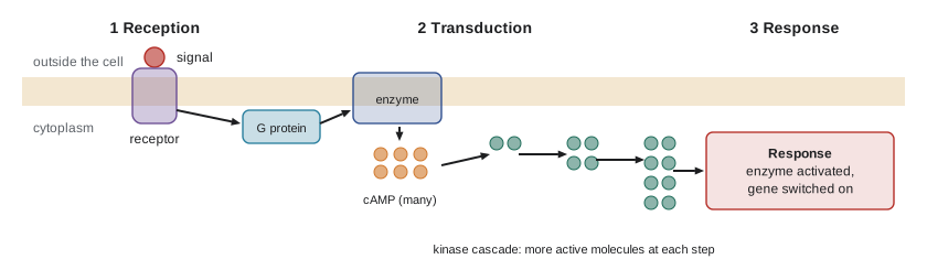
Reception is the binding of a signal molecule (a ligand) to a specific receptor protein, which changes shape. Transduction converts that shape change into a form that can produce a response, usually a relay of several molecules inside the cell. Response is the final cellular activity: an enzyme switched on, a gene expressed, a channel opened, the cell dividing or dying. Because each step can involve many molecules, signals are amplified and can be adjusted along the way.
Receptors
Where the receptor sits depends on the signal. Large or water-soluble signals, such as peptide hormones and most neurotransmitters, cannot cross the membrane and bind receptors on the cell surface. Small, hydrophobic signals, such as steroid hormones (estrogen, testosterone, cortisol), thyroid hormone and nitric oxide, cross the membrane and bind receptors inside the cell.
| Receptor type | How it works | Example |
|---|---|---|
| G protein-coupled receptors (GPCRs) | A protein that crosses the membrane seven times; the bound signal activates a G protein, which switches on an enzyme or channel | Adrenaline receptors, receptors for smell and light; targets of about a third of approved drugs |
| Receptor tyrosine kinases (RTKs) | Two receptors pair up when the signal binds and add phosphate groups to each other, starting several pathways | Insulin and growth factor receptors; often overactive in cancers |
| Ligand-gated ion channels | The signal opens a channel that lets ions through | Acetylcholine receptors at nerve–muscle junctions |
| Intracellular receptors | The signal enters the cell; the receptor–signal complex acts as a transcription factor that switches genes on or off | Estrogen, testosterone, cortisol and thyroid hormone receptors |
Receptors bind their signals reversibly, and how many are occupied depends on the signal's concentration relative to the receptor's dissociation constant, Kd, the concentration at which half the receptors are occupied. The relation has the same shape as the Michaelis–Menten curve from lesson 8:
# Fraction of receptors occupied at different hormone concentrations
Kd = 1.0          # nanomolar (a typical high-affinity hormone receptor)
for L in [0.1, 0.5, 1, 2, 5, 10, 100]:
    occ = L / (Kd + L)
    print(f"[hormone] = {L:6.1f} nM  ->  {occ*100:5.1f} % occupied")
# Hormones circulate at very low concentrations, often nanomolar or below[hormone] = 0.1 nM -> 9.1 % occupied [hormone] = 0.5 nM -> 33.3 % occupied [hormone] = 1.0 nM -> 50.0 % occupied [hormone] = 2.0 nM -> 66.7 % occupied [hormone] = 5.0 nM -> 83.3 % occupied [hormone] = 10.0 nM -> 90.9 % occupied [hormone] = 100.0 nM -> 99.0 % occupied
Transduction: second messengers and phosphorylation
Two mechanisms dominate signal transduction. The first is protein phosphorylation. Enzymes called protein kinases transfer a phosphate group from ATP to specific amino acids on target proteins, changing their shape and activity; protein phosphatases remove the phosphates, switching the proteins back. Many pathways are phosphorylation cascades in which one kinase activates the next. The human genome encodes more than 500 protein kinases, and many cancer drugs, such as imatinib for chronic myeloid leukemia, work by blocking an overactive kinase.
The second is small molecules called second messengers, produced inside the cell in response to the first messenger outside. Cyclic AMP (cAMP), made from ATP by the enzyme adenylyl cyclase, was the first discovered. Calcium ions are another: cells keep calcium in the cytosol about 10,000 times lower than outside, so opening calcium channels produces a sudden, strong signal that triggers muscle contraction, neurotransmitter release and much else. Second messengers diffuse quickly through the cytosol, spreading the signal.
Amplification: a small signal, a big response
One of the most important features of signaling pathways is amplification. When adrenaline binds a liver cell, each activated receptor activates many G proteins; each activated enzyme makes many cAMP molecules; each kinase phosphorylates many copies of the next protein. Over several steps, a handful of hormone molecules can lead to the release of millions of glucose molecules within seconds, preparing the body for fight or flight.
Worked problem
Amplifying the adrenaline signal
Suppose one adrenaline molecule bound to a receptor activates 10 G proteins; each activates one adenylyl cyclase that makes 100 cAMP; every 2 cAMP activate one protein kinase A molecule, which phosphorylates 10 phosphorylase kinase molecules; each of those activates 10 glycogen phosphorylase molecules; and each phosphorylase releases 1,000 glucose units from glycogen per minute. How many glucose units are released per minute?
Show solution
- cAMP made: 10 × 100 = 1,000.
- Protein kinase A activated: 1,000 ÷ 2 = 500.
- Phosphorylase kinase activated: 500 × 10 = 5,000.
- Glycogen phosphorylase activated: 5,000 × 10 = 50,000.
- Glucose released: 50,000 × 1,000 = 50,000,000 per minute.
Answer: About 50 million glucose units per minute from one hormone molecule, under these illustrative numbers. Real values vary, but amplification of a millionfold or more is typical.
# Amplification through a signal cascade (illustrative factors)
steps = [("receptor -> G proteins", 10),
         ("G protein -> cAMP (via adenylyl cyclase)", 100),
         ("cAMP -> protein kinase A (2 cAMP each)", 0.5),
         ("PKA -> phosphorylase kinase", 10),
         ("phosphorylase kinase -> glycogen phosphorylase", 10),
         ("phosphorylase -> glucose per minute", 1000)]
n = 1
for name, factor in steps:
    n *= factor
print(f"{name:<48} x{factor:<6} running total {n:,.0f}")receptor -> G proteins x10 running total 10 G protein -> cAMP (via adenylyl cyclase) x100 running total 1,000 cAMP -> protein kinase A (2 cAMP each) x0.5 running total 500 PKA -> phosphorylase kinase x10 running total 5,000 phosphorylase kinase -> glycogen phosphorylase x10 running total 50,000 phosphorylase -> glucose per minute x1000 running total 50,000,000
Responses and switching off
Signals produce two broad kinds of response. Some act in the cytoplasm within seconds, by activating enzymes or opening channels, like glycogen breakdown or muscle contraction. Others change gene expression in the nucleus, taking minutes to hours, like steroid hormones switching on genes for development or growth factors telling a cell to divide.
Just as important as switching signals on is switching them off. Enzymes such as phosphodiesterase break down cAMP; phosphatases remove phosphates; G proteins switch themselves off by splitting their bound GTP; receptors are taken into the cell by endocytosis (lesson 7). Caffeine partly works by blocking adenosine receptors and, at high doses, phosphodiesterase. Failure to switch off has consequences: cholera toxin locks G proteins in the on state in intestinal cells, causing massive secretion of salt and water, and mutations that lock the growth-signaling protein Ras in its active form are found in many cancers. A related form of signaling tells cells to die: apoptosis, programmed cell death, removes unneeded or damaged cells in a controlled way, and its failure also contributes to cancer (lesson 12).
The cytoskeleton
Electron microscopy and fluorescence imaging reveal that the cytoplasm is threaded by a network of protein filaments, the cytoskeleton. It gives cells their shape and mechanical strength, anchors organelles, provides tracks for transport, separates chromosomes during division and drives cell movement. Unlike a skeleton of bone, it is highly dynamic: its filaments are constantly assembled and disassembled from protein subunits, so a cell can remodel it in minutes.
| Property | Microfilaments | Intermediate filaments | Microtubules |
|---|---|---|---|
| Protein subunit | Actin | Various fibrous proteins (keratins, lamins, vimentin, neurofilaments) | Tubulin (alpha and beta dimers) |
| Diameter | About 7 nm | About 8 to 12 nm | About 25 nm (hollow) |
| Main functions | Cell shape at the surface, muscle contraction, crawling, cytoplasmic streaming, pinching cells in two | Mechanical strength; anchoring the nucleus; the nuclear lamina | Tracks for vesicles and organelles, mitotic spindle, cilia and flagella |
| Motor proteins | Myosin | None | Kinesin and dynein |
| Dynamics | Rapid assembly and disassembly | Relatively stable | Rapid growth and shrinkage (dynamic instability) |
Microtubules grow from an organizing center near the nucleus, the centrosome, which in animal cells contains a pair of centrioles. They switch unpredictably between growing and shrinking, a behavior called dynamic instability, which lets the cell search its interior and quickly rebuild its network. During cell division they form the spindle that separates chromosomes (lesson 12). Drugs that interfere with microtubules stop dividing cells, which is why paclitaxel (Taxol, first isolated from the bark of the Pacific yew) and vinca alkaloids (from the Madagascar periwinkle) are used to treat cancer, and colchicine (from the autumn crocus) is used for gout.
Motor proteins and movement
Motor proteins convert the chemical energy of ATP into movement along filaments. Kinesins generally walk along microtubules toward the cell's periphery, carrying vesicles and organelles; dyneins walk the other way, toward the center. Each step of kinesin moves about 8 nm and uses one ATP. Myosins move along actin filaments; in muscle, sliding of myosin along actin shortens the cell, and the 1950s sliding filament model explained how.
# Transport along a long nerve axon by kinesin (fast axonal transport)
speed_um_per_s = 1.0               # kinesin cargo speed, about 1 um per second
steps_per_s = speed_um_per_s * 1000 / 8   # 8 nm per step, one ATP per step
axon_m = 1.0                       # sciatic nerve: spine to foot
seconds = axon_m * 1e6 / speed_um_per_s
print(f"Steps (and ATP) per second per motor: {steps_per_s:.0f}")
print(f"Time to carry a vesicle 1 m: {seconds/86400:.1f} days")
D = 1e-12                          # a vesicle diffusing in cytoplasm, m^2/s (rough)
print(f"Diffusion over 1 m would take about {1**2/(2*D)/86400/365:,.0f} years")Steps (and ATP) per second per motor: 125 Time to carry a vesicle 1 m: 11.6 days Diffusion over 1 m would take about 15,855 years
Motor-driven transport moves a vesicle down a one-meter nerve in less than two weeks; diffusion alone would take thousands of years (lesson 7). Defects in axonal transport are linked to several neurological diseases.
Cilia and eukaryotic flagella are built around a ring of nine pairs of microtubules surrounding a central pair (the 9 + 2 arrangement), and dynein motors make them bend. Cilia beating in coordinated waves sweep mucus and trapped particles out of your airways; smoking paralyses them. A sperm cell swims with a flagellum. Crawling cells, such as white blood cells chasing bacteria and cells moving in a healing wound, push their front edge forward by assembling actin filaments and pull the rear along with myosin.
Common mistakes
MistakeHormones always enter cells to act.
InsteadWater-soluble hormones bind surface receptors and act through second messengers; only small hydrophobic signals such as steroids enter cells.
MistakeSignal transduction passes one molecule along a chain, one to one.
InsteadEach step usually activates many molecules, amplifying the signal enormously.
MistakeThe cytoskeleton is a fixed, rigid frame.
InsteadMost cytoskeletal filaments are constantly assembled and disassembled, letting cells change shape and divide.
MistakeSecond messengers are the hormones that arrive at the cell.
InsteadThe first messenger is the external signal; second messengers such as cAMP and calcium are produced inside the cell in response.
MistakeKinesin and dynein walk along actin filaments.
InsteadKinesin and dynein walk along microtubules; myosin walks along actin.
Summary and key terms
- Cells signal by direct contact, autocrine, paracrine, synaptic and endocrine (hormonal) routes; bacteria use quorum sensing.
- Signaling has three stages: reception by a specific receptor, transduction through relay molecules, and a cellular response.
- Surface receptors include GPCRs, receptor tyrosine kinases and ligand-gated channels; steroid hormones use intracellular receptors.
- Transduction uses phosphorylation by kinases (reversed by phosphatases) and second messengers such as cAMP and calcium.
- Cascades amplify signals by factors of a million or more; switching off signals is as important as switching them on.
- The cytoskeleton has actin microfilaments (7 nm), intermediate filaments (about 10 nm) and tubulin microtubules (25 nm).
- Motor proteins (myosin on actin; kinesin and dynein on microtubules) use ATP to move cargo, beat cilia and contract muscle.
- ligand
- A molecule that binds specifically to a receptor or other protein.
- receptor
- A protein that binds a specific signal molecule and changes shape to start a response.
- signal transduction
- The process by which a signal at a receptor is converted into a cellular response.
- G protein-coupled receptor
- A seven-pass membrane receptor that activates a G protein when its ligand binds.
- protein kinase
- An enzyme that transfers a phosphate group from ATP to a protein.
- second messenger
- A small molecule, such as cAMP or calcium ions, that relays a signal inside the cell.
- amplification
- The multiplication of a signal’s effect at each step of a pathway.
- cytoskeleton
- The network of protein filaments that shapes the cell and moves its contents.
- microtubule
- A hollow tube of tubulin that acts as a track for motors and forms the spindle.
- motor protein
- A protein that uses ATP to move along a cytoskeletal filament.
Practice problems
Work each problem on paper first, then reveal the answer to check.
Distinguish paracrine and endocrine signaling with an example of each.
Show answer
Paracrine signals act on nearby cells (growth factors in a wound); endocrine hormones travel in the blood to distant cells (insulin).
Name the three stages of cell signaling.
Show answer
Reception, transduction and response.
Why do steroid hormones have intracellular receptors while insulin has a surface receptor?
Show answer
Steroids are small and hydrophobic and cross the membrane; insulin is a large water-soluble protein that cannot.
A receptor has Kd = 2 nM. What fraction is occupied at 6 nM hormone?
Show answer
6 / (2 + 6) = 0.75, or 75 percent.
What is a second messenger? Give two examples.
Show answer
A small molecule made inside the cell in response to an external signal; cyclic AMP and calcium ions.
Explain how a signaling cascade amplifies a signal.
Show answer
Each activated molecule activates many molecules of the next component, so the number of active molecules multiplies at each step.
How does cholera toxin cause severe diarrhea?
Show answer
It locks G proteins in their active state in intestinal cells, keeping cAMP high, which drives massive secretion of salt and water.
Match each motor protein to its filament.
Show answer
Myosin with actin microfilaments; kinesin and dynein with microtubules.
Why are drugs that disrupt microtubules useful in cancer treatment?
Show answer
Microtubules form the mitotic spindle, so blocking them stops rapidly dividing cells.
Estimate how long kinesin moving at 1 µm/s takes to carry cargo 50 cm along an axon.
Show answer
500,000 s, about 5.8 days.
Lesson quiz · 5 questions
Score 60% or more to complete this lesson.
Create a free account to save your progress, take quizzes and earn a certificate.