
BIOL-107 · Lesson 12 of 12 · 31 min
The cell cycle, mitosis and meiosis, and capstone: a cell biology investigation
The third statement of the cell theory says every cell comes from a pre-existing cell (lesson 1). This final lesson asks how. It follows the cell cycle that copies and divides a cell, the mitosis that shares its chromosomes equally, the meiosis that makes eggs and sperm with half the chromosomes, and the controls whose failure leads to cancer. It ends with a capstone that pulls the course together: planning a small cell biology investigation, analyzing its data, and thinking about the ethics of research with human cells. Genetics (BIOL-201), developmental biology (BIOL-360) and cancer biology (BIOL-450) build on these foundations.
Why cells divide, and what they must copy
Cell division serves different purposes. A bacterium or yeast cell divides to reproduce. A fertilized human egg divides to build a body of trillions of cells. Adult tissues divide to replace cells: your gut lining renews itself every few days, and you make about two million red blood cells every second. Whatever the purpose, a dividing cell must copy its DNA exactly and share the copies evenly between two daughter cells.
The amount of DNA to copy is large. A human cell has 46 chromosomes holding about 6 billion base pairs, roughly 2 meters of DNA if stretched out, packed into a nucleus about 6 µm across. In a non-dividing cell, DNA is loosely packed as chromatin. Before division, each chromosome is copied into two identical sister chromatids joined at a constriction called the centromere, and the chromatin condenses into the compact X-shaped chromosomes seen in textbook pictures. Bacteria, with a single circular chromosome, divide by the simpler process of binary fission: the chromosome is copied, the copies move apart, and the cell pinches in two.
The eukaryotic cell cycle
The life of a dividing eukaryotic cell is a repeating cycle with two main parts: interphase, when the cell grows and copies its DNA, and the mitotic (M) phase, when it divides. Interphase takes most of the time and has three stages.
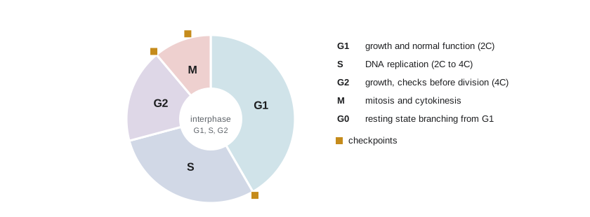
| Phase | What happens | DNA content (per nucleus) |
|---|---|---|
| G1 (gap 1) | The cell grows, makes proteins and organelles, and carries out its normal functions | 2C (one copy of each chromosome pair) |
| S (synthesis) | DNA is replicated; each chromosome becomes two sister chromatids | Rises from 2C to 4C |
| G2 (gap 2) | The cell grows further and prepares for division; checks that DNA copying is complete | 4C |
| M (mitosis and cytokinesis) | Sister chromatids are separated and the cytoplasm divides | 4C, then 2C in each daughter |
| G0 | A non-dividing state; many cells, such as most neurons and heart muscle cells, stay here permanently | 2C |
Here C is the DNA content of one complete set of chromosomes in an egg or sperm (for humans about 3 billion base pairs). A typical human cell in culture completes the cycle in about 24 hours, with M phase taking only about an hour. Early frog embryos skip the gap phases and divide every 30 minutes; some liver cells divide only about once a year.
Because the time a cell spends in a phase is proportional to the fraction of cells found in that phase in a growing population, biologists can estimate phase lengths by counting. The mitotic index is the fraction of cells in mitosis.
Worked problem
Timing phases from a microscope count
In a stained onion root tip, a student counts 400 cells: 352 in interphase, 28 in prophase, 6 in metaphase, 4 in anaphase and 10 in telophase. The cell cycle in these cells lasts about 22 hours. Estimate the mitotic index and the duration of each phase of mitosis.
Show solution
- Cells in mitosis = 28 + 6 + 4 + 10 = 48. Mitotic index = 48/400 = 0.12 (12 percent).
- Each phase’s duration ≈ (fraction of cells in that phase) × total cycle time.
- Prophase: 28/400 × 22 h ≈ 1.54 h (about 92 minutes).
- Metaphase: 6/400 × 22 h ≈ 0.33 h (20 minutes); anaphase: 4/400 × 22 h ≈ 0.22 h (13 minutes); telophase: 10/400 × 22 h ≈ 0.55 h (33 minutes).
Answer: Mitotic index 12 percent; mitosis lasts about 2.6 hours, with prophase the longest phase and anaphase the shortest. The method assumes cells are dividing steadily and not synchronized.
Mitosis: sharing chromosomes equally
Mitosis is the division of the nucleus into two genetically identical nuclei. It is a continuous process, but biologists divide it into stages for description:
| Stage | Key events |
|---|---|
| Prophase | Chromatin condenses into visible chromosomes, each with two sister chromatids; the nucleolus disappears; the mitotic spindle of microtubules begins to form from the centrosomes |
| Prometaphase | The nuclear envelope breaks up; spindle microtubules attach to protein structures called kinetochores at each centromere |
| Metaphase | Chromosomes line up in a plane across the middle of the cell (the metaphase plate), each attached to both poles |
| Anaphase | The proteins holding sister chromatids together are cut; the chromatids, now separate chromosomes, are pulled to opposite poles as kinetochore microtubules shorten |
| Telophase | Nuclear envelopes re-form around each set of chromosomes, which begin to decondense; the spindle breaks down |
| Cytokinesis | The cytoplasm divides: in animal cells a ring of actin and myosin pinches the cell in two (cleavage furrow); in plant cells vesicles build a new wall (cell plate) across the middle |
The spindle is built from the microtubules of lesson 11, and its motors and dynamic instability move the chromosomes. The result is two daughter cells with the same number and kind of chromosomes as the parent.
Growth of cell populations
When cells divide steadily, their numbers grow exponentially, doubling every generation time. After \(t\) hours with a doubling time \(t_d\), a population that started with \(N_0\) cells has \(N = N_0 \cdot 2^{t/t_d}\) cells. Exponential growth explains why a single bacterium can become billions overnight and why a small tumor can grow rapidly.
import math
# Exponential growth of a cell culture: N = N0 * 2**(t / td)
def cells(N0, t, td):
    return N0 * 2 ** (t / td)

print("E. coli, td = 20 min, from 1 cell after 8 h:", f"{cells(1, 480, 20):.2e}")
print("Human cells, td = 24 h, from 1e5 after 5 days:", f"{cells(1e5, 120, 24):.2e}")

# Estimating doubling time from two counts
N1, N2, hours = 2.0e5, 1.6e6, 72
td = hours * math.log(2) / math.log(N2 / N1)
print(f"Doubling time from counts {N1:.1e} -> {N2:.1e} in {hours} h: {td:.1f} h")E. coli, td = 20 min, from 1 cell after 8 h: 1.68e+07 Human cells, td = 24 h, from 1e5 after 5 days: 3.20e+06 Doubling time from counts 2.0e+05 -> 1.6e+06 in 72 h: 24.0 h
Real cultures do not grow forever. Bacteria in a flask show a lag phase, an exponential phase, a stationary phase when nutrients run low, and a death phase. Normal animal cells in culture stop dividing when they touch their neighbors (contact inhibition) and, after a limited number of divisions (about 40 to 60 for human fibroblasts, the Hayflick limit), stop dividing altogether, partly because the protective ends of their chromosomes, telomeres, shorten with each division.
Controlling the cycle and the origins of cancer
The cell cycle is driven by a control system based on proteins called cyclins and cyclin-dependent kinases (Cdks). Cdks are protein kinases (lesson 11) that are active only when bound to a cyclin; cyclin levels rise and fall through the cycle, switching on the events of each phase in turn. Leland Hartwell, Tim Hunt and Paul Nurse shared the 2001 Nobel Prize in Physiology or Medicine for discovering this system in yeast and sea urchin eggs. At checkpoints, the cell checks conditions before continuing: at the end of G1 (is the cell big enough, are conditions favorable, is DNA undamaged?), at the end of G2 (is DNA fully and correctly copied?), and in metaphase (are all chromosomes attached to the spindle?). The protein p53 halts the cycle when DNA is damaged and can trigger apoptosis if the damage cannot be repaired; it is mutated in about half of all human cancers.
Cancer is a disease of cells that divide when they should not. It arises when mutations accumulate in two kinds of genes: proto-oncogenes, which normally promote division and become overactive oncogenes (like a stuck accelerator), and tumor suppressor genes such as p53, which normally restrain division and are inactivated (like failed brakes). Usually several mutations are needed, which is one reason cancer risk rises with age. Cancer cells ignore contact inhibition, evade apoptosis, divide indefinitely (often by reactivating telomerase), attract a blood supply and can spread to other tissues (metastasis). Many treatments target dividing cells, which is why chemotherapy also affects fast-dividing normal tissues such as hair follicles and the gut lining.
Meiosis: halving the chromosome number
Humans have 23 pairs of chromosomes: one set of 23 from each parent. Cells with two sets are diploid (2n = 46 in humans); eggs and sperm have one set and are haploid (n = 23). Each pair of matching chromosomes, one from each parent, carrying the same genes in the same order, is a homologous pair. If eggs and sperm were made by mitosis, the chromosome number would double every generation. Meiosis prevents that: it is a special pair of divisions that turns one diploid cell into four haploid cells, each genetically different.
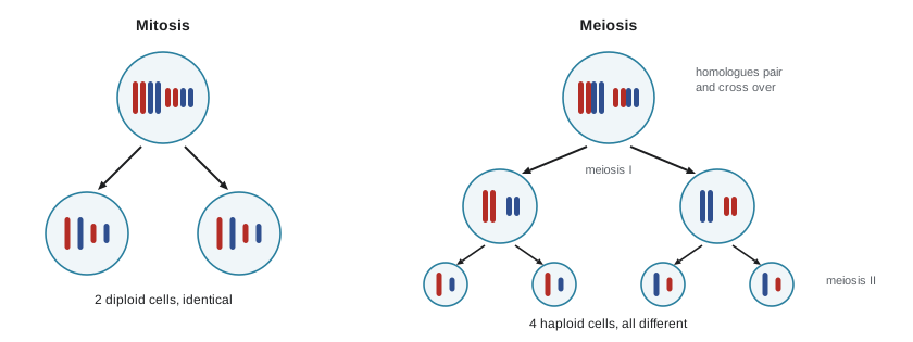
| Feature | Mitosis | Meiosis |
|---|---|---|
| Purpose | Growth, repair, asexual reproduction | Making eggs and sperm (gametes) for sexual reproduction |
| Number of divisions | One | Two (meiosis I and meiosis II) |
| Daughter cells | Two, diploid, genetically identical to the parent | Four, haploid, genetically different |
| Pairing of homologous chromosomes | No | Yes, in prophase I |
| Crossing over | Normally no | Yes, exchanges segments between homologues |
| What separates | Sister chromatids | Homologues in meiosis I; sister chromatids in meiosis II |
Meiosis creates variety in two ways. In crossing over, homologous chromosomes swap segments during prophase I, producing chromosomes with new combinations of the parents' versions of genes. In independent assortment, each pair of homologues lines up at random in metaphase I, so each gamete gets a random mix of maternal and paternal chromosomes. With 23 pairs, independent assortment alone gives \(2^{23}\), over 8 million, combinations per parent, before counting crossing over and the random meeting of egg and sperm. Mistakes in separating chromosomes, called nondisjunction, produce gametes with an extra or missing chromosome; Down syndrome, with three copies of chromosome 21, usually arises this way. Genetics (BIOL-201) follows what this means for inheritance.
# Chromosome number and DNA content through mitosis and meiosis (human, n = 23)
n = 23
rows = [("G1 (diploid cell)",        2*n, "2C"),
        ("after S phase (G2)",       2*n, "4C"),
        ("each mitotic daughter",    2*n, "2C"),
        ("after meiosis I (each)",   n,   "2C"),
        ("after meiosis II (each)",  n,   "1C")]
for stage, chromosomes, dna in rows:
    print(f"{stage:<26} chromosomes: {chromosomes:>2}   DNA: {dna}")
print("Combinations from independent assortment:", f"{2**n:,}")G1 (diploid cell) chromosomes: 46 DNA: 2C after S phase (G2) chromosomes: 46 DNA: 4C each mitotic daughter chromosomes: 46 DNA: 2C after meiosis I (each) chromosomes: 23 DNA: 2C after meiosis II (each) chromosomes: 23 DNA: 1C Combinations from independent assortment: 8,388,608
Capstone: a cell biology investigation
Science is a way of asking questions and testing answers. To finish the course, we plan and analyze an investigation of the kind you might run in a first-year laboratory, using ideas from several lessons. The data below are invented for teaching.
The question: does a microtubule-disrupting drug change the mitotic index of onion root tip cells? Lesson 11 predicts that blocking spindle microtubules should trap cells in metaphase, so more cells should appear to be in mitosis. A hypothesis is a testable explanation: the drug blocks spindle formation, arresting cells in mitosis. From it we make a prediction: treated root tips will have a higher mitotic index than untreated ones. The null hypothesis, which statistics tests, is that there is no difference.
| Element of design | In this investigation |
|---|---|
| Independent variable | Treatment: drug (low concentration) or water only |
| Dependent variable | Mitotic index: cells in mitosis ÷ total cells counted |
| Controlled variables | Onion variety, age of roots, temperature, treatment time (4 h), staining method, number of cells counted (400 per root tip) |
| Control group | Root tips treated with water only |
| Replication | Eight root tips per group, each from a different bulb |
| Blinding | Slides coded by a partner so the counter does not know the treatment |
| Safety | Drug handled with gloves according to its safety data sheet; stain and acid used in a fume hood (lesson 4) |
import numpy as np
from scipy import stats

rng = np.random.default_rng(107)
# Invented data: mitotic index (%) from 8 root tips per group, 400 cells each
control = np.round(rng.normal(11.5, 1.6, 8), 1)
treated = np.round(rng.normal(19.0, 2.4, 8), 1)
for name, x in [("control", control), ("treated", treated)]:
    print(f"{name:<8} {x}  mean {x.mean():.2f}  SD {x.std(ddof=1):.2f}")

t, p = stats.ttest_ind(treated, control, equal_var=False)   # Welch's t-test
diff = treated.mean() - control.mean()
pooled = np.sqrt((control.var(ddof=1) + treated.var(ddof=1)) / 2)
print(f"Difference in means: {diff:.2f} percentage points")
print(f"Welch t = {t:.2f}, p = {p:.2g}")
print(f"Effect size (Cohen's d) = {diff / pooled:.1f}")control [12.1 8.5 11.4 14.3 9.1 12.5 10.6 14. ] mean 11.56 SD 2.10 treated [17.9 17.5 23.1 22.2 18.2 14.2 15. 20.1] mean 18.52 SD 3.15 Difference in means: 6.96 percentage points Welch t = 5.19, p = 0.00021 Effect size (Cohen's d) = 2.6
Interpreting the output takes care. The mean and standard deviation (SD) summarize each group; the SD shows how much individual root tips vary. The t-test asks how likely a difference at least this large would be if the null hypothesis were true; a small p-value (conventionally below 0.05) means the data would be surprising if the drug had no effect, so we reject the null hypothesis. A small p-value does not prove the hypothesis, does not measure how big the effect is, and does not rule out a flaw in the design. The effect size, here the difference in means divided by the pooled SD, shows how large the difference is relative to the natural variation.
A good report would go further. Is the result consistent with the mechanism? Looking at which phase the extra cells are in would test it: if the drug blocks the spindle, the excess should be in prometaphase or metaphase, with few in anaphase. What are the limitations? Eight root tips is a small sample; counting can be biased (hence blinding); one drug concentration and one time point say nothing about dose or timing. What next? A dose–response experiment, a time course, or fluorescence microscopy of microtubules (lesson 4) to see the spindle directly.
Research ethics and human cells
Much cell biology uses human cells, and the most famous human cell line raises ethical questions every student should know. In 1951, cells were taken from a tumor of Henrietta Lacks, a 31-year-old Black woman being treated for cervical cancer at Johns Hopkins Hospital in Baltimore, without her knowledge or consent, as was common practice then. Her cells, named HeLa, were the first human cells to grow indefinitely in culture. They have been used in tens of thousands of studies and contributed to the polio vaccine, cancer research and many other advances, while her family for decades knew nothing of their use and received no benefit. In 2013 the US National Institutes of Health reached an agreement with the Lacks family giving them a say over access to HeLa genome data, and in 2023 her estate settled a lawsuit with a biotechnology company over the commercial use of her cells. Her story is now widely taught as a lesson about consent, privacy and fairness in research, especially toward communities that have been exploited in the past.
Today, research involving human participants or identifiable human biological materials in the United States is governed, for federally funded research, by the Federal Policy for the Protection of Human Subjects (the Common Rule, revised in 2018). Projects must be reviewed by an institutional review board (IRB), participants must give informed consent, and privacy must be protected. The revised rule added an option for broad consent for storing and reusing identifiable specimens. Research with tribal nations increasingly follows tribal research codes and data sovereignty principles, such as the CARE principles for Indigenous data governance.
Ethics also covers how you handle data. Record observations as you make them, keep raw data, never adjust results to fit expectations, report experiments that did not work, and credit others' work. Using AI tools to help write a lab report is governed by your institution's academic integrity policy; whatever the policy, the data and the conclusions must be your own and honestly described.
Common mistakes
MistakeInterphase is a resting phase.
InsteadDuring interphase the cell grows, carries out its functions and replicates its DNA (S phase); only G0 cells are truly non-dividing.
MistakeChromosome number doubles in S phase.
InsteadDNA content doubles, but each chromosome now consists of two sister chromatids joined at the centromere; the number of chromosomes stays the same until anaphase.
MistakeMeiosis is just mitosis done twice.
InsteadMeiosis I separates homologous chromosomes after pairing and crossing over, halving the chromosome number; only meiosis II resembles mitosis.
MistakeA p-value below 0.05 proves the hypothesis is true.
InsteadIt indicates the data would be unlikely if the null hypothesis were true; it does not prove the hypothesis, measure effect size or exclude design flaws.
MistakeCancer is caused by a single mutation.
InsteadMost cancers require several mutations in oncogenes and tumor suppressor genes, accumulated over time.
Summary and key terms
- The cell cycle has interphase (G1, S, G2) and M phase (mitosis and cytokinesis); DNA content rises from 2C to 4C in S phase.
- Phase durations can be estimated from the fraction of cells in each phase; the mitotic index is the fraction in mitosis.
- Mitosis (prophase, prometaphase, metaphase, anaphase, telophase) and cytokinesis produce two genetically identical diploid cells.
- Populations grow exponentially, N = N0 · 2^(t/td), until limited by nutrients, contact inhibition or the Hayflick limit.
- Cyclins and Cdks drive the cycle; checkpoints and proteins such as p53 halt it when something is wrong; cancer results from accumulated mutations in oncogenes and tumor suppressor genes.
- Meiosis produces four genetically different haploid gametes through two divisions, with crossing over and independent assortment.
- Good investigations use controls, replication, blinding and appropriate statistics, and research with human cells requires ethics review, consent and respect, as the HeLa story shows.
- cell cycle
- The ordered sequence of growth, DNA replication and division in a eukaryotic cell.
- sister chromatids
- The two identical copies of a replicated chromosome, joined at the centromere.
- mitosis
- Division of the nucleus into two genetically identical nuclei.
- cytokinesis
- Division of the cytoplasm to form two daughter cells.
- mitotic index
- The proportion of cells in a sample that are undergoing mitosis.
- checkpoint
- A control point in the cell cycle where conditions are checked before the cycle continues.
- tumor suppressor gene
- A gene whose protein normally restrains cell division; its loss contributes to cancer.
- meiosis
- Two successive divisions that produce four haploid cells from one diploid cell.
- homologous chromosomes
- A pair of chromosomes, one from each parent, carrying the same genes in the same order.
- crossing over
- Exchange of segments between homologous chromosomes during prophase I of meiosis.
Practice problems
Work each problem on paper first, then reveal the answer to check.
Name the stages of interphase and state what happens in each.
Show answer
G1: growth and normal function; S: DNA replication; G2: further growth and preparation for division.
A cell has 4C DNA. Which phases of the cycle could it be in?
Show answer
G2, or M phase before cytokinesis (prophase to telophase).
In a sample of 600 cells, 54 are in mitosis. If the cycle takes 20 h, how long does mitosis last?
Show answer
Mitotic index 0.09; mitosis ≈ 0.09 × 20 = 1.8 h.
Describe what happens in anaphase of mitosis.
Show answer
Sister chromatids separate and are pulled to opposite poles of the cell by shortening spindle microtubules.
How does cytokinesis differ in animal and plant cells?
Show answer
Animal cells form a cleavage furrow with an actin–myosin ring; plant cells build a cell plate from vesicles that becomes a new wall.
A culture grows from 1 × 10^4 to 8 × 10^4 cells in 60 hours. What is its doubling time?
Show answer
Three doublings (×8) in 60 h, so td = 20 h.
Explain the difference between proto-oncogenes and tumor suppressor genes in cancer.
Show answer
Proto-oncogenes promote division and become overactive oncogenes; tumor suppressor genes restrain division and are inactivated.
Give two ways meiosis generates genetic variation.
Show answer
Crossing over between homologues and independent assortment of homologous pairs at metaphase I.
A species has 2n = 16. How many chromosomes are in its gametes, and how many combinations does independent assortment allow?
Show answer
n = 8 chromosomes; 2^8 = 256 combinations.
Why is the HeLa story important for research ethics today?
Show answer
Henrietta Lacks’s cells were taken and used without consent, and her family received no say or benefit; it shaped modern rules on consent, privacy and fair treatment.
Lesson quiz · 5 questions
Score 60% or more to complete this lesson.
Create a free account to save your progress, take quizzes and earn a certificate.