
BIOL-207 · Lesson 8 of 12 · 29 min
Protein folding, targeting and modification
A polypeptide leaving the ribosome is not yet a working protein. It must fold into the right three-dimensional shape, reach the right place in the cell, and often be cut, decorated with chemical groups or joined to partners. Each of these steps can be regulated, and each can go wrong. This lesson follows a protein from the ribosome exit tunnel to its destination: how folding happens and how chaperones assist it, how sorting signals direct proteins to membranes, organelles and the outside of the cell, how post-translational modifications change protein function, and how unwanted or damaged proteins are destroyed. It is deliberately brief; BIOL-214 covers organelles and protein sorting in depth, and BIOC-310 covers protein structure in detail.
Sequence determines structure
In the late 1950s Christian Anfinsen, working with the enzyme ribonuclease A, showed that a protein unfolded by urea and a reducing agent, which broke its four disulfide bonds, could refold spontaneously into its active form when the chemicals were slowly removed. He concluded that the amino acid sequence contains all the information needed to specify the folded structure, which is the most stable arrangement under physiological conditions. This principle earned him a share of the 1972 Nobel Prize in Chemistry and underlies the modern prediction of protein structures from sequence. In 2024 the Nobel Prize in Chemistry recognized David Baker for protein design and Demis Hassabis and John Jumper for AlphaFold, a machine-learning system that predicts structures from sequences with high accuracy for most proteins.
Folding is driven largely by the hydrophobic effect: nonpolar side chains cluster in the interior, away from water, while polar and charged side chains face outward. Hydrogen bonds in the backbone create the regular secondary structures, α helices and β sheets, and these pack together into the tertiary structure. Many proteins also assemble from several polypeptide chains into a quaternary structure, as hemoglobin does from four.
Chaperones
Inside a cell, a growing polypeptide faces a crowded environment, with protein concentrations of 200 to 300 mg per mL. Partly folded chains expose hydrophobic surfaces that tend to stick to each other and aggregate. Molecular chaperones prevent this. Hsp70 proteins bind exposed hydrophobic stretches as chains emerge from the ribosome and release them in cycles driven by ATP, giving them chances to fold correctly. Chaperonins, such as the bacterial GroEL–GroES complex and the eukaryotic TRiC, form barrel-shaped chambers in which a single protein can fold in isolation. Many chaperones were first identified as heat-shock proteins, made in large amounts when cells are heated, because heat unfolds proteins and increases the need for help.
When folding fails
Misfolded proteins cause disease in two broad ways. Some mutations prevent a protein from folding properly so that it is destroyed before it can work. The most common cystic fibrosis mutation, ΔF508, deletes one phenylalanine from the CFTR chloride channel (BIOL-201); the protein misfolds, is retained in the endoplasmic reticulum and degraded. Drugs called correctors, combined with potentiators that improve channel opening, now help the mutant protein fold and reach the cell surface; the triple combination elexacaftor, tezacaftor and ivacaftor has greatly improved lung function for many people with cystic fibrosis. In the second category, misfolded proteins form toxic aggregates, as in Alzheimer, Parkinson and Huntington diseases, or are infectious, as with prions.
Sorting signals
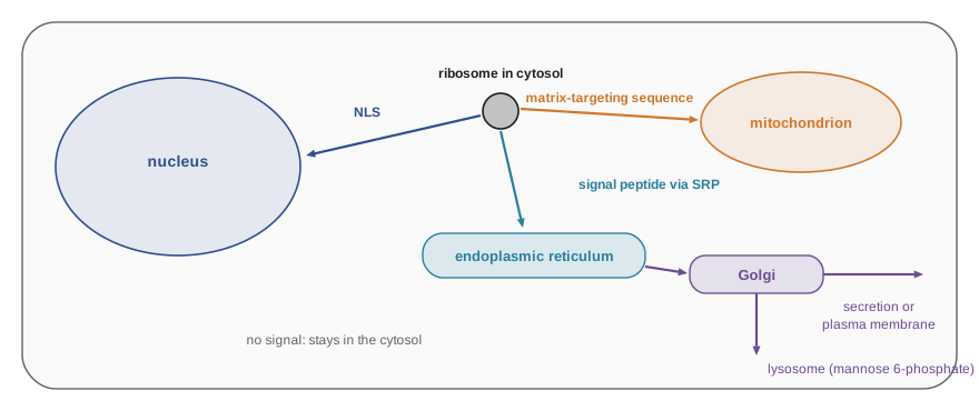
Every protein begins synthesis on a ribosome in the cytosol, yet proteins end up in the nucleus, mitochondria, chloroplasts, peroxisomes, membranes or outside the cell. They are directed by sorting signals, usually short stretches of amino acids in the protein itself, a concept for which Günter Blobel received the 1999 Nobel Prize. Proteins without any signal stay in the cytosol.
| Destination | Typical signal | Machinery | When it acts |
|---|---|---|---|
| Endoplasmic reticulum (and secretory pathway) | N-terminal signal peptide: a core of 7–15 hydrophobic residues | Signal recognition particle (SRP), SRP receptor, Sec61 translocon | During translation (co-translational) |
| Nucleus | Nuclear localization signal: a short cluster of lysines and arginines, for example PKKKRKV | Importins and nuclear pore complexes | After translation |
| Mitochondrial matrix | N-terminal amphipathic helix with positive charges | TOM and TIM translocases | After translation |
| Peroxisome | C-terminal tripeptide SKL | Peroxins | After translation |
| Stay in the ER | C-terminal KDEL | KDEL receptor returns escaped proteins | After leaving the ER |
| Lysosome | Mannose 6-phosphate added in the Golgi | Mannose 6-phosphate receptors | In the Golgi |
The secretory pathway illustrates sorting at work. As a protein destined for secretion or for a membrane begins to emerge from the ribosome, its N-terminal signal peptide is recognized by the signal recognition particle (SRP), a ribonucleoprotein that pauses translation and brings the ribosome to the SRP receptor on the endoplasmic reticulum (ER) membrane. Translation resumes and the growing chain threads through a channel, the Sec61 translocon, into the ER lumen. A signal peptidase then usually removes the signal peptide. Membrane proteins contain additional hydrophobic segments that stop transfer and become membrane-spanning helices. From the ER, proteins travel in vesicles to the Golgi apparatus, where they are further modified and sorted to the plasma membrane, lysosomes or secretory vesicles.
Signal peptides share no exact sequence, but they share a pattern: a few positively charged residues near the start, a core of hydrophobic residues, and a polar region before the cleavage site. Hydropathy plots, which average the hydrophobicity of residues in a sliding window along a sequence, reveal such segments. The scale of Kyte and Doolittle (1982) assigns each amino acid a hydropathy value, from +4.5 for isoleucine to −4.5 for arginine.
# Kyte-Doolittle hydropathy in a sliding window of 9 residues
KD = {"A": 1.8, "R": -4.5, "N": -3.5, "D": -3.5, "C": 2.5, "Q": -3.5, "E": -3.5, "G": -0.4,
      "H": -3.2, "I": 4.5, "L": 3.8, "K": -3.9, "M": 1.9, "F": 2.8, "P": -1.6, "S": -0.8,
      "T": -0.7, "W": -0.9, "Y": -1.3, "V": 4.2}

# Human preproinsulin (110 residues); the first 24 form the signal peptide
preproinsulin = ("MALWMRLLPLLALLALWGPDPAAAFVNQHLCGSHLVEALYLVCGERGFFYTPKTRREAEDLQVGQVELGGGPGAGSLQPLALEGSLQKRGIVEQCCTSICSLYQLENYCN")

w = 9
scores = [sum(KD[a] for a in preproinsulin[i:i+w]) / w for i in range(len(preproinsulin) - w + 1)]
peak = max(range(len(scores)), key=scores.__getitem__)
print("length:", len(preproinsulin), "residues")
print(f"most hydrophobic window: residues {peak+1}-{peak+w} ({preproinsulin[peak:peak+w]}), mean {scores[peak]:.2f}")
print("windows with mean > 1.6 (candidate hydrophobic segments):",
      [i + 1 for i, s in enumerate(scores) if s > 1.6][:12], "...")
print("signal peptide (residues 1-24):", preproinsulin[:24])length: 110 residues most hydrophobic window: residues 7-15 (LLPLLALLA), mean 2.76 windows with mean > 1.6 (candidate hydrophobic segments): [5, 6, 7, 8, 9, 10, 11, 35, 36] ... signal peptide (residues 1-24): MALWMRLLPLLALLALWGPDPAAA
The most hydrophobic windows fall within the first 24 residues of preproinsulin, the signal peptide, confirming what the hydropathy pattern predicts. Software used in research combines such scores with machine learning to predict signal peptides and membrane-spanning segments across entire genomes.
Proteolytic processing: insulin
Insulin, the hormone whose discovery in Toronto by Frederick Banting, Charles Best, James Collip and John Macleod in 1921 transformed the treatment of diabetes, shows how cutting a precursor creates an active protein. The gene encodes preproinsulin, 110 amino acids. Removal of the 24-residue signal peptide in the ER produces proinsulin, 86 residues, which folds and forms three disulfide bonds. In secretory vesicles, proteases called prohormone convertases cut out the middle segment, the C-peptide, leaving two chains, A (21 residues) and B (30 residues), held together by two disulfide bonds. Mature insulin and C-peptide are secreted together, so measuring C-peptide in blood indicates how much insulin a person’s own pancreas is making, which helps doctors distinguish type 1 from type 2 diabetes.
Worked problem
Accounting for the residues of preproinsulin
Preproinsulin has 110 amino acids. The signal peptide is 24 residues, the B chain 30, the A chain 21. How long is the C-peptide region removed from proinsulin, including the paired basic residues at the cut sites?
Show solution
- Proinsulin = 110 − 24 = 86 residues.
- Insulin chains = 30 + 21 = 51 residues.
- Removed from proinsulin = 86 − 51 = 35 residues.
- The C-peptide itself is 31 residues; the other 4 are the two pairs of basic residues (Arg–Arg and Lys–Arg) that mark the cut sites and are trimmed away.
Answer: 35 residues are removed from proinsulin: the 31-residue C-peptide plus 4 basic residues at the cleavage sites.
In 1978, insulin became one of the first human proteins made by recombinant DNA technology, with the A and B chains produced separately in E. coli. Today recombinant insulins, including engineered analogues with altered absorption, have replaced insulin from pig and cow pancreas. Many other proteins are made as inactive precursors and activated by cutting, including digestive enzymes such as trypsinogen, blood clotting factors and the caspases that drive programmed cell death.
Post-translational modifications
More than 400 kinds of chemical modification of proteins are known. A few are especially widespread.
| Modification | What is added | Typical residues | Main effects |
|---|---|---|---|
| Phosphorylation | Phosphate (by kinases; removed by phosphatases) | Ser, Thr, Tyr | Switches activity, binding and location on and off; central to signalling |
| Glycosylation | Sugar chains | Asn (N-linked, in the ER); Ser or Thr (O-linked, in the Golgi) | Folding, stability, cell recognition, blood group antigens |
| Acetylation | Acetyl group | Lys; protein N-terminus | Chromatin opening (on histones), stability |
| Methylation | Methyl groups | Lys, Arg | Chromatin regulation (on histones) |
| Ubiquitination | The 76-residue protein ubiquitin | Lys | Chains of ubiquitin usually mark proteins for destruction by the proteasome |
| Lipidation | Fatty acid or prenyl groups | Cys, Gly | Anchors proteins to membranes |
| Disulfide bonds | Covalent S–S bond between two cysteines | Cys | Stabilizes secreted and extracellular proteins |
Phosphorylation deserves special mention. The human genome encodes about 520 protein kinases, and roughly a third of all human proteins are phosphorylated at some point. Adding a phosphate, with its two negative charges, can change a protein’s shape, create a docking site for other proteins or alter its location. Because kinases and phosphatases act quickly and reversibly, phosphorylation is the main currency of intracellular signalling. Many cancers are driven by kinases that are stuck in the on state, and kinase inhibitors such as imatinib, which targets the BCR–ABL kinase produced by the Philadelphia chromosome (BIOL-201 lesson 9), have become important cancer drugs.
Protein degradation
Cells constantly destroy proteins: damaged ones, misfolded ones and regulatory ones whose levels must change quickly. In the ubiquitin–proteasome system, a cascade of three enzymes (E1, E2 and E3) attaches chains of ubiquitin to target proteins, and the 26S proteasome, a barrel-shaped protease complex, recognizes the chains, unfolds the target and cuts it into short peptides. Humans have more than 600 E3 ubiquitin ligases, which provide specificity by recognizing particular targets. The discovery of ubiquitin-mediated degradation earned Aaron Ciechanover, Avram Hershko and Irwin Rose the 2004 Nobel Prize in Chemistry. Proteasome inhibitors such as bortezomib are used to treat multiple myeloma, a cancer of antibody-producing cells that rely heavily on the proteasome to cope with the huge amount of protein they make.
Protein half-lives range from minutes for regulatory proteins such as the cyclins that drive the cell cycle to months or years for structural proteins such as the crystallins of the eye lens and collagen. Protein levels depend on the balance between synthesis and degradation, just as mRNA levels do (lesson 5), and the same mathematics applies.
import math

def steady_level(synth_per_hour, half_life_hours):
    return synth_per_hour * half_life_hours / math.log(2)

print("Proteins made at 1,000 molecules per hour:")
for name, h in [("cyclin-like regulator", 0.5), ("typical enzyme", 20), ("long-lived structural protein", 500)]:
    print(f"  {name:30s} half-life {h:6.1f} h -> steady state {steady_level(1000, h):10,.0f} molecules")

# How fast does a protein disappear after synthesis stops?
h = 0.5
for t in (0, 0.5, 1, 2):
print(f"t = {t:3.1f} h: {100 * 0.5 ** (t / h):5.1f}% of the regulator left")Proteins made at 1,000 molecules per hour: cyclin-like regulator half-life 0.5 h -> steady state 721 molecules typical enzyme half-life 20.0 h -> steady state 28,854 molecules long-lived structural protein half-life 500.0 h -> steady state 721,348 molecules t = 0.0 h: 100.0% of the regulator left t = 0.5 h: 50.0% of the regulator left t = 1.0 h: 25.0% of the regulator left t = 2.0 h: 6.2% of the regulator left
Worked problem
Molecular weight from sequence length
An average amino acid residue in a protein weighs about 110 daltons (Da). Estimate the mass of a 450-residue protein, and the number of codons and coding nucleotides needed, including the stop codon.
Show solution
- Mass ≈ 450 × 110 = 49,500 Da, about 50 kDa.
- Codons = 450 + 1 stop = 451.
- Coding nucleotides = 451 × 3 = 1,353.
Answer: About 50 kDa, encoded by 1,353 nucleotides of coding sequence. The mRNA is longer, because it also carries untranslated regions at both ends and a poly(A) tail.
Bringing it together
Lessons 5 to 8 have followed the flow of information from gene to working protein. In a eukaryotic cell, that path includes transcription with general factors and the CTD, capping, splicing and polyadenylation, export, scanning and initiation, elongation and termination, folding with chaperones, sorting, modification and eventually degradation. Every step offers the cell a point of control and every step can fail in disease. Lessons 9 and 10 turn to the first and most important control point: deciding which genes are transcribed.
Common mistakes
MistakeProteins need chaperones to determine their final structure.
InsteadThe sequence determines the structure; chaperones prevent aggregation and give chains chances to fold correctly.
MistakeAll proteins are made on ribosomes bound to the ER.
InsteadAll proteins begin on cytosolic ribosomes; only those with signal peptides are brought to the ER by SRP.
MistakeNuclear proteins enter the nucleus during translation.
InsteadNuclear import occurs after translation, through nuclear pores, guided by nuclear localization signals.
MistakeUbiquitin is an enzyme that digests proteins.
InsteadUbiquitin is a small protein tag; the proteasome does the digestion.
MistakeInsulin is made directly as two chains from two genes.
InsteadOne gene encodes preproinsulin; cleavage of the signal peptide and C-peptide produces the two chains.
Summary and key terms
- Anfinsen showed that sequence determines structure; the hydrophobic effect drives folding.
- Chaperones such as Hsp70 and chaperonins prevent aggregation and assist folding.
- Misfolding causes disease, either through loss of protein (as in cystic fibrosis ΔF508) or toxic aggregation.
- Sorting signals direct proteins: signal peptides to the ER via SRP, NLS to the nucleus, matrix-targeting sequences to mitochondria.
- Many proteins are activated by proteolytic cleavage, as preproinsulin becomes insulin and C-peptide.
- Phosphorylation, glycosylation, acetylation, methylation, ubiquitination and lipidation change protein function and location.
- The ubiquitin–proteasome system destroys proteins; levels reflect the balance of synthesis and degradation.
- hydrophobic effect
- The tendency of nonpolar groups to cluster away from water, a main driver of protein folding.
- molecular chaperone
- A protein that helps others fold correctly and prevents aggregation.
- signal peptide
- An N-terminal hydrophobic sequence that targets a protein to the endoplasmic reticulum.
- signal recognition particle (SRP)
- A ribonucleoprotein that binds signal peptides and brings ribosomes to the ER.
- nuclear localization signal
- A short basic sequence that directs a protein into the nucleus.
- proteolytic processing
- Activation or maturation of a protein by cutting its precursor.
- phosphorylation
- Addition of a phosphate group to Ser, Thr or Tyr by a kinase.
- glycosylation
- Addition of sugar chains to a protein.
- ubiquitin
- A small protein attached to targets, often marking them for destruction.
- proteasome
- A large protease complex that degrades ubiquitinated proteins.
Practice problems
Work each problem on paper first, then reveal the answer to check.
What did Anfinsen’s ribonuclease experiment show?
Show answer
A denatured protein can refold spontaneously, so its sequence contains the information for its structure.
Why are many chaperones also called heat-shock proteins?
Show answer
They are produced in large amounts when heat unfolds proteins and increases the need for folding help.
Describe the typical features of a signal peptide.
Show answer
A positively charged N-terminal region, a hydrophobic core of 7–15 residues and a polar region before the cleavage site.
What happens when SRP binds a signal peptide?
Show answer
Translation pauses and the ribosome is brought to the SRP receptor and translocon on the ER membrane.
Where would a protein ending in SKL be found?
Show answer
In peroxisomes.
Why does measuring C-peptide help assess a person’s own insulin production?
Show answer
C-peptide is released with endogenous insulin but is not present in injected insulin.
A protein has a half-life of 2 hours. What fraction remains 6 hours after synthesis stops?
Show answer
(1/2)³ = 1/8.
Estimate the mass of a 300-residue protein.
Show answer
About 300 × 110 = 33,000 Da (33 kDa).
What is the role of E3 ligases?
Show answer
They recognize specific target proteins and transfer ubiquitin to them, giving the system specificity.
Why are proteasome inhibitors effective against multiple myeloma?
Show answer
Myeloma cells make huge amounts of antibody protein and depend heavily on the proteasome to clear misfolded protein.
Lesson quiz · 5 questions
Score 60% or more to complete this lesson.
Create a free account to save your progress, take quizzes and earn a certificate.