
BIOL-207 · Lesson 11 of 12 · 33 min
Molecular techniques I: cutting, copying and cloning DNA
Everything in the earlier lessons was discovered with tools, and since the 1970s the tools themselves have come from molecular biology: enzymes borrowed from bacteria and viruses that cut, copy and join DNA. Recombinant DNA technology lets researchers isolate a single gene from a genome of billions of base pairs, make millions of copies of it, change it at will and express it in another organism. This lesson covers the core methods: restriction enzymes, gel electrophoresis, the polymerase chain reaction and its quantitative form, and molecular cloning in bacteria. Lesson 12 continues with sequencing and genome editing and puts the methods to work in an invented investigation.
Restriction enzymes
Bacteria defend themselves against phages with restriction enzymes, which cut DNA at specific sequences, and protect their own DNA by methylating those same sequences. In the early 1970s, Hamilton Smith, Daniel Nathans and Werner Arber showed how these enzymes work and that they could be used to cut DNA into reproducible fragments; they shared the 1978 Nobel Prize. Thousands of restriction enzymes with different specificities are now known, and hundreds are sold commercially.
Most of the enzymes used in laboratories are type II enzymes that recognize a short palindromic sequence, 4 to 8 bp long, and cut within it. A DNA palindrome reads the same 5′ to 3′ on both strands: the EcoRI site, 5′-GAATTC-3′, has the reverse complement 5′-GAATTC-3′. Some enzymes cut both strands at the same place, leaving blunt ends; others cut at staggered positions, leaving short single-stranded overhangs called sticky ends. EcoRI cuts between G and A on each strand, leaving four-nucleotide 5′ overhangs of AATT. Any two fragments cut with the same enzyme have compatible sticky ends that can pair and be joined by DNA ligase, which is the basis of cloning.
| Enzyme | Source organism | Recognition site (5′ to 3′, cut marked by /) | Ends produced |
|---|---|---|---|
| EcoRI | Escherichia coli | G/AATTC | 5′ overhang AATT |
| BamHI | Bacillus amyloliquefaciens | G/GATCC | 5′ overhang GATC |
| HindIII | Haemophilus influenzae | A/AGCTT | 5′ overhang AGCT |
| PstI | Providencia stuartii | CTGCA/G | 3′ overhang TGCA |
| EcoRV | Escherichia coli | GAT/ATC | Blunt |
| NotI | Nocardia otitidiscaviarum | GC/GGCCGC | 5′ overhang GGCC (rare cutter, 8 bp site) |
How often should an enzyme cut? In random sequence with equal base frequencies, a particular 6 bp site occurs on average once every 4⁶ = 4,096 bp, a 4 bp site once every 256 bp and an 8 bp site once every 65,536 bp. Real genomes are not random, so actual frequencies differ; NotI cuts mammalian DNA rarely because its site contains two CpG dinucleotides, which are depleted (lesson 10).
SITES = {"EcoRI": ("GAATTC", 1), "BamHI": ("GGATCC", 1), "HindIII": ("AAGCTT", 1)}

def digest(seq, enzymes, circular=False):
    cuts = sorted(i + off for e in enzymes for site, off in [SITES[e]]
                  for i in range(len(seq)) if seq.startswith(site, i))
    if not cuts:
        return [len(seq)]
    if circular:
        frags = [b - a for a, b in zip(cuts, cuts[1:])] + [len(seq) - cuts[-1] + cuts[0]]
    else:
        pts = [0] + cuts + [len(seq)]
        frags = [b - a for a, b in zip(pts, pts[1:])]
    return sorted(frags, reverse=True)

# An invented 3,000 bp plasmid built from repeats of a neutral 10-mer, with sites inserted
filler = "ACTGTCAGTA" * 300
plasmid = list(filler)
for pos, site in [(400, "GAATTC"), (1250, "GGATCC"), (2100, "GAATTC"), (2700, "AAGCTT")]:
    plasmid[pos:pos+6] = site
plasmid = "".join(plasmid)
print("plasmid length:", len(plasmid))
for combo in (["EcoRI"], ["BamHI"], ["EcoRI", "BamHI"], ["EcoRI", "BamHI", "HindIII"]):
    print(f"{'+'.join(combo):22s} fragments (bp): {digest(plasmid, combo, circular=True)}")
print("random 6-bp site expected every", 4**6, "bp")plasmid length: 3000 EcoRI fragments (bp): [1700, 1300] BamHI fragments (bp): [3000] EcoRI+BamHI fragments (bp): [1300, 850, 850] EcoRI+BamHI+HindIII fragments (bp): [850, 850, 700, 600] random 6-bp site expected every 4096 bp
Worked problem
Mapping sites from a double digest
A 5.0 kb circular plasmid gives one fragment of 5.0 kb with EcoRI, one of 5.0 kb with BamHI, and two fragments, 3.8 kb and 1.2 kb, when cut with both. What can you conclude?
Show solution
- One fragment from a circle means one cut: the plasmid has one EcoRI site and one BamHI site.
- Cutting a circle at two points gives two fragments whose sizes equal the two distances around the circle between the sites.
- So the sites are 1.2 kb apart one way around the circle, and 3.8 kb the other way.
Answer: The plasmid has single EcoRI and BamHI sites 1.2 kb apart (or equivalently 3.8 kb apart going the other way). Combining several such digests, called restriction mapping, was how DNA was mapped before sequencing became cheap.
Gel electrophoresis
DNA fragments are separated by size using gel electrophoresis. Samples are loaded into wells at one end of a slab of agarose (for fragments of a few hundred base pairs up to tens of kilobases) or polyacrylamide (for small fragments), and an electric field is applied. Because DNA carries a uniform negative charge per unit length, all fragments move toward the positive electrode, but the gel matrix slows larger fragments more. Over a useful range, the distance travelled is roughly proportional to the logarithm of fragment size. Fragments are made visible with fluorescent dyes that bind DNA. A ladder of fragments of known sizes run alongside lets researchers estimate unknown sizes from a standard curve.
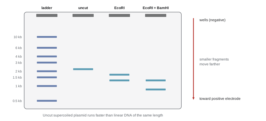
import numpy as np

# Ladder: fragment sizes (bp) and measured migration distances (mm) on one gel (illustrative)
ladder_bp = np.array([10000, 6000, 4000, 3000, 2000, 1500, 1000, 500])
ladder_mm = np.array([12.0, 17.5, 22.0, 25.5, 31.0, 35.0, 40.5, 49.0])

slope, intercept = np.polyfit(ladder_mm, np.log10(ladder_bp), 1)
fit = slope * ladder_mm + intercept
r2 = 1 - np.sum((np.log10(ladder_bp) - fit)**2) / np.sum((np.log10(ladder_bp) - np.log10(ladder_bp).mean())**2)
print(f"standard curve: log10(size) = {slope:.4f} x distance + {intercept:.3f}   (R^2 = {r2:.4f})")
for d in (20.0, 28.0, 44.0):
print(f"band at {d:4.1f} mm -> about {10 ** (slope * d + intercept):6.0f} bp")standard curve: log10(size) = -0.0345 x distance + 4.382 (R^2 = 0.9980) band at 20.0 mm -> about 4918 bp band at 28.0 mm -> about 2605 bp band at 44.0 mm -> about 731 bp
The polymerase chain reaction
In 1983 Kary Mullis, a chemist at the Cetus Corporation in California, conceived a way to copy a chosen stretch of DNA exponentially, the polymerase chain reaction (PCR). Mullis shared the 1993 Nobel Prize in Chemistry with Michael Smith of the University of British Columbia, who developed site-directed mutagenesis, the use of synthetic oligonucleotides to make precise changes in a DNA sequence. PCR needs a template, two primers that flank the target and pair with opposite strands, the four dNTPs, magnesium ions and a heat-stable DNA polymerase.
Each cycle has three steps. Denaturation at about 95 °C separates the strands. Annealing at a lower temperature, often 50 to 65 °C, lets the primers pair with their targets; the temperature is chosen a few degrees below the primers’ Tm (lesson 2). Extension at 72 °C lets the polymerase extend the primers. Early PCR used the E. coli polymerase, which was destroyed at each denaturation step and had to be replaced. The breakthrough was Taq polymerase, from Thermus aquaticus, a bacterium isolated from a hot spring in Yellowstone National Park by Thomas Brock in the 1960s. Taq survives repeated heating, so the whole reaction can run unattended in a thermal cycler.
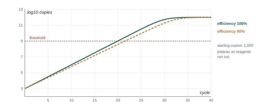
In an ideal reaction, the number of copies of the target doubles each cycle. After n cycles, starting from N₀ copies, there are N₀ × 2ⁿ copies. Real reactions have an efficiency E below 1, so each cycle multiplies copies by (1 + E). Eventually primers, dNTPs or polymerase activity run short and the reaction reaches a plateau. Even so, 30 cycles of perfect doubling multiply a target more than a billion-fold, which is why PCR can detect a few molecules of DNA in a sample, and why contamination is such a concern in PCR laboratories.
N0 = 10           # starting copies of the target
for E in (1.0, 0.95, 0.90, 0.80):
    print(f"efficiency {E:.2f}:", "  ".join(f"{n} cycles {N0*(1+E)**n:.2e}" for n in (10, 20, 30)))

# Cycles needed to reach 1e10 copies from 10 copies
import math
for E in (1.0, 0.9):
print(f"E = {E}: {math.log(1e10 / N0) / math.log(1 + E):.1f} cycles to reach 1e10 copies")efficiency 1.00: 10 cycles 1.02e+04 20 cycles 1.05e+07 30 cycles 1.07e+10 efficiency 0.95: 10 cycles 7.95e+03 20 cycles 6.32e+06 30 cycles 5.02e+09 efficiency 0.90: 10 cycles 6.13e+03 20 cycles 3.76e+06 30 cycles 2.30e+09 efficiency 0.80: 10 cycles 3.57e+03 20 cycles 1.27e+06 30 cycles 4.55e+08 E = 1.0: 29.9 cycles to reach 1e10 copies E = 0.9: 32.3 cycles to reach 1e10 copies
Primer design is the most important practical decision in PCR. Good primers are about 18 to 25 nucleotides long, with 40 to 60 per cent GC content and similar melting temperatures for the forward and reverse primer. A G or C at the 3′ end (a GC clamp) helps the polymerase start, but more than three G or C in the last five bases can promote false starts. Primers should not pair with themselves or with each other, especially at their 3′ ends, which would produce short artefacts called primer dimers.
COMP = str.maketrans("ACGT", "TGCA")
def rc(s): return s.translate(COMP)[::-1]
def tm(s): return 2 * (s.count("A") + s.count("T")) + 4 * (s.count("G") + s.count("C"))

def check(name, p):
    gc = (p.count("G") + p.count("C")) / len(p)
    clamp = sum(b in "GC" for b in p[-5:])
    dimer = any(rc(p[-4:]) in p for _ in [0])     # 3' end able to pair within the primer itself
    issues = []
    if not 18 <= len(p) <= 25: issues.append("length")
    if not 0.40 <= gc <= 0.60: issues.append("GC content")
    if p[-1] not in "GC": issues.append("no 3' G/C clamp")
    if clamp > 3: issues.append("too many G/C at 3' end")
    if dimer: issues.append("3' self-complementarity")
    print(f"{name}: {p}  len {len(p)}  GC {gc:.0%}  Tm(Wallace) {tm(p)} C  ->", ", ".join(issues) or "looks fine")

check("forward", "AGCTTGACCGGAATTCAGTC")
check("reverse", "TCCGATGGTAGCTACCAGTC")
check("poor ", "GCGCGCATATGCGC")forward: AGCTTGACCGGAATTCAGTC len 20 GC 50% Tm(Wallace) 60 C -> looks fine reverse: TCCGATGGTAGCTACCAGTC len 20 GC 55% Tm(Wallace) 62 C -> looks fine poor : GCGCGCATATGCGC len 14 GC 71% Tm(Wallace) 48 C -> length, GC content, too many G/C at 3' end, 3' self-complementarity
Reverse transcription and quantitative PCR
PCR copies DNA, but many questions concern RNA: which genes are being expressed, or whether an RNA virus is present. Reverse transcriptase (lesson 1) copies RNA into complementary DNA (cDNA), which can then be amplified; the combined method is RT-PCR. In quantitative PCR (qPCR), also called real-time PCR, a fluorescent dye or probe reports the amount of product after each cycle. The cycle at which fluorescence crosses a threshold, the threshold cycle (Ct), is lower when there is more starting template: with perfect doubling, each 1-cycle decrease in Ct means twice as much starting material. RT-qPCR was the standard test for SARS-CoV-2 during the COVID-19 pandemic, and Ct values were sometimes used as rough indicators of viral load.
For gene expression, the ΔΔCt method compares a target gene with a reference gene whose expression is assumed constant (often one encoding a housekeeping protein such as GAPDH or β-actin), in a treated sample versus a control.
Worked problem
Fold change by ΔΔCt
In control cells, a target gene has Ct = 26.0 and the reference gene Ct = 18.0. In treated cells, the target has Ct = 23.5 and the reference Ct = 18.2. What is the fold change in target expression?
Show solution
- ΔCt control = 26.0 − 18.0 = 8.0.
- ΔCt treated = 23.5 − 18.2 = 5.3.
- ΔΔCt = 5.3 − 8.0 = −2.7.
- Fold change = 2^(2.7) ≈ 6.5.
Answer: The target is about 6.5 times higher in treated cells, relative to the reference gene. The method assumes both genes amplify with close to 100 per cent efficiency; if not, efficiency-corrected calculations are needed.
import numpy as np
rng = np.random.default_rng(207)

# Three biological replicates per condition (illustrative Ct values)
ctrl_target = 26.0 + rng.normal(0, 0.12, 3); ctrl_ref = 18.0 + rng.normal(0, 0.1, 3)
trt_target  = 23.5 + rng.normal(0, 0.12, 3); trt_ref  = 18.2 + rng.normal(0, 0.1, 3)

d_ctrl = ctrl_target - ctrl_ref
d_trt = trt_target - trt_ref
ddct = d_trt - d_ctrl.mean()
fold = 2 ** (-ddct)
print("delta Ct control:", np.round(d_ctrl, 2))
print("delta Ct treated:", np.round(d_trt, 2))
print("fold change per treated replicate:", np.round(fold, 2))
print(f"mean fold change {fold.mean():.2f} (sd {fold.std(ddof=1):.2f})")delta Ct control: [8.14 7.91 7.94] delta Ct treated: [4.95 5.23 5.12] fold change per treated replicate: [8.25 6.79 7.34] mean fold change 7.46 (sd 0.74)
With three replicates per condition, the mean fold change comes out at about 7.5, a little above the single-sample estimate of 6.5, because the replicate Ct values vary by a few tenths of a cycle and each tenth of a cycle changes the result by about 7 per cent. Because qPCR is exponential, small differences in Ct matter, which is why experiments report the variation among biological replicates and use statistics rather than a single measurement.
Molecular cloning
To clone a gene is to insert it into a vector, a DNA molecule that can replicate in a host cell, and to grow many identical copies. The most common vectors are plasmids engineered with three essential features: an origin of replication, so the plasmid is copied in the host; a selectable marker, usually an antibiotic resistance gene, so that only cells carrying the plasmid survive on medium with the antibiotic; and a multiple cloning site, a short stretch containing unique sites for many restriction enzymes where an insert can be placed.
In the classic approach, the vector and the DNA to be cloned are cut with the same restriction enzyme, mixed so that their sticky ends pair, and joined with DNA ligase. The products are introduced into E. coli by transformation, using heat shock or an electric pulse to let cells take up DNA, as Griffith’s pneumococci did naturally (lesson 1). Cells are spread on plates containing the antibiotic, so that only transformed cells grow into colonies. Many vectors use blue–white screening to find colonies with an insert: the multiple cloning site lies inside a fragment of the lacZ gene (lacZα, from lesson 9). An intact lacZα lets cells make active β-galactosidase, which turns the chemical X-gal blue. An insert interrupts lacZα, so colonies carrying recombinant plasmids are white.
Modern laboratories often use methods that do not depend on restriction sites. Gibson assembly, developed at the J. Craig Venter Institute, joins fragments that share 20 to 40 bp of overlapping sequence using a mix of an exonuclease, a polymerase and ligase in a single tube, allowing many pieces to be assembled at once. Site-directed mutagenesis, building on Michael Smith’s method, uses primers containing a deliberate mismatch to introduce a chosen change into a cloned gene by PCR.
Worked problem
How many colonies to screen?
After a ligation, 30 per cent of the colonies on an ampicillin plate carry a plasmid with the desired insert. If you pick colonies at random without blue–white screening, how many must you pick to have a 95 per cent chance of finding at least one correct clone?
Show solution
- P(a colony is not correct) = 0.7.
- P(none of n colonies correct) = 0.7ⁿ; we need 0.7ⁿ ≤ 0.05.
- n ≥ ln 0.05 / ln 0.7 = −3.00 / −0.357 ≈ 8.4.
Answer: Pick at least 9 colonies. With blue–white screening, picking white colonies raises the success rate sharply, which is why it became standard.
Expressing the cloned gene to make its protein requires an expression vector with a suitable promoter and, for bacteria, a ribosome-binding site (lesson 7). Human proteins that need eukaryotic modifications, such as specific glycosylation (lesson 8), are often made in yeast, insect cells or Chinese hamster ovary cells instead of bacteria. Most therapeutic antibodies are made this way.
Common mistakes
MistakeRestriction enzymes cut DNA at random.
InsteadRestriction enzymes cut at specific recognition sequences, usually palindromes.
MistakeIn a gel, larger fragments move farther.
InsteadSmaller fragments move farther; distance is roughly proportional to the logarithm of size.
MistakeThirty cycles of PCR give 30 times as much DNA.
InsteadEach cycle can double the target, so 30 cycles give up to about 2³⁰, roughly 10⁹ times as much.
MistakeA lower Ct in qPCR means less starting template.
InsteadA lower Ct means more starting template; the threshold is reached sooner.
MistakeBlue colonies in blue–white screening contain the insert.
InsteadWhite colonies contain the insert, which disrupts lacZα; blue colonies have intact lacZα.
Summary and key terms
- Type II restriction enzymes cut at short palindromic sites, leaving sticky or blunt ends; a 6 bp site appears about every 4 kb at random.
- Double digests and restriction mapping locate sites; gel electrophoresis separates fragments by size against a ladder.
- PCR amplifies a target exponentially through cycles of denaturation, annealing and extension with heat-stable Taq polymerase.
- Copies after n cycles are N₀(1 + E)ⁿ; good primers are 18–25 nt with balanced GC and matched Tm.
- RT-PCR copies RNA via reverse transcriptase; qPCR uses Ct values, and ΔΔCt gives relative expression as 2^(−ΔΔCt).
- Cloning vectors carry an origin, a selectable marker and a multiple cloning site; blue–white screening identifies inserts.
- Gibson assembly and site-directed mutagenesis extend cloning beyond restriction sites.
- restriction enzyme
- An enzyme that cuts DNA at a specific recognition sequence.
- palindrome
- A DNA sequence identical to its reverse complement.
- sticky end
- A short single-stranded overhang left by a staggered cut.
- gel electrophoresis
- Separation of nucleic acids or proteins by size in an electric field through a gel.
- polymerase chain reaction (PCR)
- Exponential amplification of a DNA target using primers and a heat-stable polymerase.
- primer
- A short oligonucleotide that pairs with a template and provides a 3′ end for extension.
- threshold cycle (Ct)
- The qPCR cycle at which fluorescence crosses a set threshold.
- complementary DNA (cDNA)
- DNA copied from RNA by reverse transcriptase.
- vector
- A DNA molecule used to carry and replicate cloned DNA in a host.
- transformation
- Uptake of DNA by a cell, used to introduce plasmids into bacteria.
Practice problems
Work each problem on paper first, then reveal the answer to check.
Is 5′-GGATCC-3′ a palindrome? Why?
Show answer
Yes; its reverse complement is also 5′-GGATCC-3′.
How often would a 4 bp site occur in random sequence?
Show answer
About once every 4⁴ = 256 bp.
A linear 10 kb fragment has EcoRI sites at 2 kb and 7 kb. What fragments does EcoRI produce?
Show answer
2 kb, 5 kb and 3 kb.
Why does DNA move toward the positive electrode?
Show answer
Its phosphate backbone is negatively charged.
Why was Taq polymerase essential for practical PCR?
Show answer
It survives the 95 °C denaturation step, so it need not be added each cycle.
Starting with 100 copies, how many are there after 20 cycles at 100 per cent efficiency?
Show answer
100 × 2²⁰ ≈ 1.05 × 10⁸.
Two samples have Ct values of 22 and 25 for the same gene. Which had more template and by how much?
Show answer
The Ct 22 sample, about 2³ = 8 times more.
ΔΔCt = +1.5. What is the fold change?
Show answer
2^(−1.5) ≈ 0.35, a decrease to about a third.
Name the three essential features of a plasmid cloning vector.
Show answer
Origin of replication, selectable marker and multiple cloning site.
Why are colonies with inserts white in blue–white screening?
Show answer
The insert disrupts lacZα, so no active β-galactosidase forms to turn X-gal blue.
Lesson quiz · 5 questions
Score 60% or more to complete this lesson.
Create a free account to save your progress, take quizzes and earn a certificate.