
BIOL-207 · Lesson 6 of 12 · 31 min
Transcription and RNA processing in eukaryotes
Eukaryotic transcription takes place inside a nucleus, on DNA wrapped in chromatin, and produces RNAs that must be extensively modified before they can leave the nucleus and be translated. Where a bacterium makes a short-lived mRNA that ribosomes start reading immediately, a eukaryotic cell makes a precursor RNA, caps its 5′ end, cuts out internal segments and joins the rest, adds a tail of adenines to its 3′ end, and exports the finished message to the cytoplasm. Each step is a point of quality control and of regulation. This lesson describes the three eukaryotic RNA polymerases, the assembly of the transcription machinery at promoters, and the processing steps that turn a primary transcript into a mature mRNA.
Three RNA polymerases
Eukaryotes divide transcription among three nuclear RNA polymerases, each related to the bacterial enzyme but larger, with 12 to 17 subunits. Plants have two more, Pol IV and Pol V, which make small RNAs involved in silencing. Mitochondria and chloroplasts have their own polymerases, reflecting their bacterial ancestry.
| Polymerase | Main products | Share of transcription in a growing cell | Sensitivity to α-amanitin |
|---|---|---|---|
| RNA polymerase I | Large ribosomal RNAs (28S, 18S, 5.8S) from one precursor | Most RNA by mass, in the nucleolus | Insensitive |
| RNA polymerase II | All mRNAs; most small nuclear RNAs; microRNA precursors; many lncRNAs | Most genes | Very sensitive |
| RNA polymerase III | Transfer RNAs, 5S rRNA, U6 snRNA, other small RNAs | Small, abundant RNAs | Moderately sensitive |
The toxin α-amanitin, made by the death cap mushroom (Amanita phalloides), binds RNA polymerase II tightly and stops it moving along DNA. Cells of the liver, which absorb the toxin first, stop making mRNA and die over several days, which is why death-cap poisoning is so dangerous. Death caps have spread in British Columbia, where they are associated with imported trees, and in California, and they have caused fatal poisonings in both places. Differences in sensitivity to α-amanitin were how researchers first distinguished the three polymerases.
Promoters and general transcription factors
Unlike bacterial RNA polymerase with its sigma factor, eukaryotic RNA polymerase II cannot recognize promoters on its own. It relies on a set of general transcription factors (GTFs), named TFIIA, TFIIB, TFIID, TFIIE, TFIIF and TFIIH, which assemble with the polymerase at the core promoter to form the preinitiation complex. The core promoter is the stretch of about 40 bp around the start site. Many, but far from all, Pol II promoters contain a TATA box, with consensus TATAAA, about 25 to 30 bp upstream of the start site. Others use different elements, such as the initiator (Inr) at the start site or a downstream promoter element.
Assembly begins when TFIID, which includes the TATA-binding protein (TBP), binds the promoter. TBP binds in the minor groove and bends the DNA sharply. TFIIB then binds, and positions RNA polymerase II, which arrives with TFIIF. TFIIE and TFIIH join last. TFIIH has two key enzymatic activities: a helicase that opens the DNA at the start site, and a kinase that phosphorylates the carboxyl-terminal domain (CTD) of the largest polymerase subunit. The CTD is a long tail of repeats of seven amino acids, Tyr-Ser-Pro-Thr-Ser-Pro-Ser, repeated 52 times in humans. Phosphorylation of serine 5 in these repeats helps the polymerase escape the promoter; phosphorylation of serine 2 accumulates during elongation. The changing pattern of CTD phosphorylation acts as a platform that recruits capping, splicing and polyadenylation enzymes at the right moments, coupling RNA processing to transcription.
TFIIH is also part of the nucleotide excision repair system (BIOL-201 lesson 10), which is why some inherited defects in its subunits cause both repair deficiency and developmental problems. In living cells, the general factors and polymerase alone give only a low basal level of transcription. Strong expression requires activator proteins bound to enhancers, and a large co-activator complex called Mediator that relays their signals to the polymerase. Lesson 10 covers these regulatory layers.
Promoter-proximal pausing
In many animal genes, RNA polymerase II starts transcription and then pauses 20 to 60 nucleotides downstream of the start site, held by pausing factors. Release from the pause requires the kinase P-TEFb, which phosphorylates the polymerase CTD and the pausing factors. Pausing allows genes to be primed for rapid activation, as with the heat-shock genes of fruit flies, where paused polymerases can be released within minutes of a temperature rise. Pausing is now recognized as a major point of regulation across the genome, not a curiosity of a few genes.
Capping
Almost as soon as the 5′ end of a Pol II transcript emerges from the polymerase, about 20 to 30 nucleotides into synthesis, it receives a cap: a guanine nucleotide joined backwards, through an unusual 5′-to-5′ triphosphate bridge, and then methylated at position 7 of the guanine (m⁷G). The cap protects the RNA from 5′ to 3′ exonucleases, helps export it from the nucleus, and is recognized by the translation initiation factor eIF4E, which recruits the ribosome in the cytoplasm (lesson 7). Messenger RNAs that lack a cap are rapidly degraded.
Splicing
The most striking feature of eukaryotic genes, discovered in 1977 by Phillip Sharp and Richard Roberts working with adenovirus, is that their coding sequences are interrupted by non-coding segments. The segments that remain in the mature RNA are exons; the intervening segments that are removed are introns. The removal of introns and the joining of exons is splicing. In human genes, exons average about 150 nucleotides while introns average several thousand, so most of a typical gene is intron. The gene for dystrophin, which is defective in Duchenne muscular dystrophy (BIOL-201), spans about 2.2 million bp, but its mature mRNA is only about 14,000 nucleotides; transcribing it takes around 16 hours.
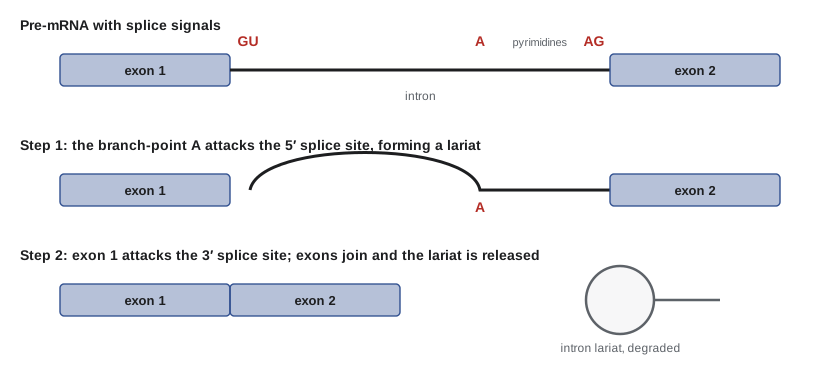
Introns are recognized by short sequences at their ends and inside them. Nearly all introns begin with GU at the 5′ splice site and end with AG at the 3′ splice site, the GU–AG rule. Upstream of the 3′ splice site lies a polypyrimidine tract (a run of C and U), and upstream of that a branch point containing a conserved adenine. Splicing occurs in two chemical steps. First, the 2′-OH of the branch-point adenine attacks the phosphate at the 5′ splice site, cutting the RNA and forming a looped structure called a lariat. Second, the newly freed 3′-OH of the upstream exon attacks the 3′ splice site, joining the two exons and releasing the intron as a lariat, which is then degraded.
These reactions are carried out by the spliceosome, a huge, dynamic complex of five small nuclear RNAs (U1, U2, U4, U5 and U6) and well over 100 proteins. U1 snRNA pairs with the 5′ splice site; U2 pairs with the branch point. Structural studies by cryo-electron microscopy have shown that the catalytic centre of the spliceosome is formed by RNA, the U6 snRNA, coordinating metal ions, making the spliceosome a ribozyme, like the ribosome. Some introns in fungi, plants and organelles can even splice themselves without any protein, which supports the idea that the spliceosome evolved from such self-splicing introns.
Worked problem
Finding the mature mRNA
A short invented pre-mRNA reads 5′-AUGGCUAAG|GUAAGUCUUACUAACCCUUUCAG|GCAUUAUGA-3′, where the vertical bars mark the intron boundaries. Identify the splice sites and write the mature sequence.
Show solution
- The intron is GUAAGUCUUACUAACCCUUUCAG: it begins with GU and ends with AG.
- Inside it, UACUAAC contains a branch-point A, and CCCUUUC is a polypyrimidine tract just upstream of the final AG.
- Remove the intron and join the exons: AUGGCUAAG + GCAUUAUGA.
Answer: The mature RNA is 5′-AUGGCUAAGGCAUUAUGA-3′, 18 nucleotides that read as six codons ending in the stop codon UGA. The sequence is invented to show the signals; real introns are usually far longer.
def splice(pre, introns):
    """Remove introns given as (start, end) positions, 0-based, end exclusive."""
    out, pos = [], 0
    for s, e in sorted(introns):
        assert pre[s:s+2] == "GU" and pre[e-2:e] == "AG", "GU-AG rule violated"
        out.append(pre[pos:s])
        pos = e
    out.append(pre[pos:])
    return "".join(out)

pre = "AUGGCUAAGGUAAGUCUUACUAACCCUUUCAGGCAUUAUGA"
mature = splice(pre, [(9, 32)])
print("pre-mRNA length:", len(pre))
print("mature mRNA:    ", mature, f"({len(mature)} nt)")
print("codons:", " ".join(mature[i:i+3] for i in range(0, len(mature), 3)))pre-mRNA length: 41 mature mRNA: AUGGCUAAGGCAUUAUGA (18 nt) codons: AUG GCU AAG GCA UUA UGA
Alternative splicing
A single gene can be spliced in more than one way. Exons can be skipped or included, alternative 5′ or 3′ splice sites can be used, introns can be retained, and mutually exclusive exons can be chosen. More than 90 per cent of human multi-exon genes produce more than one splice form, which helps explain how about 20,000 genes produce a much larger variety of proteins. Splicing choices are controlled by RNA-binding proteins that bind exonic and intronic enhancers and silencers. Many differ between tissues: neurons and muscle cells, for example, have distinctive splicing programs.
The number of possible products grows quickly. A gene with several independently chosen alternative exons can, in principle, produce many isoforms. The most famous example is the Dscam gene of the fruit fly, which has four clusters of mutually exclusive exons containing 12, 48, 33 and 2 alternatives. Choosing one exon from each cluster gives 12 × 48 × 33 × 2 = 38,016 possible proteins, more than twice the number of genes in the fly genome. Different neurons express different combinations, which helps them distinguish their own branches from those of other neurons.
from math import prod
from itertools import product

# Dscam: four clusters of mutually exclusive alternative exons
clusters = {"exon 4": 12, "exon 6": 48, "exon 9": 33, "exon 17": 2}
print("Dscam possible isoforms:", f"{prod(clusters.values()):,}")

# A smaller gene: 3 cassette exons, each independently included or skipped
cassettes = ["E2", "E4", "E6"]
forms = []
for choice in product([True, False], repeat=len(cassettes)):
    exons = ["E1"] + [c for c, keep in zip(cassettes, choice) if keep] + ["E7"]
    forms.append("-".join(exons))
print(f"\n{len(forms)} isoforms from {len(cassettes)} cassette exons:")
for f in forms:
print(" ", f)Dscam possible isoforms: 38,016 8 isoforms from 3 cassette exons: E1-E2-E4-E6-E7 E1-E2-E4-E7 E1-E2-E6-E7 E1-E2-E7 E1-E4-E6-E7 E1-E4-E7 E1-E6-E7 E1-E7
Splicing goes wrong in disease. An estimated 10 to 15 per cent or more of disease-causing point mutations affect splicing, by destroying a splice site, creating a new one or disrupting an enhancer. Spinal muscular atrophy (SMA), once the most common genetic cause of infant death, results from loss of the SMN1 gene. A nearly identical backup gene, SMN2, differs by a single base that causes exon 7 to be skipped in most transcripts, giving an unstable protein. Two treatments work by correcting this splicing. Nusinersen is an antisense oligonucleotide, injected into the spinal fluid, that blocks a splicing silencer in SMN2 intron 7. Risdiplam is a small molecule taken by mouth that stabilizes U1 snRNP binding at the exon 7 splice site. Both raise the amount of full-length SMN protein. Newborn screening for SMA, now part of provincial and state programs, matters because treatment works best before symptoms appear.
Polyadenylation and termination
The 3′ end of a Pol II transcript is not set by where the polymerase stops. Instead, a signal sequence in the RNA, AAUAAA in most cases, located 10 to 30 nucleotides upstream of the cleavage site, is recognized by the cleavage and polyadenylation specificity factor (CPSF). Along with a GU-rich element downstream, it directs cleavage of the RNA. Poly(A) polymerase then adds a tail of about 200 adenines in mammals, without any template. The tail protects the mRNA, helps export and promotes translation; its gradual shortening in the cytoplasm marks the mRNA for decay. After cleavage, the polymerase continues for a while, but the uncapped RNA it is still making is degraded by an exonuclease that chases it down and helps knock the polymerase off the template, the so-called torpedo model of termination.
| Processing step | Signal or site | Main machinery | Function |
|---|---|---|---|
| 5′ capping | First nucleotides of the transcript | Capping enzyme, methyltransferase | Protection, export, translation initiation |
| Splicing | GU at 5′ site, branch-point A, pyrimidine tract, AG at 3′ site | Spliceosome (U1, U2, U4, U5, U6 snRNPs) | Removes introns; alternative splicing makes isoforms |
| 3′ cleavage and polyadenylation | AAUAAA upstream, GU-rich element downstream | CPSF, CstF, poly(A) polymerase | Defines the 3′ end; protection and translation |
| Export | Completed, processed mRNA with bound proteins | Export receptors at nuclear pores | Delivers mRNA to ribosomes |
Export, surveillance and decay
Only properly processed mRNAs are exported through nuclear pore complexes. Splicing deposits a protein complex, the exon junction complex, about 20 to 24 nucleotides upstream of each exon–exon junction. During the first round of translation, ribosomes displace these complexes. If a ribosome reaches a stop codon more than about 50 nucleotides upstream of the last exon junction, a junction complex remains downstream, signalling that the stop codon is premature. The mRNA is then destroyed by nonsense-mediated decay. This surveillance protects cells from truncated proteins that might be harmful, and it explains why many nonsense mutations reduce mRNA levels rather than producing short proteins.
Eukaryotic mRNAs are generally more stable than bacterial ones. Half-lives in mammalian cells range from minutes for mRNAs encoding signalling proteins and transcription factors, which often carry AU-rich destabilizing elements in their 3′ untranslated regions, to more than a day for mRNAs of housekeeping proteins. Decay usually begins with shortening of the poly(A) tail, followed by removal of the cap and exonuclease digestion.
Worked problem
Timing transcription of a long gene
RNA polymerase II elongates at about 3 kb per minute in human cells. How long does it take to transcribe a 30 kb gene, and the 2.2 Mb dystrophin gene?
Show solution
- 30 kb gene: 30 / 3 = 10 minutes.
- Dystrophin: 2,200 kb / 3 kb per minute ≈ 733 minutes ≈ 12 hours.
- Measured times for dystrophin are around 16 hours, consistent with somewhat slower average elongation over such a long gene.
Answer: About 10 minutes for a typical 30 kb gene, but on the order of half a day for dystrophin. Splicing of early introns begins long before transcription ends, which is possible because splicing is coupled to transcription.
RNA editing
In some RNAs, the sequence is changed after transcription. In humans, the ADAR enzymes convert adenosine to inosine, which ribosomes read as guanosine; editing of a glutamate receptor mRNA in the brain changes one amino acid that controls calcium flow through the channel and is essential for normal function. Another enzyme, APOBEC1, converts a cytidine to uridine in apolipoprotein B mRNA in the intestine, creating a stop codon and producing a shorter protein than the liver makes from the same gene. Editing is a reminder that the sequence of a protein cannot always be read directly from the genome.
Common mistakes
MistakeEukaryotic RNA polymerase II binds promoters on its own, like a bacterial holoenzyme.
InsteadIt needs general transcription factors, beginning with TFIID binding the core promoter.
MistakeAll eukaryotic promoters have a TATA box.
InsteadOnly a minority do; many use other core promoter elements.
MistakeIntrons are removed after the mRNA reaches the cytoplasm.
InsteadSplicing occurs in the nucleus, mostly while transcription is still under way.
MistakeThe poly(A) tail is encoded in the DNA as a string of T.
InsteadThe tail is added without a template by poly(A) polymerase after cleavage.
MistakeEach gene encodes exactly one protein.
InsteadAlternative splicing allows most human multi-exon genes to make several protein isoforms.
Summary and key terms
- Eukaryotes have three nuclear RNA polymerases; Pol II transcribes protein-coding genes and is very sensitive to α-amanitin.
- Pol II needs general transcription factors; TFIID (with TBP) binds the core promoter, often at a TATA box about 30 bp upstream.
- TFIIH opens DNA and phosphorylates the CTD; CTD phosphorylation coordinates capping, splicing and polyadenylation.
- The 5′ m⁷G cap protects the mRNA and helps ribosomes find it.
- Splicing removes introns using GU–AG signals and the branch-point A; the spliceosome’s catalytic core is RNA.
- Alternative splicing produces many isoforms; splicing defects cause diseases such as spinal muscular atrophy, now treatable.
- AAUAAA directs cleavage and poly(A) addition; nonsense-mediated decay destroys mRNAs with premature stop codons.
- general transcription factors
- Proteins needed by RNA polymerase II to bind promoters and begin transcription.
- TATA box
- A core promoter element, consensus TATAAA, about 25–30 bp upstream of many eukaryotic start sites.
- carboxyl-terminal domain (CTD)
- The repeated tail of RNA polymerase II whose phosphorylation coordinates RNA processing.
- 5′ cap
- A 7-methylguanosine joined 5′ to 5′ to the start of an mRNA.
- exon
- A segment of a gene retained in the mature RNA.
- intron
- A segment of a gene removed from the RNA by splicing.
- spliceosome
- The RNA–protein complex that removes introns.
- alternative splicing
- Production of different mature RNAs from one gene by joining different exons.
- polyadenylation
- Addition of a poly(A) tail to the 3′ end of an RNA after cleavage.
- nonsense-mediated decay
- Destruction of mRNAs containing premature stop codons.
Practice problems
Work each problem on paper first, then reveal the answer to check.
Which RNA polymerase transcribes tRNA genes?
Show answer
RNA polymerase III.
Why is α-amanitin poisoning so damaging to the liver?
Show answer
It inhibits RNA polymerase II, stopping mRNA synthesis in liver cells, which absorb the toxin first.
What does TBP do when it binds the TATA box?
Show answer
It binds the minor groove and sharply bends the DNA, nucleating assembly of the preinitiation complex.
Give two functions of the 5′ cap.
Show answer
Protection from 5′ exonucleases and recruitment of the ribosome through eIF4E (also export).
State the GU–AG rule.
Show answer
Introns almost always begin with GU and end with AG.
What is a lariat?
Show answer
The looped intron formed when the branch-point A joins the 5′ end of the intron.
A gene has five cassette exons, each included or skipped independently. How many isoforms are possible?
Show answer
2⁵ = 32.
How does nusinersen treat spinal muscular atrophy?
Show answer
It blocks a splicing silencer in SMN2 so exon 7 is included, increasing full-length SMN protein.
What sequence signals 3′ cleavage and polyadenylation?
Show answer
AAUAAA about 10–30 nt upstream of the cleavage site, with a downstream GU-rich element.
Why are many nonsense mutations associated with low mRNA levels?
Show answer
Nonsense-mediated decay destroys mRNAs with premature stop codons upstream of exon junctions.
Lesson quiz · 5 questions
Score 60% or more to complete this lesson.
Create a free account to save your progress, take quizzes and earn a certificate.