
BIOL-450 · Lesson 8 of 12 · 24 min
Invasion and metastasis
Most people who die of cancer die of metastases, not of their primary tumour. A primary breast or colon cancer can usually be removed by surgery; it is the cells that have already left, travelled and settled elsewhere that defeat treatment. Metastasis is also remarkably inefficient: tumours shed huge numbers of cells into the blood, yet very few of them ever form a detectable secondary tumour. This lesson follows the steps of the metastatic cascade, the cell biology of invasion, the question of why particular cancers spread to particular organs, and the puzzle of metastatic dormancy.
The metastatic cascade
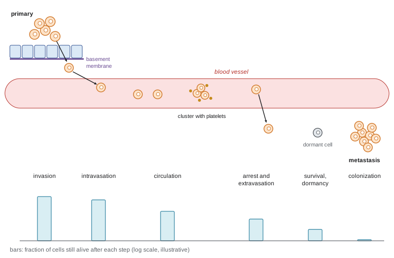
Metastasis is often described as a sequence of steps, each of which a cancer cell must survive. Cells must invade the surrounding tissue by breaking through the basement membrane and moving through the stroma; intravasate, crossing the wall of a blood or lymphatic vessel to enter circulation; survive in the circulation, where they face shear forces, loss of anchorage and immune attack; arrest in the capillaries of a distant organ and extravasate, crossing back out of the vessel; survive in the new tissue, often for long periods as single cells or tiny clusters; and finally colonize the site, proliferating into a macroscopic metastasis, which requires adapting to a foreign microenvironment. Colonization is usually the rate-limiting step.
Invasion and epithelial–mesenchymal transition
Epithelial cells are held together by adherens junctions built on E-cadherin and anchored to a basement membrane. To invade, carcinoma cells must loosen these attachments, become motile and degrade or squeeze through the matrix. A developmental program called the epithelial–mesenchymal transition (EMT) provides much of this machinery. During embryonic development EMT lets cells leave the epithelium during gastrulation and neural crest migration, as BIOL-360 described. In cancer, transcription factors such as SNAIL, SLUG, TWIST and ZEB1 repress E-cadherin and switch on mesenchymal genes such as vimentin and N-cadherin, giving cells a motile, fibroblast-like character. Loss of E-cadherin is a feature of lobular breast cancer and of diffuse gastric cancer, and germline CDH1 mutations cause hereditary diffuse gastric cancer.
EMT is better understood as a spectrum than a switch. Many invading cells occupy hybrid states that retain some epithelial features, and such hybrid cells may be the most effective at metastasis, because they can move yet still form clusters. The reverse process, mesenchymal–epithelial transition, may help disseminated cells re-establish epithelial growth at distant sites. EMT programs also link to stem-like properties and drug resistance, a connection with the cancer stem cell ideas in lesson 9. Not all invasion requires full EMT: cells can invade collectively as strands or sheets, led by specialized leader cells, and some switch between mesenchymal and amoeboid modes of movement, squeezing through gaps without degrading the matrix.
Invasion needs proteases. Matrix metalloproteinases (MMPs), many secreted by stromal cells, degrade collagen and other matrix proteins, and invasive cells form invadopodia, actin-rich protrusions that concentrate proteolytic activity. Broad MMP inhibitors were tested in large clinical trials in the 1990s and failed, partly because they were given to patients with advanced disease after invasion had already happened, and partly because some MMPs restrain tumours. The episode is often cited as a lesson in translating biology into drugs.
Through the circulation
Circulating tumour cells (CTCs) can be found in the blood of many patients with metastatic carcinomas, usually at very low numbers, from none to hundreds per millilitre among billions of blood cells. Their count predicts outcome in breast, prostate and colorectal cancer. Most CTCs die quickly, from shear stress, loss of attachment (a death called anoikis) or natural killer cells. Clusters of CTCs, though rarer than single cells, are far more likely to seed metastases, and platelets coating CTCs shield them from immune attack and help them stick to vessel walls. Experiments in mice, in which labelled cells are injected into the circulation, show that the great majority of cells reaching an organ do not grow into metastases; estimates often quoted suggest that fewer than one in a thousand, and sometimes fewer than one in ten thousand, cells entering the blood will eventually form a macroscopic tumour.
# Metastatic inefficiency: chaining step-by-step survival probabilities (illustrative values from experimental ranges).
import numpy as np
steps = [('survive the circulation', 0.10), ('arrest and extravasate', 0.30),
         ('survive as single cells in the organ', 0.05), ('colonize: grow into a micrometastasis', 0.10),
         ('progress to a macroscopic metastasis', 0.20)]
p = 1.0
for name, s in steps:
    p *= s
    print(f"{name:42s} step {s:4.2f}  cumulative {p:.1e}")
shed_per_day = 1e5                      # cells shed into the blood per day by a large tumour (illustrative)
print(f"expected metastases founded per day: {shed_per_day * p:.1f}")
rng = np.random.default_rng(8)
days = 365
founded = rng.poisson(shed_per_day * p, days).sum()
print(f"over one year of shedding (simulated): about {founded} founding events")survive the circulation step 0.10 cumulative 1.0e-01 arrest and extravasate step 0.30 cumulative 3.0e-02 survive as single cells in the organ step 0.05 cumulative 1.5e-03 colonize: grow into a micrometastasis step 0.10 cumulative 1.5e-04 progress to a macroscopic metastasis step 0.20 cumulative 3.0e-05 expected metastases founded per day: 3.0 over one year of shedding (simulated): about 1060 founding events
The numbers are illustrative but within the ranges reported in mouse experiments. They show two things at once. Metastasis is spectacularly inefficient for any one cell, and yet a tumour that sheds cells every day for months has a high chance of founding metastases, which is why removing a primary tumour early matters so much and why the absence of visible metastases at diagnosis does not guarantee there are none.
Detecting CTCs is technically demanding because they are so rare. The first system cleared by the US FDA, in 2004, captured cells carrying the epithelial marker EpCAM with magnetic beads and counted those that also stained for cytokeratins and lacked the white blood cell marker CD45. Counts of five or more CTCs per 7.5 millilitres of blood predicted shorter survival in metastatic breast cancer. Newer methods sort cells by size, deformability or a wider panel of markers, because cells that have undergone EMT may lose EpCAM and be missed. Sequencing single CTCs, or cell-free tumour DNA released into blood (lesson 10), offers a liquid biopsy that can track a cancer's evolution without repeated tissue sampling.
Seed and soil: organotropism
Metastases are not randomly distributed. Breast cancer spreads to bone, liver, lung and brain; prostate cancer overwhelmingly to bone; colorectal cancer first to the liver; uveal melanoma to the liver; and lung cancer to brain, bone, liver and adrenal glands. Paget's seed and soil hypothesis, based on autopsy records of breast cancer patients in 1889, proposed that cancer cells grow only in hospitable organs. In the 1920s James Ewing argued instead that anatomy explains the pattern: cells lodge in the first capillary bed downstream. Both are partly right. Colorectal cancer spreads to the liver largely because intestinal blood drains there through the portal vein, but prostate cancer's preference for bone, and breast cancer's subtypes' different preferences, reflect biology.
Molecular studies by Joan Massagué and others identified gene sets that mediate organ-specific metastasis in breast cancer models, and work by David Lyden showed that tumours prepare distant sites before cells arrive, creating pre-metastatic niches through secreted factors and extracellular vesicles, including exosomes whose surface integrins help determine which organs they target. Bone metastases illustrate the soil at work. Breast cancer cells in bone secrete parathyroid hormone-related protein, which stimulates osteoblasts to produce RANKL, activating osteoclasts that dissolve bone. Bone breakdown releases growth factors such as TGF-β stored in the matrix, which stimulate more tumour growth, a vicious cycle. Drugs that block osteoclasts, such as bisphosphonates and the RANKL antibody denosumab, reduce fractures and other skeletal complications in patients with bone metastases.
| Primary cancer | Common metastatic sites | Notes |
|---|---|---|
| Breast | Bone, liver, lung, brain | Pattern varies by subtype; HER2-positive and triple-negative more often brain |
| Prostate | Bone (mostly bone-forming lesions) | PSMA-PET improves detection |
| Colorectal | Liver, lung, peritoneum | Portal venous drainage favours the liver; some liver metastases are resectable |
| Lung | Brain, bone, liver, adrenal glands | Brain metastases common in EGFR- and ALK-driven disease |
| Melanoma (skin) | Lymph nodes, lung, liver, brain | Uveal melanoma spreads mainly to the liver |
| Pancreas | Liver, peritoneum, lung | Often metastatic at diagnosis |
Dormancy and late relapse
Some cancers recur years or decades after apparently successful treatment. Estrogen receptor-positive breast cancer is the classic example: a substantial share of recurrences happen more than five years after diagnosis, sometimes after twenty years. Disseminated tumour cells can be found in the bone marrow of patients with no visible metastases, and many apparently survive in a non-dividing or slowly dividing state. Dormancy can operate at the level of single cells, arrested by signals from their niche, such as those from bone marrow and blood vessel walls, or at the level of tiny micrometastases in which proliferation is balanced by death through immune attack or lack of blood supply. What awakens dormant cells is not well understood; ageing, inflammation and changes in the niche have all been implicated. Extended endocrine therapy, for up to ten years in some patients, is based partly on the long tail of late recurrence.
When does metastasis begin?
The textbook model was linear: a primary tumour grows, acquires further mutations and, late in its evolution, spawns metastases. Genomic comparisons of primary tumours and metastases from the same patient have complicated the picture. In some cancers, metastatic clones share most of their mutations with a dominant clone in the primary, consistent with late spread. In others, such as some breast and colorectal cancers, metastases appear to have separated early, while the primary tumour was still small, and evolved in parallel. Spread can also occur from one metastasis to another. These findings matter for how we interpret the absence of detectable metastases and why systemic therapy after surgery, called adjuvant therapy, reduces recurrence in many cancers: it targets cells that have already left.
Treating metastatic disease
Treatment of metastatic disease is usually systemic: chemotherapy, hormonal therapy, targeted drugs and immunotherapy. For a limited number of metastases, a state called oligometastatic disease, surgery or stereotactic radiotherapy to each site can sometimes give long-term control, and resection of colorectal liver metastases cures a meaningful fraction of selected patients. Brain metastases pose special problems because the blood–brain barrier excludes many drugs, though newer targeted agents such as osimertinib for EGFR-mutant lung cancer and tucatinib for HER2-positive breast cancer penetrate better. Lesson 11 returns to systemic therapy and resistance.
For Maya, mediastinal lymph node involvement means her cancer has begun the cascade: cells have invaded lymphatics and established themselves in a regional node, though not in distant organs. The risk that undetectable cells have already spread further is the reason her treatment includes systemic therapy rather than local treatment alone.
Worked problem
Reasoning about metastatic inefficiency
A tumour sheds 1 x 10 to the power 5 cells into the blood each day. Suppose 10 per cent survive the circulation, 30 per cent of those extravasate, 5 per cent of those survive in the organ, 10 per cent of survivors form micrometastases and 20 per cent of micrometastases progress. How many macroscopic metastases are founded per day, and per 100 days?
Show solution
- Overall probability per shed cell = 0.10 x 0.30 x 0.05 x 0.10 x 0.20 = 3 x 10 to the power -5.
- Per day: 1 x 10 to the power 5 x 3 x 10 to the power -5 = 3 founding events.
- Per 100 days: about 300.
- Even if each step were ten times less efficient in some patients, months of shedding could still seed metastases, which argues for early treatment.
Answer: About 3 per day, or about 300 per 100 days, under these illustrative assumptions.
Common mistakes
MistakeAssuming most circulating tumour cells form metastases.
InsteadMetastasis is highly inefficient; almost all CTCs die or never grow.
MistakeTreating EMT as an all-or-nothing switch.
InsteadEMT is a spectrum; hybrid states may be the most metastatic.
MistakeExplaining all metastatic patterns by blood flow alone.
InsteadAnatomy matters, as for colorectal cancer and the liver, but organ biology and pre-metastatic niches also determine where cells grow.
MistakeAssuming metastasis always happens late.
InsteadGenomic studies show that in some cancers metastases separate early and evolve in parallel.
MistakeConcluding a patient is cured because no metastases are visible.
InsteadDormant disseminated cells can cause recurrence years later; adjuvant therapy targets them.
Summary and key terms
- Metastasis causes most cancer deaths and proceeds through invasion, intravasation, circulation, extravasation, survival and colonization.
- EMT, driven by SNAIL, TWIST and ZEB1, loosens epithelial adhesion and promotes motility; hybrid states are common.
- Circulating tumour cells are rare and mostly die; clusters and platelet-coated cells are more likely to seed metastases.
- Metastasis is extremely inefficient per cell, yet sustained shedding makes metastasis likely.
- Organotropism reflects both anatomy and the seed and soil, including pre-metastatic niches and organ-specific programs.
- Disseminated cells can lie dormant for years, explaining late relapse, especially in estrogen receptor-positive breast cancer.
- Metastasis can begin early in some cancers, which supports the use of adjuvant systemic therapy.
- metastatic cascade
- The sequence of steps by which cancer cells spread from a primary tumour to form distant tumours.
- intravasation
- Entry of cancer cells into blood or lymphatic vessels.
- extravasation
- Exit of cancer cells from vessels into a distant tissue.
- epithelial–mesenchymal transition
- A program by which epithelial cells lose adhesion and gain motile, mesenchymal traits.
- E-cadherin
- The adhesion protein of epithelial adherens junctions, often lost during invasion.
- anoikis
- Cell death triggered by loss of attachment to the matrix.
- circulating tumour cell
- A cancer cell found in the bloodstream.
- organotropism
- The tendency of a cancer to metastasize to particular organs.
- pre-metastatic niche
- A site in a distant organ prepared by tumour-derived factors before cancer cells arrive.
- dormancy
- A state in which disseminated cancer cells survive without detectable growth.
- adjuvant therapy
- Systemic treatment given after removal of the primary tumour to reduce recurrence.
Practice problems
Work each problem on paper first, then reveal the answer to check.
List the main steps of the metastatic cascade.
Show answer
Invasion, intravasation, survival in circulation, arrest and extravasation, survival in the new organ, colonization.
Which step of the cascade is usually rate-limiting?
Show answer
Colonization, growth into a macroscopic metastasis.
Name two transcription factors that drive EMT.
Show answer
SNAIL, SLUG, TWIST or ZEB1.
Why did broad MMP inhibitors fail in trials?
Show answer
They were given to patients with advanced disease, and some MMPs restrain tumours.
Why are CTC clusters more dangerous than single CTCs?
Show answer
They survive better and seed metastases far more efficiently.
Why does colorectal cancer spread first to the liver?
Show answer
Intestinal blood drains through the portal vein to the liver.
Describe the vicious cycle of breast cancer bone metastasis.
Show answer
Tumour PTHrP increases RANKL and osteoclast activity; bone breakdown releases TGF-β, stimulating more tumour growth.
Why is extended endocrine therapy used in some breast cancers?
Show answer
Estrogen receptor-positive breast cancer can recur many years later from dormant cells.
Lesson quiz · 5 questions
Score 60% or more to complete this lesson.
Create a free account to save your progress, take quizzes and earn a certificate.