
BIOL-450 · Lesson 10 of 12 · 28 min
Diagnosis, staging and genomic profiling
Everything in the first nine lessons becomes practical at the moment a cancer is found. A physician must decide whether a shadow on a scan is cancer, what kind it is, how far it has spread and which of the many possible treatments the tumour's biology makes worthwhile. This lesson follows that sequence: screening to find cancers before symptoms appear, the biopsy and pathology that establish a diagnosis, staging, and the genomic and protein tests that now guide treatment for most advanced cancers, including the sequencing that found Maya's KRAS G12C mutation and the mismatch repair testing in Daniel's family.
Screening: finding cancer early
Screening tests people without symptoms, aiming to find cancers, or precancers, at a stage when treatment is more effective. The principles were set out for the World Health Organization by James Wilson and Gunnar Jungner in 1968: the disease should be an important health problem with a recognizable early stage, there should be an acceptable test and an effective treatment for early disease, and the benefits should outweigh the harms. Proving benefit is harder than it looks, because three biases make screening appear better than it is. Lead-time bias: diagnosing a cancer earlier lengthens survival measured from diagnosis even if death occurs at the same time. Length bias: slowly growing tumours spend longer in the detectable pre-symptomatic window, so screening preferentially finds the less dangerous ones. Overdiagnosis: some screen-detected cancers would never have caused symptoms in the person's lifetime, yet are treated, with all the harms of treatment. Only randomized trials that measure deaths from the cancer, not survival time after diagnosis, avoid these biases.
# Lead-time bias: screening that finds a cancer earlier but does not change the date of death still "improves" survival.
import numpy as np
rng = np.random.default_rng(10)
n = 10000
onset = rng.uniform(50, 70, n)                       # age at which the tumour becomes detectable by screening
preclinical = rng.gamma(4, 0.75, n)                  # years from screen-detectable to symptoms
death = onset + preclinical + rng.gamma(2, 1.5, n)   # date of death, the same with or without screening
dx_symptoms = onset + preclinical
dx_screen = onset + rng.uniform(0, 1, n) * preclinical   # screening finds it partway through the preclinical phase
for label, dx in (('diagnosed at symptoms', dx_symptoms), ('diagnosed by screening', dx_screen)):
    surv = death - dx
    print(f"{label:24s} mean survival {surv.mean():4.1f} y, alive 5 y after diagnosis {np.mean(surv > 5):5.1%}")
print(f"mean age at death in both groups: {death.mean():.1f} y (unchanged)")diagnosed at symptoms mean survival 3.0 y, alive 5 y after diagnosis 15.3% diagnosed by screening mean survival 4.5 y, alive 5 y after diagnosis 35.3% mean age at death in both groups: 66.0 y (unchanged)
Breast, cervical, colorectal and, more recently, lung cancer screening have all been shown in randomized trials or strong observational evidence to reduce deaths. Cervical screening has shifted from cytology (the Pap test) to testing for high-risk HPV, which is more sensitive and allows longer intervals; self-collected vaginal samples are increasingly offered. Colorectal screening uses the faecal immunochemical test (FIT), which detects human hemoglobin, followed by colonoscopy if positive, or colonoscopy directly; removing adenomas during colonoscopy prevents cancers as well as finding them. Lung cancer screening with low-dose computed tomography (CT) reduced lung cancer deaths by 20 per cent compared with chest X-rays in the US National Lung Screening Trial, reported in 2011, and by about a quarter in men in the European NELSON trial. Prostate-specific antigen (PSA) testing is the most debated: it reduces prostate cancer deaths modestly but carries a high rate of overdiagnosis, which active surveillance of low-risk cancers and MRI before biopsy have reduced.
In Canada, screening programs are run by the provinces and territories, guided by the Canadian Task Force on Preventive Health Care and by provincial agencies. The Task Force's 2016 guideline recommends annual low-dose CT for up to three consecutive years in adults aged 55 to 74 with at least 30 pack-years of smoking who smoke or quit within the past 15 years, and some provinces, such as Ontario, select people with a risk-prediction model instead of pack-years alone. Maya, at 58 with about 20 pack-years who quit 15 years before her diagnosis, would not have met the 30 pack-year criterion, though a risk-model program might have included her. Organized colorectal programs offer FIT every one or two years from age 50, and several provinces, including British Columbia and Ontario, let people self-refer for mammography from age 40.
Newer approaches look for cancer signals in blood. Multi-cancer early detection tests analyse cell-free DNA for methylation patterns that indicate both the presence of cancer and its probable tissue of origin. They can detect many cancer types for which no screening exists, but their sensitivity for stage I cancers is low, false positives lead to investigations, and whether they reduce cancer deaths has not yet been established in randomized trials.
Diagnosis: biopsy and pathology
Imaging can raise suspicion, but cancer is diagnosed by examining tissue. A sample is obtained by needle biopsy, endoscopic biopsy, surgical excision or, for blood cancers, from blood and bone marrow. For a lung nodule like Maya's, options include CT-guided needle biopsy and bronchoscopy; endobronchial ultrasound lets a bronchoscopist sample mediastinal lymph nodes through the airway wall, which in Maya's case provided both the diagnosis and the staging. The pathologist examines thin sections stained with hematoxylin and eosin to identify the tumour type from its architecture and cytology, and to assign a grade, a measure of how abnormal the cells look and how fast they divide.
Immunohistochemistry (IHC) uses antibodies to show which proteins tumour cells express. It identifies a tumour's lineage when the appearance is ambiguous, for example TTF-1 and napsin A in lung adenocarcinoma, p40 in squamous cell carcinoma, CDX2 in intestinal tumours and specific keratin patterns; it reports therapeutic targets such as estrogen and progesterone receptors and HER2 in breast cancer; and it assesses mismatch repair proteins and PD-L1. HER2 status illustrates how tests are calibrated against treatments: a tumour scoring 3+ by IHC, or 2+ with amplification on in situ hybridization, is HER2-positive and treated with trastuzumab and related drugs, and since 2022 tumours scoring 1+ or 2+ without amplification, called HER2-low, can also benefit from the antibody–drug conjugate trastuzumab deruxtecan.
Staging: how far has it spread?
Staging describes a cancer's anatomical extent, which is the strongest single predictor of outcome for most solid tumours and the foundation of treatment choice. The TNM system, maintained by the Union for International Cancer Control and the American Joint Committee on Cancer, scores T for the size and local extent of the primary tumour, N for spread to regional lymph nodes and M for distant metastasis. Combinations are grouped into stages I to IV. Stage I usually means a small tumour confined to its organ, stages II and III indicate larger tumours or regional node involvement, and stage IV means distant metastasis. Clinical staging uses examination, imaging and biopsies before treatment; pathological staging uses the surgical specimen. Staging rules are revised periodically as evidence accumulates; the ninth edition of the lung cancer TNM classification, which subdivides mediastinal nodal and metastatic categories more finely, came into use in 2025.
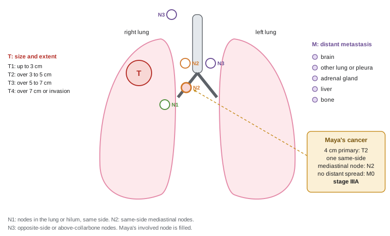
Maya's tumour was 4 centimetres across with one involved mediastinal node on the same side and no distant spread on FDG-PET-CT and brain MRI, a combination placing her in stage III. The details matter for treatment, which lesson 11 describes: a multidisciplinary tumour board judged that her disease was best treated without surgery.
From genes to treatment: molecular profiling
For many cancers, the next step after diagnosis and staging is molecular profiling, which looks for alterations that predict response to specific drugs. In non-small cell lung cancer, guidelines in both Canada and the United States call for testing every advanced non-squamous tumour for a set of actionable alterations, because each has a matched targeted therapy.
| Alteration in lung adenocarcinoma | Approximate frequency | Matched therapy (examples) |
|---|---|---|
| EGFR activating mutations (exon 19 deletion, L858R) | About 15 per cent in North America; about 40 to 50 per cent in East Asia | Osimertinib |
| KRAS G12C | About 12 per cent, mostly in people who smoked | Sotorasib, adagrasib |
| ALK fusions | About 3 to 5 per cent | Alectinib, lorlatinib |
| ROS1 fusions | About 1 to 2 per cent | Crizotinib, entrectinib, repotrectinib |
| BRAF V600E | About 1 to 2 per cent | Dabrafenib with trametinib |
| MET exon 14 skipping | About 3 per cent | Capmatinib, tepotinib |
| RET fusions | About 1 to 2 per cent | Selpercatinib, pralsetinib |
| ERBB2 (HER2) mutations | About 2 to 3 per cent | Trastuzumab deruxtecan |
| NTRK fusions | Under 1 per cent | Larotrectinib, entrectinib |
Testing one gene at a time would use up scarce tissue, so most laboratories now use next-generation sequencing (NGS) panels that read hundreds of cancer genes at once, often from DNA and RNA together, because fusions are detected more reliably in RNA. Sequencing is usually done on the tumour and, ideally, on a matched normal sample such as blood, so that inherited variants can be distinguished from somatic ones. A typical report lists each variant with its variant allele fraction (VAF), the proportion of sequencing reads carrying it, along with copy number changes, fusions, the tumour mutational burden (TMB) and microsatellite instability (MSI) status. Knowledge bases such as OncoKB and the ESMO Scale for Clinical Actionability of molecular Targets (ESCAT) rank alterations by the strength of evidence that they predict benefit from a drug.
Interpreting VAF requires knowing the tumour's purity, the fraction of cells in the sample that are cancer cells, and its copy number at that locus. In a sample that is 60 per cent tumour, a heterozygous mutation present in every cancer cell, in a region with two copies, will appear in about 30 per cent of reads, because the normal cells contribute only wild-type copies. A VAF much lower than expected suggests a subclonal mutation, and a VAF much higher suggests that the mutant allele has been duplicated or the wild-type allele lost, as often happens for TP53.
# From variant allele fraction (VAF) to cancer cell fraction (CCF), allowing for purity and copy number.
def ccf(vaf, purity, tumour_cn=2, normal_cn=2, mutant_copies=1):
    # expected VAF for a clonal mutation = purity*m / (purity*CN_t + (1-purity)*CN_n); invert for the fraction of cancer cells
    total = purity * tumour_cn + (1 - purity) * normal_cn
    return vaf * total / (purity * mutant_copies)
purity = 0.60
calls = [('KRAS G12C', 0.31, 2, 1), ('TP53 R273L', 0.58, 2, 2),   # TP53: wild-type allele lost, mutant duplicated
         ('STK11 Q37*', 0.29, 2, 1), ('PIK3CA E545K', 0.07, 2, 1)]
for gene, vaf, cn, m in calls:
    c = ccf(vaf, purity, cn, 2, m)
    status = 'clonal (truncal)' if c > 0.8 else 'subclonal'
print(f"{gene:13s} VAF {vaf:4.2f} copies {cn}, mutant copies {m} -> CCF {min(c, 1.0):4.2f} {status}")KRAS G12C VAF 0.31 copies 2, mutant copies 1 -> CCF 1.00 clonal (truncal) TP53 R273L VAF 0.58 copies 2, mutant copies 2 -> CCF 0.97 clonal (truncal) STK11 Q37* VAF 0.29 copies 2, mutant copies 1 -> CCF 0.97 clonal (truncal) PIK3CA E545K VAF 0.07 copies 2, mutant copies 1 -> CCF 0.23 subclonal
Maya's report, in this illustrative version, lists KRAS G12C at a VAF consistent with every tumour cell carrying it, a TP53 mutation with loss of the other allele, and a small subclonal PIK3CA mutation. The PIK3CA subclone is unlikely to be treated directly, but it is worth noting, because PI3K pathway activation is one of the routes by which tumours escape KRAS inhibitors. Her tumour's TMB was moderately high, as is typical of smoking-related lung cancers, and PD-L1 IHC showed staining on 60 per cent of tumour cells. Both are relevant to immunotherapy, discussed in lesson 12. Some panels also flag a possible germline variant; when a pathogenic variant appears in a gene such as BRCA2 at a VAF near 50 per cent in both tumour and normal samples, the patient should be referred for genetic counselling and confirmatory testing.
Tumour-agnostic biomarkers and Daniel's family
A few biomarkers predict benefit regardless of where the cancer started. In 2017 the FDA granted the first approval based on a biomarker rather than a tumour site: pembrolizumab, a PD-1 antibody, for any solid tumour with high microsatellite instability or mismatch repair deficiency (MSI-H or dMMR) that had progressed on standard treatment. Approvals followed for NTRK fusions (larotrectinib in 2018), for high TMB of at least 10 mutations per megabase, for BRAF V600E with dabrafenib and trametinib, and for HER2-positive solid tumours with trastuzumab deruxtecan. Health Canada and other regulators have taken varied approaches to tumour-agnostic indications. The logic connects directly to earlier lessons: tumours with defective mismatch repair accumulate thousands of mutations, many producing abnormal peptides that T cells can recognize once the PD-1 brake is released.
Daniel's colorectal cancer, introduced in lesson 3, was tested by IHC as part of universal mismatch repair screening, and it showed loss of MSH2 and MSH6 proteins. Unlike sporadic MLH1 loss, loss of MSH2 is not usually caused by promoter methylation, so germline testing followed and found the MSH2 variant that confirmed Lynch syndrome. The same result has a second meaning: if his cancer recurs or spreads, its dMMR status predicts a strong chance of benefit from immune checkpoint inhibitors, and for some localized dMMR rectal cancers, PD-1 blockade alone has produced complete responses that let patients avoid surgery and radiation, a striking result from a small trial reported in 2022 and still being confirmed in larger studies.
Liquid biopsy
Tumours shed fragments of DNA into the blood, called circulating tumour DNA (ctDNA), a fraction of the total cell-free DNA that comes mostly from dying blood cells. Sequencing ctDNA offers a liquid biopsy: a blood test that can find actionable mutations when tissue is insufficient, track response, and detect resistance mutations as they emerge, without repeated invasive biopsies. Several ctDNA panels are approved by the FDA and used in Canada, often run alongside tissue testing at diagnosis to shorten the time to treatment.
The main limitation is sensitivity. The fraction of cell-free DNA derived from the tumour can be below 0.1 per cent, especially in early-stage cancers and in tumours, such as some brain cancers, that shed little DNA. Detecting a mutation at that level requires deep sequencing and error-correction methods, and a negative result does not exclude a mutation. A second pitfall is clonal hematopoiesis, from lesson 9: mutations in blood cells, common in older people, appear in cell-free DNA and can be mistaken for tumour mutations unless white blood cells are sequenced too. The most promising emerging use is detection of minimal residual disease: after surgery for colon cancer, patients with detectable ctDNA have a much higher risk of recurrence, and trials are testing whether ctDNA results can guide who needs adjuvant chemotherapy.
Worked problem
Will a liquid biopsy detect the mutation?
A ctDNA assay sequences 5,000 tumour-informative DNA molecules covering KRAS codon 12 in a blood sample. The tumour fraction of cell-free DNA is 0.2 per cent, and the mutation is clonal and heterozygous. The assay calls a mutation if at least 3 mutant molecules are seen. What is the chance of detection?
Show solution
- Expected fraction of molecules carrying the mutation = tumour fraction x 0.5 = 0.001.
- Expected mutant molecules = 5,000 x 0.001 = 5.
- With a Poisson mean of 5, the probability of fewer than 3 mutant molecules is P(0) + P(1) + P(2) = 0.0067 + 0.0337 + 0.0842 = 0.125.
- So the chance of detection is about 1 - 0.125 = 0.875; at a tumour fraction of 0.05 per cent the expected count falls to 1.25 and detection becomes unlikely.
Answer: About 88 per cent; sensitivity depends on how many molecules are sampled and on the tumour fraction, not only on the sequencing technology.
Common mistakes
MistakeJudging a screening test by survival after diagnosis.
InsteadLead-time and length bias inflate survival; only deaths from the cancer in randomized trials show benefit.
MistakeAssuming every screen-detected cancer needed treatment.
InsteadOverdiagnosis is common for some cancers, notably prostate and thyroid.
MistakeReading a VAF of 30 per cent as a subclonal mutation.
InsteadIn a sample with 60 per cent tumour cells, a clonal heterozygous mutation gives a VAF near 30 per cent.
MistakeTreating a negative ctDNA result as proof there is no mutation.
InsteadLow tumour shedding can make a liquid biopsy falsely negative; tissue testing may still be needed.
MistakeIgnoring germline findings on a tumour panel.
InsteadA variant at about 50 per cent VAF in tumour and normal samples may be inherited and should lead to genetic counselling.
Summary and key terms
- Screening finds cancers earlier, but lead-time bias, length bias and overdiagnosis mean benefit must be shown by reductions in deaths.
- Breast, cervical, colorectal and lung cancer screening reduce deaths; eligibility rules differ between Canada and the United States.
- Diagnosis requires tissue; histology, grade and immunohistochemistry define tumour type and targets such as HER2 and hormone receptors.
- TNM staging describes tumour size, nodal spread and metastasis, and drives treatment choice.
- Next-generation sequencing panels detect actionable alterations; variant allele fraction must be read in light of purity and copy number.
- Tumour-agnostic biomarkers include MSI-H/dMMR, high TMB, NTRK fusions, BRAF V600E and HER2 positivity.
- Liquid biopsy of ctDNA can find mutations, track resistance and detect residual disease, but sensitivity is limited at low tumour fractions.
- lead-time bias
- Apparent survival gain from earlier diagnosis without any delay in death.
- length bias
- The tendency of screening to detect slower-growing, less dangerous tumours.
- overdiagnosis
- Diagnosis of a cancer that would never have caused symptoms or death.
- immunohistochemistry
- Staining tissue with antibodies to show which proteins cells express.
- TNM staging
- A system describing tumour size and extent, lymph node spread and distant metastasis.
- next-generation sequencing panel
- A test that sequences many cancer genes at once.
- variant allele fraction
- The proportion of sequencing reads carrying a variant.
- tumour purity
- The fraction of cells in a sample that are cancer cells.
- tumour mutational burden
- The number of somatic mutations per megabase of sequenced DNA.
- circulating tumour DNA
- DNA fragments released by tumour cells into the blood.
- minimal residual disease
- Cancer cells remaining after treatment at levels below clinical detection.
Practice problems
Work each problem on paper first, then reveal the answer to check.
Explain length bias in one sentence.
Show answer
Slowly growing cancers spend longer in the detectable pre-symptomatic period, so screening finds them preferentially.
What did the National Lung Screening Trial show?
Show answer
Low-dose CT reduced lung cancer deaths by 20 per cent compared with chest X-ray.
Which IHC markers support lung adenocarcinoma and squamous cell carcinoma?
Show answer
TTF-1 and napsin A for adenocarcinoma; p40 for squamous cell carcinoma.
What do T, N and M stand for?
Show answer
Primary tumour size and extent, regional lymph nodes, distant metastasis.
In a sample of 50 per cent purity, what VAF is expected for a clonal heterozygous mutation in a diploid region?
Show answer
About 25 per cent.
Name three tumour-agnostic biomarkers.
Show answer
MSI-H/dMMR, high TMB, NTRK fusions, BRAF V600E or HER2 positivity.
Why is clonal hematopoiesis a problem for liquid biopsy?
Show answer
Blood cell mutations in cell-free DNA can be mistaken for tumour mutations.
Why did Daniel have germline testing after his IHC result?
Show answer
Loss of MSH2 and MSH6 is not usually due to methylation, suggesting Lynch syndrome.
Lesson quiz · 5 questions
Score 60% or more to complete this lesson.
Create a free account to save your progress, take quizzes and earn a certificate.