
BIOL-450 · Lesson 3 of 12 · 25 min
Tumour suppressors and the two-hit hypothesis
If oncogenes are the accelerators of cancer, tumour suppressor genes are the brakes. They encode proteins that restrain the cell cycle, repair or report DNA damage, trigger death in damaged cells, or hold cells in place within a tissue. Cancer requires losing these restraints, and unlike oncogenes, tumour suppressors usually act recessively at the level of the cell: both copies must be inactivated before the brake fails. This lesson explains how that principle was discovered, how it accounts for inherited cancer syndromes, and how the best known tumour suppressors, RB, p53, BRCA1 and BRCA2, PTEN and APC, keep cells in check.
Evidence that suppressor genes exist
Two lines of evidence pointed to genes whose loss causes cancer. In the 1960s and 1970s, Henry Harris fused cancer cells with normal cells and found that the hybrids often did not form tumours in animals, as though the normal cell supplied something the cancer cell had lost. Later, families with dominantly inherited cancer syndromes suggested that a single inherited defect could predispose to cancer, yet tumours arose only in some cells and only after delay, implying that something else had to happen in each tumour.
Knudson's two-hit hypothesis
Retinoblastoma is a rare cancer of the developing retina in young children. In 1971, Alfred Knudson compared children with retinoblastoma in both eyes, often with several tumours and a family history, to children with a single tumour in one eye and no family history. The bilateral cases were diagnosed earlier. Knudson showed that the age at diagnosis in bilateral cases fitted a model in which one more random event was needed, while unilateral sporadic cases fitted a model needing two events. He proposed that retinoblastoma requires two mutational hits in the same retinal cell. In hereditary cases, one hit is inherited and present in every cell, so only one more somatic event is needed in any of the millions of retinal cells, making tumours likely, multiple and early. In sporadic cases, both hits must occur somatically in the same cell, which is rare, so tumours are single and later.
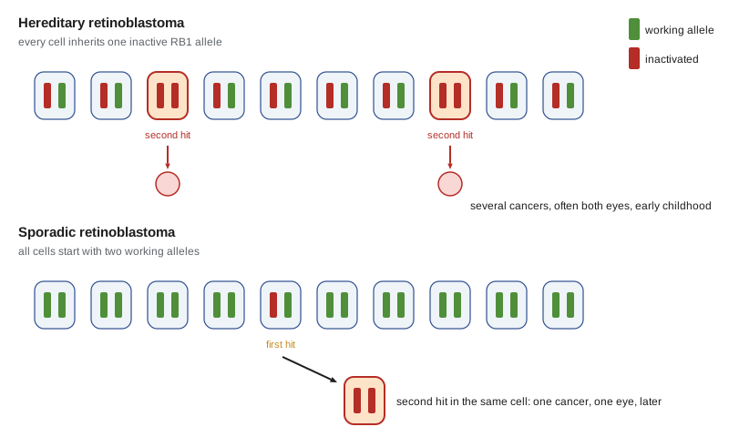
The gene, RB1 on chromosome 13q14, was cloned in 1986. Its product, the retinoblastoma protein RB, was the first tumour suppressor characterized. The two-hit hypothesis became the defining property of tumour suppressor genes and explained a puzzle: an inherited cancer syndrome appears dominant in families, because inheriting one bad allele makes cancer highly likely, yet the gene behaves recessively in cells, because both alleles must be lost.
# Knudson's model: fraction of children diagnosed by age t with one hit needed (hereditary) or two (sporadic).
import numpy as np
cells = 3e5          # susceptible retinal precursor cells (illustrative)
mu = 1.7e-7          # rate of inactivating an RB1 allele, per allele per month (illustrative)
months = np.array([6, 12, 24, 36, 48, 60])
# hereditary: one more hit in any cell. P(no tumour) = exp(-cells * mu * t)
p_her = 1 - np.exp(-cells * mu * months)
# sporadic: two sequential hits in the same cell; expected tumours ~ cells * (2*mu*t)*(mu*t)/2 for small rates
lam_spor = cells * (mu * months) ** 2
p_spor = 1 - np.exp(-lam_spor)
for t, a, b in zip(months, p_her, p_spor):
    print(f"{t:2d} months: hereditary {a:5.1%}   sporadic {b:.5%}")
print(f"mean tumours per eye in hereditary cases by 60 months: {cells * mu * 60:.1f}")
print("log-log slope of sporadic risk vs age:", round(np.polyfit(np.log(months), np.log(lam_spor), 1)[0], 2))6 months: hereditary 26.4% sporadic 0.00003% 12 months: hereditary 45.8% sporadic 0.00012% 24 months: hereditary 70.6% sporadic 0.00050% 36 months: hereditary 84.1% sporadic 0.00112% 48 months: hereditary 91.4% sporadic 0.00200% 60 months: hereditary 95.3% sporadic 0.00312% mean tumours per eye in hereditary cases by 60 months: 3.1 log-log slope of sporadic risk vs age: 2.0
The simulated curves reproduce Knudson's observation. With one hit remaining, most carriers develop a tumour within a few years, with about three tumours per eye on average, while with two hits needed the risk is tiny and rises with the square of age, the slope of 2 on a log-log plot. The parameter values are illustrative, chosen to give realistic proportions, not measured rates.
Loss of heterozygosity
How is the second hit delivered? Sometimes by an independent point mutation, but often by loss of a large chromosome region containing the normal allele, through deletion, mitotic recombination or loss of a whole chromosome followed by duplication of the remaining copy. Tumour DNA then shows loss of heterozygosity (LOH): markers near the gene that were heterozygous in the patient's normal tissue appear homozygous in the tumour. Mapping regions of recurrent LOH in tumours became a standard way to locate new tumour suppressor genes. Second hits can also be epigenetic: methylation of the promoter of the remaining allele silences it without changing its sequence, as happens to MLH1 in many sporadic colorectal cancers and to CDKN2A in many tumour types (lesson 9).
RB and the restriction point
RB controls the restriction point in late G1, the decision to commit to division covered in BIOL-214. In its hypophosphorylated form, RB binds E2F transcription factors and represses genes needed for S phase. Growth signals raise cyclin D levels; cyclin D with CDK4 or CDK6 phosphorylates RB, then cyclin E with CDK2 completes the job, releasing E2F and committing the cell to replicate its DNA. The RB pathway is disrupted in almost every cancer, though rarely by RB1 mutation itself outside retinoblastoma, osteosarcoma and small cell lung cancer. More often cyclin D is amplified, CDK4 is amplified or mutated, or the CDK inhibitor p16INK4a (encoded by CDKN2A) is deleted. Any of these frees E2F. That convergence explains why CDK4/6 inhibitors such as palbociclib work in hormone receptor-positive breast cancer, where the RB pathway is intact but driven hard by cyclin D, and do not work when RB itself is lost.
p53, guardian of the genome
TP53 is the most frequently mutated gene in human cancer, altered in roughly half of all tumours. Its protein, p53, is a transcription factor that responds to DNA damage, oncogene activation, hypoxia and other stresses. Normally p53 is kept at low levels by MDM2, a ubiquitin ligase that marks it for degradation; MDM2 is itself a p53 target gene, forming a negative feedback loop. Damage signalling through the kinases ATM, ATR, CHK1 and CHK2 phosphorylates p53 and MDM2, stabilizing p53. Activated p53 switches on p21, a CDK inhibitor that halts the cell cycle; genes for DNA repair; and pro-apoptotic genes such as PUMA, NOXA and BAX. Depending on the strength and duration of the signal and the cell type, the result is pause and repair, permanent arrest in senescence, or death. David Lane called p53 the guardian of the genome.
Unlike most tumour suppressors, TP53 is usually inactivated by missense mutations clustered in the DNA-binding domain, at hotspots such as R175, R248 and R273. Many mutant p53 proteins act as dominant negatives, poisoning the tetramer formed with any remaining normal p53, and some gain new oncogenic functions. Other tumours inactivate p53 indirectly, by amplifying MDM2 or by losing p14ARF, which normally restrains MDM2 in response to oncogene signalling. Viruses target p53 too: the HPV E6 protein marks p53 for degradation, a story for lesson 9. Germline TP53 mutations cause Li–Fraumeni syndrome, with a very high lifetime risk of sarcomas, breast cancer, brain tumours, adrenocortical carcinoma and leukemia, often in childhood or early adulthood.
BRCA1, BRCA2 and inherited breast and ovarian cancer
BRCA1 and BRCA2 are tumour suppressors needed for homologous recombination, the error-free repair of DNA double-strand breaks using the sister chromatid as a template. Women who inherit a pathogenic variant in either gene have a high lifetime risk of breast cancer, roughly 55 to 72 per cent for BRCA1 and 45 to 69 per cent for BRCA2 in large cohort studies, and an elevated risk of ovarian cancer; men carrying BRCA2 variants have higher risks of breast and prostate cancer, and both genes raise pancreatic cancer risk. Tumours in carriers show loss of the remaining normal allele, just as the two-hit model predicts. Loss of homologous recombination leaves cells dependent on other, more error-prone repair, which is both a cause of genomic chaos and a vulnerability that PARP inhibitors exploit, the subject of lesson 4.
Gatekeepers and caretakers
Kenneth Kinzler and Bert Vogelstein distinguished two kinds of tumour suppressor. Gatekeepers directly control cell proliferation or death in a tissue; losing them lets a clone expand. APC is the classic gatekeeper of the colon. Its protein is part of the destruction complex that degrades β-catenin; without APC, β-catenin accumulates, enters the nucleus and drives Wnt target genes, keeping cells in a proliferative, crypt-base state. Germline APC mutations cause familial adenomatous polyposis, with hundreds to thousands of colonic polyps and near-certain colorectal cancer without surgery, and somatic APC mutations start most sporadic colorectal cancers. Caretakers, by contrast, maintain genome integrity; losing them does not directly cause growth but raises the mutation rate, so that gatekeeper and oncogene mutations accumulate faster. BRCA1, BRCA2 and the mismatch repair genes are caretakers.
The colorectal cancer model of Eric Fearon and Vogelstein, proposed in 1990, combined these ideas: APC loss initiates a small adenoma, KRAS mutation drives growth of a larger adenoma, and loss of TP53 and of chromosome 18q accompanies the transition to carcinoma. The order varies among tumours, but the model made multistep carcinogenesis concrete and remains a teaching staple.
Haploinsufficiency and the limits of two hits
Not every tumour suppressor obeys the two-hit rule strictly. For some, losing one copy reduces protein enough to give a growth advantage, a situation called haploinsufficiency. Examples include PTEN, in which a modest drop in dosage measurably increases tumour risk in mice, and CDKN1B (p27). Others, such as some genes on large deleted regions, may act together, with partial loss of several neighbouring genes. The two-hit hypothesis remains the default, but dosage matters.
PTEN deserves a closer look because it links this lesson to the last. As the phosphatase that converts PIP3 back to PIP2, PTEN opposes PI3K signalling. Germline PTEN mutations cause PTEN hamartoma tumour syndromes, including Cowden syndrome, with risks of breast, thyroid and endometrial cancers. Somatic PTEN loss is common in glioblastoma, prostate and endometrial cancer, where it leaves AKT signalling switched on without any PI3K mutation.
| Gene | Main function | Inherited syndrome | Typical sporadic cancers |
|---|---|---|---|
| RB1 | Restrains E2F at the G1 restriction point | Hereditary retinoblastoma | Retinoblastoma, osteosarcoma, small cell lung |
| TP53 | Stress response: arrest, repair, apoptosis | Li–Fraumeni syndrome | About half of all cancers |
| BRCA1, BRCA2 | Homologous recombination repair | Hereditary breast and ovarian cancer | Some breast, ovarian, prostate, pancreatic |
| APC | Destruction of β-catenin (Wnt pathway) | Familial adenomatous polyposis | Most colorectal cancers |
| PTEN | Opposes PI3K by removing PIP3 | PTEN hamartoma tumour syndromes | Glioblastoma, prostate, endometrial |
| CDKN2A | Encodes p16INK4a and p14ARF | Familial melanoma (some families) | Melanoma, pancreatic, many others |
| MLH1, MSH2 | DNA mismatch repair | Lynch syndrome | Colorectal, endometrial (some) |
The invented family introduced in lesson 1 shows how this plays out clinically. Daniel, 44, has colorectal cancer; his mother had endometrial cancer at 50 and his uncle colorectal cancer at 46. Genetic testing finds a pathogenic MSH2 variant, confirming Lynch syndrome. His tumour shows loss of the remaining MSH2 allele and the high mutation burden that follows, a link to lesson 4. His relatives can now be offered testing and, if they carry the variant, colonoscopy from a young age.
Worked problem
Recognizing loss of heterozygosity
At a marker near the APC gene, a patient's blood DNA has two alleles, 152 and 160 base pairs long. Her colorectal tumour DNA shows only the 152 base pair allele, and sequencing of APC in the tumour finds a truncating mutation on the chromosome carrying the 152 allele. Explain what happened.
Show solution
- Normal tissue is heterozygous at the marker, so the patient inherited two different chromosome copies.
- The tumour shows only one allele: it has lost the chromosome region carrying the 160 base pair allele, which is loss of heterozygosity.
- The remaining chromosome carries a truncating APC mutation, which is the other hit.
- With one copy mutated and the other lost, no functional APC remains, consistent with the two-hit model.
Answer: One hit is the truncating APC mutation and the second is loss of the region carrying the normal allele, shown by LOH at the nearby marker.
Common mistakes
MistakeSaying tumour suppressor syndromes are recessive because the gene acts recessively.
InsteadThey are inherited dominantly in families, because one inherited allele makes a second somatic hit very likely, even though both alleles are lost in the tumour.
MistakeAssuming the second hit is always a point mutation.
InsteadSecond hits are often deletions, mitotic recombination, chromosome loss or promoter methylation.
MistakeTreating TP53 like other tumour suppressors with only truncating mutations.
InsteadTP53 is usually hit by missense mutations, many of which act as dominant negatives or gain new functions.
MistakeExpecting CDK4/6 inhibitors to work in RB-null tumours.
InsteadThey act by preventing RB phosphorylation, so they need functional RB.
MistakeCalling every tumour suppressor a gatekeeper.
InsteadCaretakers such as BRCA and mismatch repair genes protect the genome rather than restraining growth directly.
Summary and key terms
- Tumour suppressor genes restrain growth, repair or report damage, or trigger death; both alleles usually must be inactivated.
- Knudson's two-hit hypothesis explained hereditary and sporadic retinoblastoma and defines tumour suppressor behaviour.
- Second hits come from mutation, loss of heterozygosity or promoter methylation.
- RB controls the G1 restriction point; the pathway is disrupted in nearly all cancers, often upstream through cyclin D, CDK4/6 or p16.
- p53 integrates stress signals to cause arrest, repair, senescence or apoptosis and is mutated in about half of cancers.
- BRCA1, BRCA2 and mismatch repair genes are caretakers; APC is a gatekeeper of the colon.
- Haploinsufficiency shows that dosage matters for some suppressors, such as PTEN.
- tumour suppressor gene
- A gene whose product restrains cell growth or protects the genome, and whose loss promotes cancer.
- two-hit hypothesis
- Knudson's proposal that both alleles of a tumour suppressor must be inactivated for a tumour to form.
- loss of heterozygosity
- Loss of one parental allele in a region, so that tumour DNA appears homozygous where normal DNA is heterozygous.
- restriction point
- The point in late G1 after which a cell is committed to complete the cycle, controlled by RB and E2F.
- p53
- A stress-activated transcription factor that triggers cell cycle arrest, repair, senescence or apoptosis.
- MDM2
- A ubiquitin ligase that targets p53 for degradation and is itself induced by p53.
- dominant negative
- A mutant protein that interferes with the function of the normal protein produced from the other allele.
- gatekeeper
- A tumour suppressor that directly controls proliferation or death in a tissue, such as APC.
- caretaker
- A tumour suppressor that maintains genome integrity, such as BRCA1 or MLH1.
- haploinsufficiency
- A situation in which one functional copy of a gene is not enough for normal function.
- Li–Fraumeni syndrome
- An inherited cancer syndrome caused by germline TP53 mutations.
Practice problems
Work each problem on paper first, then reveal the answer to check.
Why are bilateral retinoblastomas diagnosed earlier than unilateral ones?
Show answer
Every retinal cell already carries one hit, so only one more event is needed.
In Knudson's model, how does sporadic retinoblastoma risk scale with age for small rates?
Show answer
With the square of age.
Give two mechanisms of loss of heterozygosity.
Show answer
Deletion of the region, mitotic recombination, or chromosome loss with duplication.
How does p16INK4a loss affect RB?
Show answer
CDK4/6 becomes more active and phosphorylates RB, releasing E2F.
Why do many TP53 mutations act dominantly?
Show answer
p53 works as a tetramer, and mutant subunits poison complexes with normal subunits.
What does APC do in the Wnt pathway?
Show answer
It helps the destruction complex degrade β-catenin.
Distinguish gatekeeper and caretaker genes with one example each.
Show answer
Gatekeeper: APC controls proliferation. Caretaker: BRCA2 maintains the genome.
Why does Daniel's diagnosis matter for his relatives?
Show answer
They can be tested for the MSH2 variant and offered early, frequent colonoscopy if positive.
Lesson quiz · 5 questions
Score 60% or more to complete this lesson.
Create a free account to save your progress, take quizzes and earn a certificate.