
BIOL-420 · Lesson 7 of 12 · 26 min
Vision
Vision is the best understood of the senses and the one that occupies the largest share of the primate brain: roughly a third of human cerebral cortex is involved in processing visual information. It is also a showcase for the principles of this course. Phototransduction illustrates G-protein signalling with enormous amplification; the retina is a layered circuit that computes contrast and motion before any signal leaves the eye; the primary visual cortex builds new features such as orientation from simpler inputs; and higher areas divide the work into parallel streams. This lesson follows light from photon to perception.
Phototransduction
Light is detected by photoreceptors at the back of the retina: about 120 million rods, which are extremely sensitive and serve vision in dim light, and about 6 million cones, which work in daylight and come in three types sensitive to short, medium and long wavelengths, the basis of colour vision. Cones are concentrated in the fovea, a small pit at the centre of gaze where acuity is highest; rods dominate the periphery, which is why a faint star is easier to see slightly to one side of where you look.
Photoreceptors work backwards compared with most sensory cells. In the dark, cyclic GMP holds open cation channels in the outer segment, letting a steady dark current of sodium and calcium flow in, so the cell is depolarized to about −40 mV and releases glutamate continuously. Light hyperpolarizes it. The cascade is a classic G-protein pathway with enormous amplification. A photon isomerizes 11-cis retinal, bound to the protein opsin (in rods, rhodopsin), to all-trans retinal. The activated rhodopsin catalyses the activation of hundreds of molecules of the G protein transducin; each transducin activates a phosphodiesterase that hydrolyses cyclic GMP rapidly; the fall in cyclic GMP closes cation channels, and the cell hyperpolarizes, reducing glutamate release. A rod can detect a single photon, closing a few per cent of its open channels and producing a current change of about 1 picoampere, as Denis Baylor and colleagues showed in 1979.
Termination is just as important as activation. Rhodopsin kinase phosphorylates active rhodopsin, and the protein arrestin caps it; transducin hydrolyses its bound GTP; and the fall in calcium during light activates guanylyl cyclase to resynthesize cyclic GMP. Calcium feedback underlies light adaptation, which lets cones operate over about eight orders of magnitude of light intensity, from moonlight to bright sun, by continually adjusting their sensitivity. Defects in phototransduction proteins cause inherited retinal diseases such as retinitis pigmentosa, and gene therapy for one of them, a form of Leber congenital amaurosis caused by RPE65 mutations, was approved by the US Food and Drug Administration in 2017 and by Health Canada in 2020 as the first gene therapy for an inherited disease.
The retinal circuit
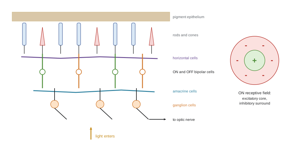
The retina is part of the brain, an outgrowth of the embryonic diencephalon, and it does substantial processing. Information flows vertically from photoreceptors to bipolar cells to retinal ganglion cells, whose axons form the optic nerve, and laterally through horizontal cells in the outer retina and amacrine cells in the inner retina. Light must pass through all these layers to reach the photoreceptors, an apparently backward arrangement that reflects how the eye develops; where the axons exit there are no photoreceptors, creating the blind spot.
Bipolar cells come in two broad classes. OFF bipolar cells express ionotropic glutamate receptors, so they are depolarized by glutamate and therefore by darkness. ON bipolar cells express the metabotropic receptor mGluR6, which closes a cation channel (TRPM1) when glutamate binds, so they are hyperpolarized by glutamate and depolarized when light reduces glutamate release. This sign inversion splits the visual signal into parallel ON and OFF channels from the first synapse, so increments and decrements of light are each signalled by an increase in firing. The two channels stay separate through the retina, the thalamus and into cortex.
Centre–surround receptive fields
In 1953 Stephen Kuffler, recording from cat retinal ganglion cells, found that each cell responds to light in a small circular region, the receptive field centre, and is opposed by light in a surrounding annulus. An ON-centre cell fires more when light falls on its centre and less when light falls on its surround; an OFF-centre cell does the opposite. Uniform illumination of both centre and surround produces only a weak response. The surround comes largely from lateral inhibition through horizontal cells. Ganglion cells therefore signal local contrast, differences in light between nearby points, rather than absolute brightness. This is efficient, because in natural scenes neighbouring points tend to have similar intensities, so transmitting differences removes redundancy, and it explains illusions such as Mach bands, in which bright and dark bands appear at the edges of luminance ramps.
A centre–surround receptive field is well described as a difference of two Gaussian functions: a narrow, strong excitatory centre minus a broad, weaker inhibitory surround. Applying this filter to an image enhances edges.
# Difference-of-Gaussians receptive field applied to a 1-D luminance profile with a step and a ramp.
import numpy as np
x = np.arange(-30, 31)                       # receptive field positions (arbitrary units)
def gauss(s): g = np.exp(-x**2 / (2 * s**2)); return g / g.sum()
dog = gauss(2.0) - gauss(6.0)                # narrow excitatory core minus equally weighted broad surround
print("DoG weights near the middle:", np.round(dog[27:34] * 100, 1))
print("sum of weights (response to uniform light):", round(dog.sum(), 3))

# luminance: dark plateau, a sharp step up, plateau, then a gentle ramp down
lum = np.concatenate([np.full(60, 20.0), np.full(60, 80.0), np.linspace(80, 40, 60), np.full(60, 40.0)])
resp = np.convolve(lum, dog, mode='same')
for i in list(range(44, 78, 3)) + list(range(110, 200, 8)):
    r = resp[i]
    bar = '#' * int(r / 1.5) if r > 0 else '-' * int(-r / 1.5)
print(f"pos {i:3d} lum {lum[i]:5.1f} response {r:+6.2f} {bar}")DoG weights near the middle: [ 0.6 5.8 11. 13.3 11. 5.8 0.6] sum of weights (response to uniform light): 0.0 pos 44 lum 20.0 response -0.29 pos 47 lum 20.0 response -1.11 pos 50 lum 20.0 response -3.39 -- pos 53 lum 20.0 response -8.31 ----- pos 56 lum 20.0 response -14.47 --------- pos 59 lum 20.0 response -3.99 -- pos 62 lum 80.0 response +14.10 ######### pos 65 lum 80.0 response +10.60 ####### pos 68 lum 80.0 response +4.68 ### pos 71 lum 80.0 response +1.65 # pos 74 lum 80.0 response +0.47 pos 77 lum 80.0 response +0.10 pos 110 lum 80.0 response +0.08 pos 118 lum 80.0 response +0.92 pos 126 lum 75.9 response +0.34 pos 134 lum 70.5 response +0.01 pos 142 lum 65.1 response +0.00 pos 150 lum 59.7 response -0.00 pos 158 lum 54.2 response -0.00 pos 166 lum 48.8 response -0.02 pos 174 lum 43.4 response -0.46 pos 182 lum 40.0 response -0.77 pos 190 lum 40.0 response -0.05 pos 198 lum 40.0 response -0.00
The filter gives zero response on uniform plateaus, because centre and surround cancel, and responds strongly at the step, positively on the bright side and negatively on the dark side. Along the linear ramp the response is again close to zero, but it deviates at the corners where the ramp begins and ends, which is where people see the bright and dark Mach bands. In the real retina there are about 20 types of ganglion cell in mammals, each tiling the retina and extracting a different feature: ON and OFF transient and sustained responses, colour opponency, and direction of motion. Direction-selective ganglion cells, for example, owe their selectivity to asymmetric inhibition from starburst amacrine cells.
The pathway to cortex
Ganglion cell axons leave the eye in the optic nerve. At the optic chiasm, axons from the nasal half of each retina cross, so the left side of the brain receives information about the right visual field from both eyes. Most axons go to the lateral geniculate nucleus (LGN) of the thalamus; others go to the superior colliculus, which orients the eyes and head; to the pretectum, which controls the pupillary reflex; and, from a special class of ganglion cells containing the photopigment melanopsin, to the suprachiasmatic nucleus, which sets the circadian clock (lesson 10). The LGN has six layers in primates: magnocellular layers carry fast, high-contrast-sensitivity, colour-blind signals from large parasol ganglion cells; parvocellular layers carry slower, finer, red–green colour-opponent signals from midget ganglion cells; and koniocellular cells between the layers carry blue–yellow signals.
Primary visual cortex
From the LGN, axons project to primary visual cortex (V1, also called striate cortex) in the occipital lobe. In the late 1950s and 1960s David Hubel and Torsten Wiesel recorded from V1 in cats and monkeys and found that most neurons respond not to spots but to bars or edges of a particular orientation. Simple cells have elongated receptive fields with separate ON and OFF subregions, as if built by lining up several LGN centre–surround fields; complex cells respond to a correctly oriented bar anywhere within a larger receptive field, as if combining several simple cells. Neurons are organized in columns: neurons in a column perpendicular to the cortical surface share orientation preference, and preference rotates smoothly across the surface, interrupted by pinwheel centres. Inputs from the two eyes are segregated into alternating ocular dominance columns. Hubel and Wiesel shared the 1981 Nobel Prize, with Roger Sperry.
Orientation tuning is quantified by recording responses to gratings at many orientations. Tuning curves are well fitted by a circular function, the von Mises function, because orientation repeats every 180 degrees:
where \(\theta_0\) is the preferred orientation, \(A\) the peak response above baseline \(b\), and \(\kappa\) sets the tuning width.
# Fit a von Mises orientation tuning curve to synthetic spike counts from an invented V1 neuron.
import numpy as np
rng = np.random.default_rng(5)
angles = np.arange(0, 180, 15)                         # degrees
def vm(theta, b, A, kappa, th0):
    return b + A * np.exp(kappa * (np.cos(np.deg2rad(2 * (theta - th0))) - 1))
true = (4.0, 30.0, 2.0, 62.0)
counts = np.array([rng.poisson(vm(a, *true)) for a in angles for _ in range(10)]).reshape(len(angles), 10)
mean = counts.mean(axis=1)
best, best_err = None, np.inf                         # coarse grid search on kappa and preferred angle
for th0 in np.arange(0, 180, 1.0):
    for kappa in np.arange(0.5, 6.0, 0.1):
        shape = np.exp(kappa * (np.cos(np.deg2rad(2 * (angles - th0))) - 1))
        X = np.column_stack([np.ones_like(shape), shape])
        coef, *_ = np.linalg.lstsq(X, mean, rcond=None)   # linear fit of b and A
        err = np.sum((X @ coef - mean) ** 2)
        if err < best_err:
            best, best_err = (coef[0], coef[1], kappa, th0), err
b, A, kappa, th0 = best
half = np.rad2deg(np.arccos(1 + np.log(0.5) / kappa)) / 2   # half-width at half height
print("orientation  mean spikes")
for a, m in zip(angles, mean): print(f"   {a:3d} deg   {m:5.1f}")
print(f"fit: preferred {th0:.0f} deg, peak {A:.1f} above baseline {b:.1f}, kappa {kappa:.1f}, half-width {half:.0f} deg")orientation mean spikes
0 deg 5.5
15 deg 8.1
30 deg 14.2
45 deg 25.2
60 deg 32.6
75 deg 27.8
90 deg 17.8
105 deg 8.7
120 deg 5.7
135 deg 4.7
150 deg 3.6
165 deg 4.5
fit: preferred 62 deg, peak 29.5 above baseline 3.1, kappa 1.7, half-width 27 degDevelopment and critical periods
Hubel and Wiesel also showed that closing one eye of a kitten during a critical period in the first few months of life permanently shifted cortical neurons to respond almost entirely to the open eye, and the deprived eye's ocular dominance columns shrank. The same closure in an adult cat had little effect. This experience-dependent competition, explained in part by Hebbian and BCM plasticity (lesson 6), has a direct clinical counterpart: amblyopia, reduced vision in one eye from childhood misalignment (strabismus), unequal focus or cataract, must be treated early, by correcting the problem and patching the stronger eye. Research on reopening plasticity in adults, by manipulating inhibition, perineuronal nets or neuromodulation, and on binocular treatments using video games, has expanded options for older children and adults.
Beyond V1: parallel streams
Beyond V1, visual information flows through more than 30 cortical areas in primates. In 1982 Leslie Ungerleider and Mortimer Mishkin proposed two broad streams: a ventral stream through V4 to inferotemporal cortex, specialized for recognizing objects (what), and a dorsal stream through MT (V5) to parietal cortex, specialized for spatial location and motion (where), later reinterpreted by Melvyn Goodale and David Milner as vision for action (how). Neurons in MT are selective for direction and speed of motion; damage to MT causes akinetopsia, an inability to perceive motion. In inferotemporal cortex, neurons respond to complex shapes, and patches such as the fusiform face area respond selectively to faces; damage causes prosopagnosia, an inability to recognize faces. Receptive fields grow larger and selectivity more complex along the ventral hierarchy, a design that inspired convolutional neural networks (CMPUT-466), whose internal representations, when trained on object recognition, resemble those recorded along the primate ventral stream.
| Stage | Typical receptive field property | Example deficit when damaged |
|---|---|---|
| Photoreceptors | Light intensity at a point; three cone types for colour | Colour blindness from cone opsin gene variants |
| Retinal ganglion cells | Centre–surround contrast; ON and OFF; about 20 types | Glaucoma: progressive loss of ganglion cells and visual field |
| LGN | Centre–surround; magno, parvo and konio channels | Field loss on the opposite side |
| V1 | Orientation, ocular dominance, spatial frequency | Cortical blindness in the corresponding field; blindsight in some cases |
| MT (V5) | Direction and speed of motion | Akinetopsia (motion blindness) |
| Inferotemporal cortex | Complex shapes, objects, faces | Object agnosia, prosopagnosia |
Vision is also actively constructed. Perception depends on expectations and context, as illusions show; feedback connections from higher to lower areas are at least as numerous as feedforward ones; and the eyes move about three times a second in saccades, so the brain must stitch together a stable world from brief snapshots, suppressing vision during each saccade. Some patients with damage to V1 can point to or discriminate stimuli they report not seeing, a phenomenon called blindsight, probably supported by pathways through the superior colliculus and LGN that bypass V1.
Worked problem
Localizing a visual field defect
An invented patient has lost vision in the right half of the visual field of both eyes (right homonymous hemianopia), with the central few degrees spared. Where is the lesion most likely to be, and why is central vision spared?
Show solution
- Loss of the same half-field in both eyes means the lesion is behind the optic chiasm, where fibres from both eyes for one visual hemifield travel together.
- The right visual field is represented in the left hemisphere, so the lesion is on the left: optic tract, LGN, optic radiation or V1.
- Macular sparing is typical of occipital lesions from stroke of the posterior cerebral artery, because the occipital pole representing the fovea often has a dual blood supply.
- The likely site is the left primary visual cortex.
Answer: A lesion of the left occipital cortex (V1), probably vascular, with macular sparing from dual blood supply to the foveal representation.
Common mistakes
MistakeSaying light depolarizes photoreceptors.
InsteadLight hyperpolarizes vertebrate photoreceptors by closing cyclic GMP-gated channels.
MistakeThinking the retina simply transmits a pixel image.
InsteadThe retina extracts contrast, motion and colour through about 20 parallel ganglion cell channels.
MistakeAssuming each eye projects only to the opposite hemisphere.
InsteadEach hemisphere receives the opposite visual field from both eyes, because nasal fibres cross at the chiasm.
MistakeExpecting monocular deprivation to have the same effect at any age.
InsteadIts strong effects are confined to a developmental critical period.
MistakeTreating the dorsal and ventral streams as completely separate.
InsteadThey interact extensively; the division describes emphasis, not isolation.
Summary and key terms
- Photoreceptors are depolarized in the dark; light triggers a rhodopsin–transducin–phosphodiesterase cascade that closes cyclic GMP-gated channels.
- Rods detect single photons; cones support colour and adapt over a vast range of intensities.
- ON and OFF bipolar cells, using mGluR6 and ionotropic receptors, split signals into parallel channels.
- Ganglion cells have centre–surround receptive fields that signal local contrast, well modelled as a difference of Gaussians.
- V1 neurons are orientation selective and organized in columns; monocular deprivation in a critical period shifts ocular dominance.
- Ventral and dorsal streams emphasize object recognition and spatial vision for action.
- phototransduction
- Conversion of light into an electrical signal in photoreceptors.
- rhodopsin
- The rod photopigment of opsin bound to 11-cis retinal.
- dark current
- The steady inward current through cyclic GMP-gated channels in photoreceptors in darkness.
- receptive field
- The region of sensory space in which a stimulus changes a neuron's activity.
- centre–surround
- A receptive field organization in which the centre and an annular surround have opposite effects.
- lateral inhibition
- Inhibition of neighbouring cells that sharpens contrast.
- orientation selectivity
- Preferential response of a neuron to edges or bars at a particular angle.
- ocular dominance column
- A cortical column dominated by input from one eye.
- critical period
- A developmental window when experience has strong, lasting effects on neural circuits.
- ventral stream
- The occipitotemporal pathway specialized for object recognition.
Practice problems
Work each problem on paper first, then reveal the answer to check.
What happens to glutamate release from a rod when light is absorbed?
Show answer
It decreases, because the rod hyperpolarizes.
Why are ON bipolar cells depolarized by light?
Show answer
Light reduces glutamate release, and glutamate acting on mGluR6 normally closes their cation channels.
A ganglion cell gives only a weak response to uniform illumination. Why?
Show answer
Its centre and surround have opposing effects that nearly cancel.
Where do fibres from the nasal hemiretinae cross?
Show answer
At the optic chiasm.
What is the difference between simple and complex cells in V1?
Show answer
Simple cells have separate ON and OFF subregions; complex cells respond to an oriented bar anywhere in a larger field.
Why must amblyopia be treated in early childhood?
Show answer
The visual cortex is most plastic during a critical period, after which recovery is much harder.
Damage to area MT causes what deficit?
Show answer
Akinetopsia, impaired perception of motion.
Which ganglion cells project to the suprachiasmatic nucleus, and why?
Show answer
Melanopsin-containing intrinsically photosensitive ganglion cells, to entrain the circadian clock to daylight.
Lesson quiz · 5 questions
Score 60% or more to complete this lesson.
Create a free account to save your progress, take quizzes and earn a certificate.