
BIOL-420 · Lesson 6 of 12 · 25 min
Synaptic plasticity
In 1949 the psychologist Donald Hebb, working at McGill University, proposed that when one cell repeatedly helps to fire another, some growth process or metabolic change strengthens the connection between them. The idea, often paraphrased as cells that fire together wire together, gave neuroscience a candidate mechanism for learning and memory: experience changes synaptic strength, and the changed synapses store the memory. Two decades later Hebb's postulate found a physical counterpart. This lesson covers long-term potentiation and depression, the molecular logic that makes them Hebbian, their dependence on the precise timing of spikes, the slower homeostatic processes that keep networks stable, and the evidence linking all of this to learning.
Discovery of long-term potentiation
In 1966 Terje Lømo, working in Per Andersen's laboratory in Oslo, noticed that brief high-frequency stimulation of the perforant path, the input from entorhinal cortex to the dentate gyrus of the hippocampus, produced a lasting increase in the response to later single stimuli. With Tim Bliss he described it in detail in 1973, in rabbits under anaesthesia, where the enhancement lasted for hours, and in awake animals for days to weeks. Long-term potentiation (LTP) is now usually studied at the synapses from CA3 Schaffer collaterals onto CA1 pyramidal neurons in hippocampal slices, where a tetanus of 100 pulses at 100 hertz, or bursts patterned on the hippocampal theta rhythm, potentiates the synapses for as long as the slice survives.
LTP at CA1 synapses has three properties that make it attractive as a memory mechanism. It is input-specific: only the synapses that were active during induction are potentiated, not other synapses on the same cell. It is cooperative: a weak stimulus that activates few fibres fails to induce LTP, while a stronger one that activates many succeeds, because enough depolarization is needed. And it is associative: a weak input that would not potentiate on its own will do so if it is active at the same time as a strong input to the same cell, linking the two, which is exactly what Hebb's rule requires for association.
The NMDA receptor as a coincidence detector
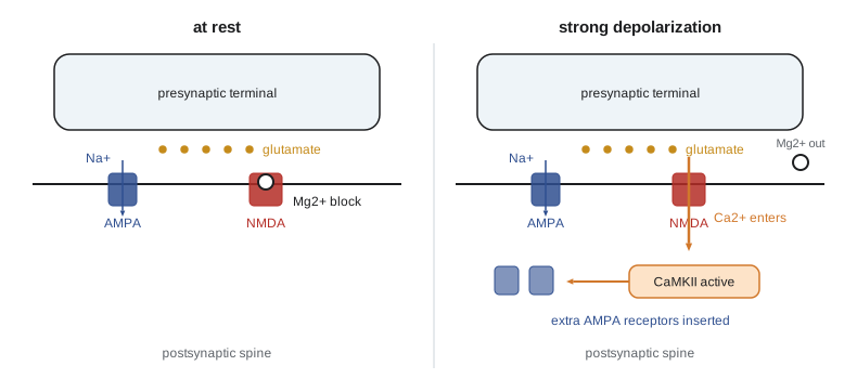
All three properties follow from the NMDA receptor. At resting potential its channel is blocked by magnesium ions, so glutamate binding alone admits little current. If the postsynaptic membrane is depolarized at the same time, by many coactive inputs or by a back-propagating action potential, the magnesium is expelled and calcium flows in. The NMDA receptor therefore opens only when presynaptic release (glutamate) and postsynaptic depolarization coincide: a molecular AND gate that implements Hebb's rule. Blocking NMDA receptors with the antagonist APV prevents the induction of LTP without affecting ordinary transmission through AMPA receptors, and Richard Morris showed in 1986 that infusing APV into the hippocampus of rats prevented them from learning the location of a hidden platform in a water maze.
Mechanisms of expression
Calcium entering through NMDA receptors activates calcium/calmodulin-dependent protein kinase II (CaMKII), which is extraordinarily abundant in spines. CaMKII is a ring of 12 subunits; once activated, a subunit can phosphorylate its neighbour at threonine 286, which keeps the kinase active even after calcium falls, a molecular switch that can outlast the trigger. Active CaMKII phosphorylates AMPA receptors, increasing their conductance, and drives insertion of additional AMPA receptors into the synapse from intracellular pools and from lateral diffusion in the membrane. The dendritic spine enlarges as its actin cytoskeleton is remodelled. Early LTP, lasting an hour or two, depends on these modifications of existing proteins.
Some synapses initially contain NMDA receptors but no AMPA receptors. They are silent at rest because NMDA receptors are blocked, and LTP can unsilence them by inserting AMPA receptors, which explains some cases in which potentiation appeared as a reduction in failures. Whether LTP also involves increased presynaptic release was debated for years; at CA1 synapses the consensus is that postsynaptic AMPA receptor trafficking is the main mechanism, while at other synapses, such as mossy fibre synapses onto CA3 neurons, LTP is presynaptic and independent of NMDA receptors.
Late LTP, lasting hours to days or longer, requires new gene transcription and protein synthesis. Strong stimulation activates protein kinase A and the transcription factor CREB, which turns on genes for synaptic proteins and structural growth. How do newly made proteins find the specific synapses that were potentiated? The synaptic tagging and capture hypothesis of Frey and Morris (1997) proposes that weak potentiation sets a local tag at the active synapses, which then capture plasticity-related proteins made in response to strong stimulation elsewhere on the cell. The persistence of memory over years despite constant protein turnover remains a puzzle; candidates for the maintenance mechanism have included the atypical kinase PKMzeta and self-sustaining interactions between CaMKII and the NMDA receptor subunit GluN2B.
Long-term depression
If synapses could only strengthen, networks would saturate. Long-term depression (LTD) weakens synapses. At CA1 synapses it is induced by prolonged low-frequency stimulation, for example 900 pulses at 1 hertz, which produces a modest, prolonged rise in postsynaptic calcium. Modest calcium preferentially activates protein phosphatases such as calcineurin and protein phosphatase 1, which dephosphorylate AMPA receptors and promote their removal by endocytosis. The direction of plasticity thus depends on the amount and time course of calcium: large, brief rises give LTP, while modest, prolonged rises give LTD. In the cerebellum, a different form of LTD at parallel fibre to Purkinje cell synapses, induced when parallel fibre input coincides with climbing fibre input, is central to motor learning (lesson 9).
The BCM theory, proposed by Elie Bienenstock, Leon Cooper and Paul Munro in 1982, generalizes this: below a modification threshold, postsynaptic activity weakens active synapses; above it, activity strengthens them; and the threshold itself slides up after periods of high activity and down after low activity. The sliding threshold keeps plasticity balanced, and the theory successfully predicted features of how visual cortex responds when one eye is deprived of vision during development.
Spike-timing-dependent plasticity
Hebb's rule speaks of one cell taking part in firing another, which implies causality and therefore order. In 1997 Henry Markram and colleagues, and in 1998 Guo-qiang Bi and Mu-ming Poo, showed that the relative timing of pre- and postsynaptic spikes determines the sign of plasticity. If a presynaptic spike precedes a postsynaptic spike by up to about 20 milliseconds, the synapse strengthens; if the order is reversed, it weakens. The magnitude falls off roughly exponentially with the interval. This is spike-timing-dependent plasticity (STDP). Mechanistically, a presynaptic spike that shortly precedes a back-propagating action potential lets glutamate-bound NMDA receptors be unblocked at the right moment, producing large calcium entry, while the reverse order produces smaller calcium entry and engages other pathways, including endocannabinoid signalling at some synapses.
where \(\Delta t = t_{\text{post}} - t_{\text{pre}}\). STDP lets a neuron learn which inputs predict its firing: inputs that consistently arrive just before the cell fires are strengthened, so the cell comes to respond earlier to them, while inputs that arrive after it fires are weakened. Applied in networks, STDP can make neurons learn sequences and become selective for repeating patterns embedded in noise.
# STDP: learning window and its effect on synapses driven by correlated or random inputs.
import numpy as np
rng = np.random.default_rng(3)
A_plus, A_minus, tau_p, tau_m = 0.010, 0.0105, 20.0, 20.0   # ms; slight depression bias
def dw(delta):                         # delta = t_post - t_pre in ms
    return A_plus * np.exp(-delta / tau_p) if delta > 0 else -A_minus * np.exp(delta / tau_m)
print("learning window:")
for d in (-60, -30, -10, -5, 5, 10, 30, 60):
    print(f"  t_post - t_pre = {d:+4d} ms -> dw = {dw(d):+.4f}")

# 20 inputs: 10 'leading' inputs fire about 5 ms before the postsynaptic spike, 10 fire at random times
T, n_post = 1000.0, 400
post_times = np.sort(rng.uniform(0, T * 50, n_post))
w = np.full(20, 0.5)
for tp in post_times:
    for i in range(20):
        if i < 10:
            tpre = tp - rng.normal(5.0, 2.0)            # leading input
        else:
            tpre = tp + rng.uniform(-50, 50)           # uncorrelated input
        w[i] = np.clip(w[i] + dw(tp - tpre), 0, 1)
print("\nmean weight of leading inputs:      ", round(w[:10].mean(), 2))
print("mean weight of uncorrelated inputs: ", round(w[10:].mean(), 2))learning window: t_post - t_pre = -60 ms -> dw = -0.0005 t_post - t_pre = -30 ms -> dw = -0.0023 t_post - t_pre = -10 ms -> dw = -0.0064 t_post - t_pre = -5 ms -> dw = -0.0082 t_post - t_pre = +5 ms -> dw = +0.0078 t_post - t_pre = +10 ms -> dw = +0.0061 t_post - t_pre = +30 ms -> dw = +0.0022 t_post - t_pre = +60 ms -> dw = +0.0005 mean weight of leading inputs: 1.0 mean weight of uncorrelated inputs: 0.51
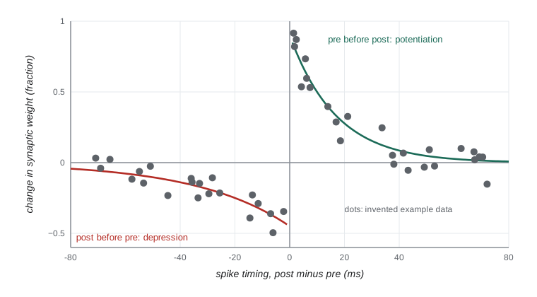
Inputs that fire just before the cell are driven to the maximum weight, while uncorrelated inputs end close to where they started: their random timing produces potentiation and depression that roughly cancel, with a slight bias toward depression built into the window. The window varies across synapses and brain regions, and neuromodulators reshape it: dopamine, acetylcholine and noradrenaline can convert timing-dependent depression into potentiation or extend the window. That interaction suggests how reward could reinforce recently active synapses, solving part of the problem of linking a delayed reward to the activity that caused it (lesson 10).
Homeostatic plasticity
Hebbian plasticity is a positive-feedback process: stronger synapses make firing more likely, which strengthens synapses further. Without a counterweight, activity would run away or fall silent. In 1998 Gina Turrigiano and colleagues showed that when activity in cultured cortical networks is blocked with tetrodotoxin for two days, the amplitudes of miniature excitatory currents increase, and when activity is increased by blocking inhibition, they decrease. All of a neuron's excitatory synapses are scaled up or down by the same factor, preserving their relative strengths while restoring the cell's average firing rate toward a set point. This synaptic scaling, along with homeostatic changes in intrinsic excitability and in inhibition, stabilizes networks over hours to days.
| Form of plasticity | Time scale | Induction | Main role |
|---|---|---|---|
| Short-term facilitation and depression | Milliseconds to seconds | Residual calcium, vesicle depletion | Temporal filtering |
| NMDA-dependent LTP | Hours to lifetime (with protein synthesis) | Coincident pre- and postsynaptic activity, large calcium rise | Strengthening specific synapses; association |
| NMDA-dependent LTD | Hours or longer | Low-frequency activity, modest calcium rise | Weakening; preventing saturation |
| STDP | Hours or longer | Precise spike order within about 20 ms | Learning causal and temporal structure |
| Synaptic scaling | Hours to days | Prolonged change in average activity | Stabilizing firing rates |
| Structural plasticity | Days to lifetime | Learning, sensory experience | Forming and removing synapses and spines |
From synapses to memory
Is LTP really how memories are stored? Several kinds of evidence point that way. Learning blocked by NMDA receptor antagonists in the hippocampus is the kind that depends on the hippocampus, such as spatial learning. Mice with mutations that prevent CaMKII autophosphorylation have impaired LTP and impaired spatial learning. Inhibitory avoidance learning in rats produces LTP-like changes at CA1 synapses that occlude further electrically induced LTP. In 2014 Sadegh Nabavi and colleagues used optogenetics in rats to create an association between a light-driven input to the amygdala and a shock, erased it by inducing LTD with low-frequency light, and restored it by inducing LTP, about as direct a demonstration of the synaptic theory of memory as has been achieved. Imaging of spines in living mice shows that motor learning produces new spines whose survival correlates with retention of the skill.
Memory involves more than individual synapses. Neurons with higher excitability at the time of learning are preferentially recruited into the ensemble that stores a memory, a process called memory allocation. The sparse set of neurons active during learning, sometimes called an engram, can be tagged genetically and later reactivated optogenetically to evoke the memory, as Susumu Tonegawa's group showed in 2012 (lesson 10).
Worked problem
Predicting the outcome of a pairing protocol
In an invented experiment, a weak input W to a CA1 neuron is stimulated at 1 hertz for 15 minutes, once alone and once paired with strong depolarization of the neuron through the recording pipette (holding at 0 mV). A second input S is not stimulated during either protocol. Predict what happens to W and S in each case, and what APV would do.
Show solution
- Low-frequency stimulation alone gives modest calcium entry through partly blocked NMDA receptors: expect LTD at W.
- Pairing with depolarization relieves the magnesium block, so each glutamate release admits large calcium: expect LTP at W.
- S was inactive in both protocols, so by input specificity it should be unchanged.
- APV blocks NMDA receptors, so it should prevent both the LTD and the LTP at W, without blocking baseline AMPA responses.
Answer: Alone: LTD at W; paired: LTP at W; S unchanged in both; APV prevents both forms at W.
Common mistakes
MistakeSaying the NMDA receptor carries most of the fast EPSP.
InsteadAMPA receptors carry most of the fast EPSP; NMDA receptors are largely blocked at rest and act as coincidence detectors.
MistakeAssuming LTP and LTD use completely different receptors.
InsteadAt CA1 both depend on NMDA receptors; the amount and time course of calcium determine the direction.
MistakeThinking Hebbian plasticity alone keeps networks stable.
InsteadHebbian plasticity is destabilizing; homeostatic mechanisms such as synaptic scaling are needed.
MistakeAssuming all LTP is postsynaptic.
InsteadAt some synapses, such as hippocampal mossy fibres, LTP is presynaptic and NMDA-independent.
MistakeEquating any change in synaptic strength with a memory.
InsteadLinking plasticity to memory requires showing necessity and sufficiency for learning, as in occlusion and optogenetic experiments.
Summary and key terms
- Hebb proposed that coactive cells strengthen their connections; LTP, discovered in 1966 and described in 1973, provides a mechanism.
- CA1 LTP is input-specific, cooperative and associative because NMDA receptors detect coincident glutamate and depolarization.
- Calcium activates CaMKII, which phosphorylates and inserts AMPA receptors; late LTP needs gene expression and synaptic tagging.
- Modest prolonged calcium activates phosphatases and produces LTD; the BCM sliding threshold balances plasticity.
- STDP strengthens synapses when pre precedes post within about 20 ms and weakens them for the reverse order.
- Synaptic scaling and other homeostatic mechanisms stabilize network activity.
- Pharmacological, genetic and optogenetic evidence links synaptic plasticity to learning and memory.
- Hebbian plasticity
- Strengthening of a synapse when presynaptic activity contributes to postsynaptic firing.
- long-term potentiation
- A lasting increase in synaptic strength after patterned activity.
- long-term depression
- A lasting decrease in synaptic strength after patterned activity.
- input specificity
- Restriction of plasticity to the synapses that were active during induction.
- associativity
- Potentiation of a weak input when it is active together with a strong input.
- CaMKII
- A calcium/calmodulin-dependent kinase whose autophosphorylation helps induce and maintain LTP.
- silent synapse
- A synapse with NMDA but no functional AMPA receptors.
- synaptic tagging
- Marking of recently active synapses so they capture plasticity-related proteins.
- spike-timing-dependent plasticity
- Plasticity whose sign and size depend on the relative timing of pre- and postsynaptic spikes.
- synaptic scaling
- Multiplicative adjustment of all of a neuron's synapses to keep its firing rate near a set point.
Practice problems
Work each problem on paper first, then reveal the answer to check.
Why is LTP associative?
Show answer
A weak input active together with a strong input shares the depolarization that unblocks its NMDA receptors.
What does APV do to baseline transmission and to LTP induction?
Show answer
Little to baseline AMPA-mediated transmission; it blocks LTP induction.
What property of CaMKII lets it act as a molecular switch?
Show answer
Autophosphorylation at threonine 286 keeps it active after calcium falls.
A presynaptic spike occurs at 50 ms and a postsynaptic spike at 40 ms. Which way does the synapse change under STDP?
Show answer
It weakens, because the postsynaptic spike came first (Δt = −10 ms).
What experimental manipulation revealed synaptic scaling?
Show answer
Blocking activity in cultures for two days with tetrodotoxin, which increased miniature EPSC amplitudes.
In BCM theory, what happens to the modification threshold after a period of high activity?
Show answer
It slides upward, making potentiation harder and depression easier.
How can silent synapses be unsilenced?
Show answer
By LTP-driven insertion of AMPA receptors.
What did Nabavi and colleagues show in 2014?
Show answer
That optogenetically induced LTD erased, and LTP restored, a conditioned fear association.
Lesson quiz · 5 questions
Score 60% or more to complete this lesson.
Create a free account to save your progress, take quizzes and earn a certificate.