
BIOL-420 · Lesson 1 of 12 · 27 min
Neurons, glia and the nervous system
A human brain contains roughly 86 billion neurons and a comparable number of glial cells, wired together by something like a hundred trillion synapses. Out of that tissue come perception, movement, memory, emotion and thought. Neurobiology asks how. This course starts where animal physiology (BIOL-350) left off: you already know how a resting potential arises, how an action potential is generated and how a chemical synapse transmits a signal. Here we go deeper, from the molecular structure of channels to the circuits that compute, the plasticity that stores memories, and the disorders and technologies that make neuroscience one of the most consequential sciences of this century.
This first lesson surveys the cells of the nervous system and how they are organized: the special shape and internal machinery of neurons, the several kinds of glia and what they really do, the barrier that protects the brain, and the overall plan of central and peripheral nervous systems. It closes with the levels of explanation, from molecules to behaviour, that the rest of the course moves between.
The neuron doctrine
In the late nineteenth century two great anatomists disagreed about the basic structure of nervous tissue. Camillo Golgi, who had invented a silver stain that blackened a few cells completely, believed nerve cells fused into a continuous network, a reticulum. Santiago Ramón y Cajal, using Golgi's own stain, argued that neurons were separate cells that communicated at points of contact, and that signals flowed in one direction, from dendrites through the cell body to the axon. They shared the Nobel Prize in 1906 and disagreed in their lectures. Electron microscopy in the 1950s settled the question in Cajal's favour: a narrow synaptic cleft about 20 nanometres wide separates most neurons. The neuron doctrine, that the neuron is the structural and functional unit of the nervous system, remains the foundation of the field, with two qualifications. Electrical synapses (gap junctions) do connect some neurons directly, and glia turn out to be active partners rather than inert packing.
The shape of a neuron
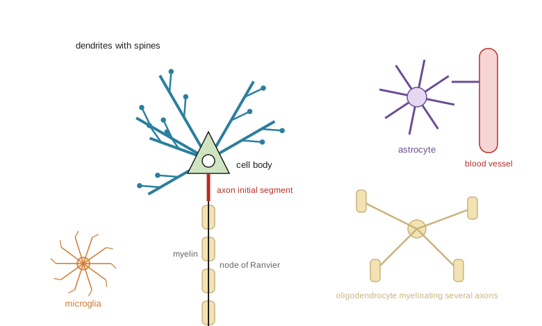
Neurons are the most polarized cells in the body. A typical vertebrate neuron has a cell body (soma) containing the nucleus and most of the protein synthesis machinery; several dendrites, branching processes that receive most synaptic input; and a single axon that carries output, sometimes over enormous distances. The axon of a motor neuron that controls a toe muscle runs from the lumbar spinal cord to the foot, more than a metre in a tall adult, from a cell body only about 50 micrometres across. If the cell body were the size of a tennis ball, the axon would be a garden hose several kilometres long.
The axon begins at the axon initial segment, a specialized stretch about 20 to 60 micrometres long packed with voltage-gated sodium channels anchored by the scaffold protein ankyrin-G. This is where action potentials normally start, which is why the initial segment is described as the neuron's decision point. Along the axon, myelinated segments alternate with nodes of Ranvier, and at its end the axon branches into terminals that form synapses on other cells. Dendrites of many neurons are studded with spines, tiny protrusions that each receive one excitatory synapse and whose size and number change with learning (lesson 6).
Neuron shape is tied to function. Cerebellar Purkinje cells have flat, fan-like dendritic trees that receive about 200,000 synaptic inputs each. Pyramidal cells of the cerebral cortex have a long apical dendrite reaching toward the surface and a skirt of basal dendrites, and send their axons to distant targets. Interneurons, which make up around a fifth of cortical neurons, keep their axons local and are mostly inhibitory. Retinal bipolar cells have short, simple processes and often do not fire action potentials at all. Neuroscientists classify neurons by morphology, by the transmitter they release, by their electrical firing patterns, by their connections and, increasingly, by the genes they express. Single-cell transcriptomic atlases of the mouse brain published in 2023 identified more than 5,000 transcriptomic cell types, a number that would have astonished Cajal.
The cytoskeleton and axonal transport
Because almost all proteins are made in or near the cell body, a neuron must ship materials along its axon continuously. Three cytoskeletal systems support this. Microtubules run lengthwise down the axon as polarized tracks, all with their plus ends pointing away from the soma; in dendrites their orientation is mixed. Neurofilaments, the neuronal intermediate filaments, set axon calibre, and larger axons conduct faster. Actin filaments are concentrated in growth cones, spines and a periodic lattice of rings spaced about 190 nanometres apart beneath the axonal membrane, discovered by super-resolution microscopy in 2013.
Axonal transport has a fast and a slow component. Fast anterograde transport, driven by kinesin motors walking toward microtubule plus ends, carries membrane-bound organelles such as synaptic vesicle precursors and mitochondria at about 200 to 400 millimetres per day. Fast retrograde transport, driven by dynein toward the cell body, returns worn-out components and carries signals such as neurotrophins from the terminals, telling the cell body that its target is alive. Slow transport moves cytoskeletal and cytosolic proteins at roughly 0.2 to 8 millimetres per day. Retrograde transport is also a route of entry: rabies virus, tetanus toxin and herpes simplex virus all travel up axons to reach the central nervous system, and neuroscientists exploit modified rabies viruses to trace which neurons connect to which.
# How long does axonal transport take along a long motor axon?
axon_mm = 1000          # spinal cord to foot, about 1 m
rates_mm_per_day = {
    "fast anterograde (kinesin)": 300,
    "fast retrograde (dynein)": 200,
    "slow component b": 6,
    "slow component a": 1,
}
for name, r in rates_mm_per_day.items():
    days = axon_mm / r
    print(f"{name:28s} {r:6.1f} mm/day -> {days:7.1f} days ({days/30.4:5.1f} months)")

# diffusion alone would be hopeless: time ~ L^2 / (2D)
D = 1e-8                 # m^2/s, a typical protein in cytoplasm
L = 1.0                  # m
t = L**2 / (2 * D)
print(f"\ndiffusion of a protein over 1 m: about {t/3.15e7:.0f} years")fast anterograde (kinesin) 300.0 mm/day -> 3.3 days ( 0.1 months) fast retrograde (dynein) 200.0 mm/day -> 5.0 days ( 0.2 months) slow component b 6.0 mm/day -> 166.7 days ( 5.5 months) slow component a 1.0 mm/day -> 1000.0 days ( 32.9 months) diffusion of a protein over 1 m: about 2 years
The comparison with diffusion shows why active transport is essential. Diffusion time grows with the square of distance, so a protein that diffuses across a cell body in seconds would need years to drift down a metre of axon. It also explains why long axons are vulnerable. Defects in motors or in the microtubule tracks cause disease: mutations in a kinesin gene underlie some hereditary spastic paraplegias, defects in dynein and its partner dynactin cause motor neuron disease, and the longest axons, to the feet, are the first affected in many peripheral neuropathies, including the neuropathy caused by some cancer chemotherapy drugs that stabilize microtubules.
Glia: more than glue
The name glia comes from the Greek for glue, which reflects nineteenth-century assumptions about their role. Glia are now known to be essential for development, signalling, metabolism, immunity and repair. The proportion of glia varies by region; the old claim that glia outnumber neurons ten to one is wrong, and careful counts with the isotropic fractionator method give a ratio close to one to one in the human brain overall.
| Cell type | Location | Main functions |
|---|---|---|
| Astrocytes | Central nervous system | Take up glutamate and potassium, supply lactate, regulate blood flow, form part of the blood–brain barrier, shape synapses |
| Oligodendrocytes | Central nervous system | Myelinate segments of up to about 50 axons each; provide metabolic support to axons |
| Schwann cells | Peripheral nervous system | Myelinate a single segment of one axon or ensheath several small unmyelinated axons; guide regeneration |
| Microglia | Central nervous system | Resident immune cells; survey tissue, remove debris and prune synapses |
| Oligodendrocyte precursor cells | Central nervous system | Divide throughout life and generate new oligodendrocytes |
| Ependymal cells | Lining of ventricles | Move cerebrospinal fluid with cilia; line the ventricles |
Astrocytes are star-shaped cells whose fine processes contact thousands of synapses and wrap around blood vessels with structures called end-feet. They clear glutamate from synapses through transporters, preventing excitotoxic build-up; buffer the potassium released during neural activity; store glycogen and, according to the astrocyte–neuron lactate shuttle hypothesis, supply lactate to active neurons; and help couple local blood flow to neural activity, which is the physiological basis of the functional MRI signal (lesson 11). Astrocytes release signalling molecules of their own, and the concept of the tripartite synapse, two neurons plus an astrocyte, captures their role, although how much astrocytes contribute to fast information processing is still debated.
Myelinating glia wrap axons in many layers of compacted membrane. In the central nervous system each oligodendrocyte myelinates segments of many axons; in the periphery each Schwann cell myelinates one segment of one axon. Myelin raises the resistance and lowers the capacitance of the axonal membrane, so current spreads farther between nodes and conduction becomes saltatory, as BIOL-350 showed. Myelin is not fixed: oligodendrocyte precursor cells keep generating new oligodendrocytes in adults, and learning a motor skill in mice requires new myelin, a form of plasticity beyond the synapse. Demyelinating diseases include multiple sclerosis, an autoimmune attack on central myelin (BIOL-380), and Guillain–Barré syndrome, an acute autoimmune attack on peripheral nerves.
Microglia arise from yolk-sac progenitors that colonize the brain during embryonic development and are the brain's resident macrophages. Their processes move constantly, surveying the tissue every few hours. They engulf dead cells and debris, respond to injury and infection and, during development, prune excess synapses tagged by complement proteins. Genetic variants affecting microglia, such as variants in TREM2, raise the risk of Alzheimer's disease, which has shifted attention toward the brain's immune system in neurodegeneration (lesson 12).
The blood–brain barrier
In most tissues, small molecules leak from capillaries through gaps between endothelial cells. In the brain, endothelial cells are sealed by tight junctions, have few transport vesicles and express efflux pumps such as P-glycoprotein that eject many foreign molecules. Together with pericytes and astrocyte end-feet, they form the blood–brain barrier. It keeps the neural environment stable, protecting neurons from fluctuations in ions, transmitters and toxins in the blood. Essential nutrients cross on dedicated carriers: glucose on GLUT1, amino acids on specific transporters.
The barrier is a major obstacle for medicine. Most large molecules, including antibodies, and a large fraction of small-molecule drugs do not reach the brain in useful amounts. Strategies to cross it include designing small lipid-soluble drugs, exploiting carriers (levodopa, the precursor of dopamine, crosses on an amino acid transporter while dopamine itself does not), engineering antibodies that hitch a ride on the transferrin receptor, and temporarily opening the barrier with focused ultrasound and microbubbles, which has been tested in early clinical trials. A few regions, the circumventricular organs, lack a full barrier so that they can sense blood-borne signals; the area postrema, for example, triggers vomiting in response to toxins.
Organization of the nervous system
The nervous system has a central division, the brain and spinal cord, and a peripheral division of nerves and ganglia. The peripheral system includes the somatic system, with sensory neurons from skin, muscles and joints and motor neurons to skeletal muscle, and the autonomic system, with sympathetic and parasympathetic divisions that BIOL-350 described, plus the enteric nervous system of the gut, which contains about half a billion neurons and can operate with considerable independence.
Within the brain, broad regions include the brainstem (medulla, pons and midbrain), which controls breathing, heart rate and arousal; the cerebellum, important for coordination and motor learning; the diencephalon, with the thalamus relaying most sensory information to the cortex and the hypothalamus regulating homeostasis and hormones; and the cerebrum, with the cerebral cortex, basal ganglia, hippocampus and amygdala. The cerebral cortex is a sheet 2 to 4 millimetres thick, folded to fit inside the skull, with six layers in most regions; input from the thalamus arrives mainly in layer 4, and layer 5 sends output to subcortical targets. Grey matter contains cell bodies and synapses; white matter contains myelinated axon tracts.
Comparative neurobiology has been central to the field. The squid giant axon allowed the first intracellular recordings; the sea slug Aplysia revealed the cellular basis of simple learning (lesson 10); the nematode C. elegans has exactly 302 neurons, whose complete wiring diagram was published in 1986; the fruit fly Drosophila, with about 140,000 neurons, now has a complete brain connectome (lesson 11); and the mouse provides genetic tools for dissecting mammalian circuits. Human data come from patients, imaging, recordings made during neurosurgery and post-mortem tissue.
Levels of explanation
Explanations in neuroscience span many levels: molecules (channels, receptors), cells (the neuron and its computation), synapses and circuits (networks of tens to millions of neurons), systems (vision, movement, memory) and behaviour. David Marr argued in 1982 that understanding a system such as vision requires three complementary levels: the computational problem being solved, the algorithm and representation that solve it, and the physical implementation in neurons. A good explanation links levels: for example, how the voltage dependence of one receptor, the NMDA receptor, lets synapses detect coincident activity, which lets circuits store associations, which supports a form of memory you can measure in behaviour. This course moves up and down those levels deliberately.
Worked problem
Estimating why the axon initial segment fires first
An invented pyramidal neuron has sodium channel density of about 50 channels per square micrometre in the soma and about 2,500 per square micrometre in the axon initial segment. If firing threshold is reached when inward sodium current exceeds outward current by a fixed amount, why does the action potential begin in the initial segment rather than the soma?
Show solution
- Sodium current per unit membrane area is proportional to channel density when the channels are opened by the same depolarization.
- The initial segment has about 50 times the density of the soma, so it produces far more inward current per unit area for the same voltage change.
- The initial segment is also thin, so its small area has a small capacitance to charge, and current from the soma depolarizes it efficiently.
- Threshold is therefore reached first in the initial segment, and the spike then propagates both down the axon and back into the soma and dendrites.
Answer: High sodium channel density and small capacitance make the initial segment the lowest-threshold site, so spikes start there and back-propagate into the cell body.
Common mistakes
MistakeSaying glia outnumber neurons ten to one in the human brain.
InsteadCareful counts give a ratio close to one to one overall, with large regional differences.
MistakeTreating glia as passive support cells.
InsteadGlia regulate synapses, ions, blood flow, myelination, immunity and synaptic pruning.
MistakeAssuming most proteins are made in the axon.
InsteadMost are made in or near the soma and carried by transport, although some local translation occurs in axons and dendrites.
MistakeThinking any drug in the blood reaches the brain.
InsteadThe blood–brain barrier excludes most large molecules and many small ones.
MistakeAssuming all neurons fire action potentials.
InsteadSome, such as many retinal bipolar cells, signal with graded potentials only.
Summary and key terms
- The neuron doctrine holds that neurons are discrete cells communicating at synapses; gap junctions and active glia qualify it.
- Neurons are highly polarized, with dendrites, soma, an axon initial segment where spikes start, and axon terminals.
- Kinesin and dynein carry cargo along polarized microtubules; diffusion would be far too slow over long axons.
- Astrocytes, oligodendrocytes, Schwann cells and microglia regulate the chemical environment, conduction, immunity and synapses.
- The blood–brain barrier protects neurons but blocks many drugs; carriers, engineered antibodies and focused ultrasound are ways around it.
- The nervous system is organized into central and peripheral divisions, and explanations link molecules, cells, circuits, systems and behaviour.
- neuron doctrine
- The principle that the neuron is the discrete structural and functional unit of the nervous system.
- axon initial segment
- The first part of the axon, rich in sodium channels, where action potentials usually begin.
- dendritic spine
- A small protrusion of a dendrite that receives a single excitatory synapse.
- axonal transport
- Motor-driven movement of materials along axonal microtubules in both directions.
- astrocyte
- A star-shaped glial cell that regulates ions, transmitters, metabolism and blood flow.
- oligodendrocyte
- A central glial cell that myelinates segments of many axons.
- microglia
- The resident immune cells of the central nervous system.
- blood–brain barrier
- The tight-junction-sealed endothelium and associated cells that restrict passage from blood to brain.
- white matter
- Brain and spinal cord tissue made mainly of myelinated axon tracts.
- connectome
- A complete map of the neurons and synaptic connections in a nervous system or region.
Practice problems
Work each problem on paper first, then reveal the answer to check.
Why could Cajal argue for separate neurons using Golgi's own stain?
Show answer
The stain filled only a few whole cells, revealing their boundaries and endings as separate from neighbours.
A 1.2 m axon carries a cargo at 300 mm per day. How long does the trip take?
Show answer
1,200 / 300 = 4 days.
Name two functions of astrocytes at synapses.
Show answer
Glutamate uptake and potassium buffering (also lactate supply and synapse formation).
How does a single oligodendrocyte differ from a single Schwann cell?
Show answer
An oligodendrocyte myelinates segments of many central axons; a Schwann cell myelinates one segment of one peripheral axon.
Why is levodopa given for Parkinson's disease rather than dopamine?
Show answer
Levodopa crosses the blood–brain barrier on an amino acid transporter; dopamine does not.
What does the area postrema do, and why does it lack a full barrier?
Show answer
It detects blood-borne toxins and triggers vomiting, so it must sample the blood directly.
Which cortical layer receives most thalamic input in sensory cortex?
Show answer
Layer 4.
State Marr's three levels of analysis.
Show answer
Computational (what problem), algorithmic (how, with what representation) and implementational (physical substrate).
Lesson quiz · 5 questions
Score 60% or more to complete this lesson.
Create a free account to save your progress, take quizzes and earn a certificate.