
BIOC-200 · Lesson 6 of 12 · 23 min
Carbohydrates
▶ Video coming soon A video version of this lesson is on the way.
Carbohydrates are the most abundant biological molecules on Earth. Every year plants and algae turn more than 100 billion tonnes of carbon dioxide into sugars by photosynthesis, and most of that carbon ends up in cellulose, the tough fibre of plant cell walls. Carbohydrates fuel cells, store energy as starch and glycogen, give structure to plants, fungi and insects, form the backbone of DNA and RNA, and coat the surfaces of cells with chains that act as identification tags. This lesson describes the structure of simple sugars, how they form rings, how they are linked into larger molecules and what those molecules do.
Monosaccharides
The simplest carbohydrates are monosaccharides, or simple sugars. They have the general formula (CH₂O)n, which gave carbohydrates their name, hydrates of carbon, though the name is chemically misleading. A monosaccharide is a chain of three to seven carbon atoms carrying a hydroxyl group on every carbon but one; that one carries a carbonyl group. If the carbonyl is at the end of the chain, as an aldehyde, the sugar is an aldose; if it is in the middle, as a ketone, the sugar is a ketose. Sugars are also named by their number of carbons: trioses have three, pentoses five and hexoses six. Glucose is an aldohexose, fructose a ketohexose and ribose an aldopentose.
The smallest sugars are the trioses glyceraldehyde and dihydroxyacetone, which reappear as intermediates in glycolysis. Glyceraldehyde has one chiral carbon and so exists as D and L forms. Larger sugars have several chiral carbons, and a sugar is labelled D or L according to the arrangement at the chiral carbon farthest from the carbonyl group: if its hydroxyl points to the right in the standard Fischer projection, the sugar belongs to the D series. Nearly all sugars in living things are D sugars, just as nearly all amino acids in proteins are L. An aldohexose has four chiral carbons and therefore 2⁴ = 16 possible stereoisomers, of which glucose, galactose and mannose are the most common in nature. Sugars that differ at only one chiral carbon, such as glucose and galactose, are called epimers.
Sugars form rings
In water, sugars with five or more carbons exist mainly as rings rather than open chains. A hydroxyl group near the far end of the chain reacts with the carbonyl group at the other end to form a hemiacetal (from an aldehyde) or a hemiketal (from a ketone). Glucose usually closes between carbon 1 and the oxygen on carbon 5, forming a six-membered ring of five carbons and one oxygen called a pyranose, after the compound pyran. Fructose often forms a five-membered furanose ring between carbon 2 and the oxygen on carbon 5.
Ring closure creates a new chiral centre at the former carbonyl carbon, called the anomeric carbon. Its new hydroxyl group can point down, below the ring as usually drawn, giving the alpha (α) anomer, or up, giving the beta (β) anomer. In solution the two anomers interconvert through the open chain, a process called mutarotation. At equilibrium in water at room temperature D-glucose is about 36% α-glucopyranose and 64% β-glucopyranose, with less than 0.01% in the open-chain form. The β form is favoured because all of its bulky groups sit in the roomier equatorial positions of the chair-shaped ring.
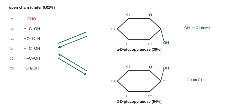
The tiny open-chain fraction matters. Its aldehyde group can be oxidized, so glucose is called a reducing sugar, and older tests for sugar in urine relied on glucose reducing copper ions. The same reactivity lets glucose react slowly and without enzymes with the amino groups of proteins. In the blood, a fraction of hemoglobin carries glucose attached in this way, and the share of this glycated hemoglobin, measured as HbA1c, reflects the average blood glucose over the previous two to three months. It is the standard test for monitoring diabetes.
Modified sugars
Many important sugars carry chemical modifications. In deoxy sugars a hydroxyl is replaced by hydrogen; 2-deoxyribose, which lacks the oxygen on carbon 2, is the sugar of DNA. In amino sugars a hydroxyl is replaced by an amino group, often acetylated; N-acetylglucosamine is the unit of chitin and part of bacterial cell walls. Sugar acids, such as glucuronic acid, carry a carboxyl group. Sugar phosphates, with a phosphate ester on a hydroxyl group, are the forms in which sugars move through metabolism: glucose 6-phosphate, fructose 1,6-bisphosphate and ribose 5-phosphate are all central intermediates, and the negative charge of the phosphate keeps them from leaking out through the cell membrane.
The glycosidic bond and disaccharides
Sugars are linked by glycosidic bonds. The anomeric hydroxyl of one sugar condenses with a hydroxyl group of another, releasing water and forming an acetal link. The bond is named by the anomeric form of the first sugar and the carbons it joins. Once the anomeric carbon is in a glycosidic bond, it can no longer open into a chain, so it is locked in the α or β form.
Three disaccharides are common in food. Maltose, from the digestion of starch, is two glucose units joined α(1→4). Lactose, the sugar of milk, is galactose joined β(1→4) to glucose. Sucrose, table sugar from sugar cane and sugar beet, is glucose and fructose joined through both of their anomeric carbons, α1→β2. Because neither anomeric carbon of sucrose is free, sucrose is not a reducing sugar, which makes it unusually stable and is one reason plants use it to transport sugar.
The body digests disaccharides with specific enzymes on the lining of the small intestine: maltase, sucrase and lactase. Most mammals stop making lactase after weaning, and so do most adults worldwide. In people whose lactase activity falls, undigested lactose passes to the large intestine, where bacteria ferment it and produce gas and discomfort; this is lactose intolerance. In populations with a long history of dairy farming, including much of northern Europe and some groups in East Africa and the Middle East, mutations that keep the lactase gene switched on into adulthood became common within the last ten thousand years, one of the clearest examples of recent human evolution.
Polysaccharides for storage
Polysaccharides are long chains of monosaccharides, sometimes thousands of units long. Unlike proteins and nucleic acids they have no information-carrying sequence; most are built from one or two kinds of sugar repeated over and over. They can be branched or unbranched, and their properties depend critically on which glycosidic bonds they contain.
Plants store glucose as starch, which comes in two forms. Amylose is an unbranched chain of glucose units linked α(1→4); the α linkage gives the chain a natural curve, and it coils into a helix with about six residues per turn. Iodine slips into the centre of this helix and turns it deep blue, the basis of the classic starch test. Amylopectin, the larger part of most starches, has the same α(1→4) chains with branches attached by α(1→6) bonds about every 24 to 30 residues. Animals store glucose as glycogen, which has the same bonds as amylopectin but is more highly branched, with a branch every 8 to 12 residues. Glycogen is stored as granules mainly in the liver, where it supplies glucose to the blood, and in muscle, where it fuels contraction.
Branching has a purpose. The enzymes that release glucose from glycogen work from the ends of the chains, and every branch adds another end. A highly branched glycogen molecule with thousands of ends can release glucose very quickly when adrenaline or glucagon signals a need, and it is more soluble than an unbranched chain of the same size. Storing glucose as a polymer also solves an osmotic problem. The glycogen in a liver cell, if released as free glucose, would raise the internal glucose concentration to about 0.4 M, enough to draw in water until the cell burst; packed into a few large molecules it has almost no osmotic effect.
Polysaccharides for structure
Cellulose, the main structural material of plants, is also an unbranched chain of glucose, but its units are linked β(1→4). That one change in the anomeric configuration transforms the polymer. Each glucose unit is flipped relative to its neighbour, the chain is straight rather than curved, and its hydroxyl groups form hydrogen bonds with those of neighbouring chains. Dozens of chains pack side by side into crystalline microfibrils that are, weight for weight, as strong as steel. Cotton is almost pure cellulose, and wood is about half.
Humans, like nearly all animals, cannot digest cellulose because we make no enzyme that breaks β(1→4) bonds between glucose units. Cows, sheep, termites and other herbivores depend on microorganisms in their guts that make cellulases. For people, cellulose is dietary fibre: it adds bulk, is partly fermented by bacteria in the colon, and helps the gut work normally.
Chitin, the second most abundant polysaccharide, is a β(1→4) chain of N-acetylglucosamine. It forms the exoskeletons of insects and crustaceans and the cell walls of fungi. Bacterial cell walls are built of peptidoglycan, chains of alternating N-acetylglucosamine and N-acetylmuramic acid cross-linked by short peptides; lysozyme in tears and saliva defends against bacteria by cutting these chains, and penicillin, as Lesson 5 noted, blocks the enzyme that makes the cross-links. Glycosaminoglycans such as hyaluronic acid, which lubricates joints, and heparin, a natural anticoagulant, are long, negatively charged polysaccharides that attract water and form gels in connective tissue.
| Polysaccharide | Units and bonds | Branching | Role |
|---|---|---|---|
| Amylose (starch) | Glucose, α(1→4) | None | Energy store in plants |
| Amylopectin (starch) | Glucose, α(1→4) and α(1→6) | Every 24–30 units | Energy store in plants |
| Glycogen | Glucose, α(1→4) and α(1→6) | Every 8–12 units | Energy store in animals |
| Cellulose | Glucose, β(1→4) | None | Plant cell walls |
| Chitin | N-acetylglucosamine, β(1→4) | None | Exoskeletons, fungal walls |
Sugars as signals
Carbohydrates are attached to many proteins and lipids, forming glycoproteins and glycolipids. Most proteins on the outer surface of cells and most proteins secreted from cells carry sugar chains, attached either to the nitrogen of asparagine (N-linked) or to the oxygen of serine or threonine (O-linked). These chains help proteins fold, protect them from proteases, and serve as markers recognized by other cells. Proteins called lectins bind specific sugar structures; they help white blood cells stick to blood vessel walls at sites of inflammation, and the influenza virus uses a lectin called hemagglutinin to attach to sialic acid on the cells of the airways.
The ABO blood groups are a familiar example. Red blood cells carry a short sugar chain on their surface. In people of group A an enzyme adds N-acetylgalactosamine to the end of the chain; in group B a slightly different form of the enzyme adds galactose; group O lacks a working enzyme and leaves the chain unfinished. People make antibodies against the sugars they lack, which is why blood transfusions must be matched.
A worked example: how much energy is stored?
Worked problem
Glycogen and fat as energy stores
A 70 kg person stores about 100 g of glycogen in the liver and 400 g in muscle. Complete oxidation of glucose releases about 17 kJ per gram (about 2,870 kJ/mol), and glycogen stored in cells carries roughly 2 g of water per gram. How much energy is stored as glycogen, and how much body mass does it occupy? Compare with storing the same energy as fat, which releases about 37 kJ per gram and is stored nearly without water.
Show solution
- Total glycogen = 100 + 400 = 500 g. Energy = 500 g × 17 kJ/g = 8,500 kJ, about 2,000 kilocalories, roughly a day of energy needs.
- Stored mass including water = 500 g × (1 + 2) = 1,500 g, or 1.5 kg.
- The same 8,500 kJ as fat needs 8,500/37 = 230 g, essentially all of it fat with little water.
- Glycogen therefore stores energy about six times less compactly: 1,500 g against 230 g. A typical person storing 15 kg of fat would need about 100 kg of hydrated glycogen to hold the same energy.
Answer: About 8,500 kJ is stored as glycogen in about 1.5 kg of tissue mass; fat stores the same energy in about 0.23 kg.
A program: anomers and storage
The program first uses the measured equilibrium fractions of glucose anomers to calculate the free energy difference between them. Then it models how quickly a glycogen molecule can release glucose: it builds trees with different branching intervals out to the same total size and counts the chain ends available to the releasing enzyme, which works only at ends.
import math

# 1. Equilibrium between the anomers of glucose at 25 C
R, T = 8.314, 298.15
alpha, beta = 0.36, 0.64
K = beta / alpha
dG = -R * T * math.log(K)
print(f"K = beta/alpha = {K:.2f}, delta G (alpha to beta) = {dG / 1000:.2f} kJ/mol")

# 2. Branching: how many chain ends does a molecule of a given size have?
def ends(total_units, branch_every):
    # Each chain segment holds branch_every units before a new branch starts.
    # A tree in which every segment carries one branch doubles its ends each tier.
    units, tiers, segments = 0, 0, 1
    while units + segments * branch_every <= total_units:
        units += segments * branch_every
        segments *= 2
        tiers += 1
    return segments // 2, tiers

print()
print("branch every   chain ends   tiers   (molecule of about 50,000 glucose units)")
for b in (12, 24, 48, 1000):
    e, t = ends(50000, b)
print(f"{b:12d} {e:12,d} {t:7d}")K = beta/alpha = 1.78, delta G (alpha to beta) = -1.43 kJ/mol
branch every chain ends tiers (molecule of about 50,000 glucose units)
12 2,048 12
24 1,024 11
48 512 10
1000 16 5The anomer equilibrium corresponds to a free energy difference of only about 1.4 kJ/mol, less than a single hydrogen bond, which is why both anomers are present in quantity. The branching model is simple, since real glycogen grows outward in tiers of about twelve units with branches on most of its inner chains, but it shows the trend clearly: a molecule branched every 12 units has thousands of ends, while one branched every 1,000 units has a handful. More ends mean more places for enzymes to work at once, and so a faster release of glucose when it is needed.
Common mistakes
MistakeGlucose in solution is mostly an open chain with an aldehyde group.
InsteadMore than 99.99% of glucose is in the α or β pyranose ring; the open chain is a tiny but reactive fraction.
MistakeStarch and cellulose differ because they are made of different sugars.
InsteadBoth are made only of glucose; they differ in the glycosidic bond, α(1→4) in starch and β(1→4) in cellulose.
MistakeAll disaccharides are reducing sugars.
InsteadSucrose is not, because both of its anomeric carbons are tied up in the glycosidic bond.
MistakeThe α and β anomers are different sugars that never interconvert.
InsteadIn solution they interconvert through the open chain (mutarotation) until they reach equilibrium.
MistakeGlycogen is branched only to make the molecule larger.
InsteadBranching multiplies the chain ends where enzymes add and release glucose, making storage and release fast and the molecule more soluble.
MistakeHumans can digest any glucose polymer.
InsteadWe have no enzyme that breaks β(1→4) bonds, so cellulose passes through as dietary fibre.
Summary and key terms
- Monosaccharides are aldoses or ketoses with three to seven carbons; nearly all natural sugars belong to the D series.
- Pentoses and hexoses close into furanose or pyranose rings, forming α and β anomers that interconvert in solution.
- Glycosidic bonds link sugars into disaccharides and polysaccharides, and the type of bond determines the polymer's shape and digestibility.
- Starch and glycogen store glucose with α(1→4) chains and α(1→6) branches; cellulose and chitin are straight β(1→4) chains that form strong fibres.
- Sugar chains on proteins and lipids act as recognition signals, as in the ABO blood groups and virus attachment.
- Aldose
- A monosaccharide whose carbonyl group is an aldehyde at the end of the chain.
- Ketose
- A monosaccharide whose carbonyl group is a ketone within the chain.
- Anomeric carbon
- The carbon that was the carbonyl carbon in the open chain and becomes chiral when the ring forms.
- Pyranose
- A six-membered sugar ring containing five carbons and one oxygen.
- Glycosidic bond
- The bond joining the anomeric carbon of one sugar to an oxygen of another molecule.
- Reducing sugar
- A sugar with a free anomeric carbon that can open to an aldehyde and be oxidized.
- Glycogen
- The highly branched glucose polymer that stores energy in animals.
Practice problems
Work each problem on paper first, then reveal the answer to check.
Classify fructose, ribose and glyceraldehyde by carbonyl type and number of carbons.
Show answer
Fructose is a ketohexose, ribose an aldopentose and glyceraldehyde an aldotriose.
How many stereoisomers does an aldopentose have?
Show answer
It has three chiral carbons, so 2³ = 8.
Which disaccharide is formed by galactose β(1→4) glucose, and in which food is it found?
Show answer
Lactose, the sugar of milk.
Why does iodine stain amylose blue but not cellulose?
Show answer
Iodine fits inside the helix formed by α(1→4)-linked amylose; straight β(1→4) cellulose chains form no helix.
A person's HbA1c rises from 5.5% to 8%. What does that suggest?
Show answer
Their average blood glucose over the previous two to three months has risen, as in poorly controlled diabetes.
Why are sugar phosphates useful as metabolic intermediates?
Show answer
The charged phosphate keeps them from crossing the membrane, and phosphorylation primes them for later reactions.
Lesson quiz · 5 questions
Score 60% or more to complete this lesson.
Create a free account to save your progress, take quizzes and earn a certificate.