
BIOC-200 · Lesson 3 of 12 · 27 min
Amino acids and the peptide bond
▶ Video coming soon A video version of this lesson is on the way.
Proteins do almost everything in a cell. They catalyse reactions, carry oxygen, pump ions across membranes, copy DNA, fight infections, contract muscles and hold tissues together. Yet every protein in every organism on Earth is built from the same set of twenty amino acids, joined end to end in a chain. The variety of proteins comes entirely from the order of those amino acids, in the same way that the variety of books comes from the order of twenty-six letters. To understand proteins we therefore start with the letters: what an amino acid is, how its charges change with pH, what makes each of the twenty different, and how they are linked by peptide bonds.
The general structure of an amino acid
An amino acid has a central carbon atom, called the alpha carbon (Cα), with four different groups attached: an amino group (–NH₂), a carboxyl group (–COOH), a hydrogen atom and a side chain, written R. The side chain is the only part that differs from one amino acid to the next. In glycine it is just a hydrogen atom; in alanine a methyl group; in tryptophan a large two-ring indole. Because the alpha carbon carries four different groups in all amino acids except glycine, it is a chiral centre, and the amino acid exists as two mirror-image forms called L and D.
Living things use almost exclusively L-amino acids to build proteins. The choice is arbitrary in the sense that a mirror-image world built from D-amino acids would work just as well, but once early life settled on one hand, every enzyme and ribosome evolved to fit it. D-amino acids do occur in nature in special places: in the cell walls of bacteria, in some antibiotics such as gramicidin, and in a few peptides made by frogs and cone snails. Their presence in bacterial cell walls is one reason the walls resist ordinary protein-digesting enzymes.
Amino acids are zwitterions
Amino acids are usually drawn with an –NH₂ and a –COOH group, but that neutral form hardly exists in water. The carboxyl group is a weak acid with a pKa near 2, and the amino group is a weak base whose conjugate acid has a pKa near 9 or 10. At pH 7 the carboxyl group has lost its proton and the amino group has gained one, so the molecule carries both a negative and a positive charge. A molecule with both charges and no net charge is called a zwitterion, from the German for hybrid ion. The zwitterion explains why amino acids are crystalline solids that melt or decompose only above 200 °C and dissolve well in water but poorly in organic solvents: they behave like salts.
The charge of an amino acid depends on pH, and it can be worked out with the Henderson–Hasselbalch equation from Lesson 2. At pH 1, well below both pKa values, both groups are protonated: the carboxyl is neutral and the amino group is positive, so the net charge is +1. At pH 12, above both pKa values, both groups have lost protons: the carboxylate is negative and the amino group is neutral, so the net charge is −1. Between the two, at the isoelectric point (pI), the average net charge is exactly zero. For an amino acid without an ionizable side chain the pI is the average of the two pKa values; for glycine, with pKa values of 2.34 and 9.60, the pI is 5.97.
The twenty side chains
The twenty standard amino acids are usually grouped by the chemistry of their side chains, because the side chains decide how a protein folds and what it can do. Each has a three-letter abbreviation and a one-letter code; the one-letter codes are used to write protein sequences compactly. The grouping below is the common one, though some amino acids sit on the boundary between groups.
Nonpolar, aliphatic side chains: glycine (Gly, G), alanine (Ala, A), valine (Val, V), leucine (Leu, L), isoleucine (Ile, I), methionine (Met, M) and proline (Pro, P). These hydrocarbon side chains avoid water and tend to be buried in the core of a folded protein. Glycine, with only a hydrogen, is the smallest and most flexible. Proline is unusual: its side chain loops back and bonds to its own amino nitrogen, forming a ring that makes the chain rigid at that point. Methionine contains sulfur in a thioether and is the first amino acid of nearly every newly made protein.
Aromatic side chains: phenylalanine (Phe, F), tyrosine (Tyr, Y) and tryptophan (Trp, W). Their flat rings are largely hydrophobic, although the hydroxyl group of tyrosine and the ring nitrogen of tryptophan can form hydrogen bonds. Tyrosine and tryptophan absorb ultraviolet light strongly near 280 nm, which is why biochemists estimate protein concentrations by measuring absorbance at that wavelength.
Polar, uncharged side chains: serine (Ser, S), threonine (Thr, T), cysteine (Cys, C), asparagine (Asn, N) and glutamine (Gln, Q). Serine and threonine carry hydroxyl groups, which are common sites for the attachment of phosphate groups that switch proteins on and off. Asparagine and glutamine are the amides of aspartate and glutamate. Cysteine carries a thiol (–SH) group; two cysteines can be oxidized to form a disulfide bond, a covalent cross-link that is especially common in proteins that work outside the cell, such as antibodies and digestive enzymes.
Positively charged (basic) side chains: lysine (Lys, K), arginine (Arg, R) and histidine (His, H). Lysine ends in an amino group with pKa about 10.5 and arginine in a guanidinium group with pKa about 12.5, so both are positive at pH 7. Histidine's imidazole ring has a pKa near 6, close to physiological pH, so it can gain or lose a proton easily. That makes histidine the most common amino acid at the active sites of enzymes that move protons around.
Negatively charged (acidic) side chains: aspartate (Asp, D) and glutamate (Glu, E). Their side-chain carboxyl groups have pKa values near 4, so at pH 7 they are negatively charged. They often bind metal ions and positive groups on other molecules, and they line the surfaces of proteins where they interact with water.
| Group | Amino acids (one-letter codes) | Typical location in a folded protein |
|---|---|---|
| Nonpolar, aliphatic | G, A, V, L, I, M, P | Buried in the core |
| Aromatic | F, Y, W | Mostly buried; absorb at 280 nm |
| Polar, uncharged | S, T, C, N, Q | Surface or active sites; S and T are phosphorylated |
| Positively charged | K, R, H | Surface; bind DNA and negative groups |
| Negatively charged | D, E | Surface; bind metal ions and positive groups |
Beyond the twenty, a few others are built into proteins by special routes. Selenocysteine, with selenium in place of the sulfur of cysteine, occurs in a handful of human enzymes such as glutathione peroxidase and is sometimes called the twenty-first amino acid. Many more amino acids are made by modifying side chains after a protein has been made: hydroxyproline in collagen, gamma-carboxyglutamate in blood-clotting factors and phosphoserine in signalling proteins are examples. Other amino acids, such as ornithine and citrulline in the urea cycle, are important metabolites but are never built into proteins.
Nine of the twenty are essential amino acids for adults: the human body cannot make them and they must come from food. They are histidine, isoleucine, leucine, lysine, methionine, phenylalanine, threonine, tryptophan and valine. The rest can be made from intermediates of metabolism, a theme that returns in Lesson 12.
Ionizable side chains and the charge of a protein
Seven of the side chains can gain or lose protons: aspartate, glutamate, histidine, cysteine, tyrosine, lysine and arginine. Together with the amino group at one end of the protein chain and the carboxyl group at the other, these groups set the net charge of a protein at any pH. A protein with many lysines and arginines, such as the histones that package DNA, is strongly positive at pH 7, which suits it for binding the negatively charged phosphate backbone of DNA. A protein rich in aspartate and glutamate, such as pepsin in the stomach, has a low isoelectric point.
| Group | Approximate pKa | Charge at pH 7 |
|---|---|---|
| α-carboxyl | 2.0–2.4 | −1 |
| Aspartate side chain | 3.9 | −1 |
| Glutamate side chain | 4.1 | −1 |
| Histidine side chain | 6.0 | Mostly 0 (about 9% positive) |
| Cysteine side chain | 8.3 | Mostly 0 |
| α-amino | 9.0–9.8 | +1 |
| Lysine side chain | 10.5 | +1 |
| Tyrosine side chain | 10.1 | 0 |
| Arginine side chain | 12.5 | +1 |
The pKa values in proteins can differ from those in free amino acids by one or two units, because neighbouring charges and buried positions change how easily a group gives up its proton. This sensitivity is useful: enzymes tune the pKa of key side chains so that they act as acids or bases at exactly the pH of the cell.
The isoelectric point of a protein matters in the laboratory. A protein is least soluble at its pI, where it has no net charge to repel its neighbours, so adjusting the pH to the pI can make it precipitate. In isoelectric focusing, proteins migrate through a pH gradient in an electric field until each reaches the pH equal to its pI and stops, which separates proteins that differ in charge by a single amino acid.
The peptide bond
Amino acids are joined by peptide bonds. The carboxyl group of one amino acid condenses with the amino group of the next, releasing a molecule of water and forming an amide linkage, –CO–NH–. A chain of two amino acids is a dipeptide, a few is an oligopeptide, and a long chain is a polypeptide. A protein is one or more polypeptides folded into a working shape; the dividing line between a peptide and a protein is usually put at about 50 amino acids. Each amino acid in the chain is called a residue, because it is what remains after water has been removed.
A polypeptide has direction. One end carries a free amino group and is called the N-terminus; the other carries a free carboxyl group and is the C-terminus. Sequences are always written from the N-terminus to the C-terminus, which is also the direction in which ribosomes make proteins. So the tripeptide written Gly-Ala-Ser, or GAS, has glycine at the N-terminus and serine at the C-terminus, and it is a different molecule from Ser-Ala-Gly.
Forming a peptide bond in water is thermodynamically uphill; the equilibrium lies far toward the free amino acids, and hydrolysis releases about 10 kJ per mole. But the hydrolysis is extremely slow without a catalyst: the half-life of a peptide bond in neutral water at 25 °C is several hundred years. This combination is ideal for life. Proteins are stable for as long as the cell needs them, yet protein-digesting enzymes called proteases can cut them in seconds when the time comes. The cell pays for making peptide bonds with energy from ATP and GTP, as Lesson 9 explains.
The peptide bond is planar
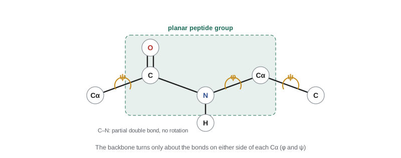
The peptide bond has a property that shapes every protein: it is flat. The lone pair of electrons on the nitrogen is shared with the carbonyl group, so the C–N bond has about 40% double-bond character. It is shorter than an ordinary C–N single bond (0.133 nm rather than 0.145 nm), and the six atoms of the peptide group, the Cα of one residue, the C and O of its carbonyl, the N and H of the next residue and that residue's Cα, lie in one plane. Rotation about the C–N bond is blocked. Almost all peptide bonds are in the trans form, with the two alpha carbons on opposite sides; the cis form is common only before proline.
Because the peptide groups are rigid plates, a polypeptide backbone can change shape only by rotating about the two single bonds on either side of each alpha carbon. These rotation angles are called phi (φ, about the N–Cα bond) and psi (ψ, about the Cα–C bond). Many combinations of φ and ψ make atoms collide, so only certain regions are allowed. A Ramachandran plot, introduced by G. N. Ramachandran in 1963, maps the allowed regions; the two largest correspond to the alpha helix and the beta sheet, which Lesson 4 describes.
Peptides with jobs of their own
Not every chain of amino acids is a large protein. Many short peptides are hormones, signals or defences. Oxytocin and vasopressin are nine residues long; insulin is two chains of 21 and 30 residues joined by disulfide bonds; glucagon has 29 residues. Glutathione, a tripeptide of glutamate, cysteine and glycine, is the main antioxidant inside cells, where its thiol group mops up reactive oxygen species. The artificial sweetener aspartame is the methyl ester of the dipeptide aspartyl-phenylalanine, about 200 times sweeter than sucrose. Peptide antibiotics made by bacteria and fungi, and the antimicrobial peptides of the human skin and gut, kill microbes by punching holes in their membranes.
A worked example: the charge of a peptide
Worked problem
Net charge and isoelectric point of a tripeptide
Estimate the net charge of the tripeptide Lys-Gly-Asp at pH 7.0, and its isoelectric point. Use pKa values of 9.0 for the N-terminal amino group, 10.5 for the lysine side chain, 3.9 for the aspartate side chain and 2.1 for the C-terminal carboxyl group.
Show solution
- List the ionizable groups: the N-terminal –NH₃⁺ (pKa 9.0) and the lysine side chain (10.5) can each carry +1; the aspartate side chain (3.9) and the C-terminal –COOH (2.1) can each carry −1. Glycine adds nothing.
- At pH 7.0 both basic groups are more than a unit below their pKa values, so they are almost fully protonated: +1 each, total about +2.
- Both acidic groups are well above their pKa values, so they are almost fully deprotonated: −1 each, total −2. The net charge is close to 0.
- For the exact value, the fraction protonated of a basic group is 1/(1 + 10^(pH − pKa)) and the fraction deprotonated of an acidic group is 1/(1 + 10^(pKa − pH)). At pH 7.0 the N-terminus contributes +0.990, lysine +0.9997, aspartate −0.9999 and the C-terminus −1.0000, a net charge of about −0.01.
- The pI lies between the two pKa values that bracket the neutral form. Starting from the fully protonated peptide (+2) and removing protons in order of increasing pKa: after the C-terminus (2.1) the charge is +1; after aspartate (3.9) it is 0; after the N-terminus (9.0) it is −1. The zero-charge form exists between pKa 3.9 and 9.0, so pI ≈ (3.9 + 9.0)/2 = 6.45.
Answer: The net charge at pH 7.0 is almost exactly zero (about −0.01), and the isoelectric point is about 6.5.
A program: the charge of any peptide
The program below calculates the net charge of a peptide at any pH from its one-letter sequence, using the same fractions as the worked example. It then finds the isoelectric point by bisection: it narrows a pH range, step by step, until the charge is zero. Finally it applies the method to three real sequences: the hormone oxytocin, a lysine-rich stretch of a histone, and an acidic stretch like those found in the stomach enzyme pepsin.
# pKa values of ionizable groups (typical values for free amino acids)
PKA_BASIC = {"Nterm": 9.0, "K": 10.5, "R": 12.5, "H": 6.0}
PKA_ACIDIC = {"Cterm": 2.1, "D": 3.9, "E": 4.1, "C": 8.3, "Y": 10.1}

def net_charge(seq, ph):
    groups_b = ["Nterm"] + [aa for aa in seq if aa in PKA_BASIC]
    groups_a = ["Cterm"] + [aa for aa in seq if aa in PKA_ACIDIC]
    plus = sum(1 / (1 + 10 ** (ph - PKA_BASIC[g])) for g in groups_b)
    minus = sum(1 / (1 + 10 ** (PKA_ACIDIC[g] - ph)) for g in groups_a)
    return plus - minus

def isoelectric_point(seq):
    lo, hi = 0.0, 14.0
    for _ in range(60):                 # bisection: charge falls as pH rises
        mid = (lo + hi) / 2
        if net_charge(seq, mid) > 0:
            lo = mid
        else:
            hi = mid
    return (lo + hi) / 2

print("Lys-Gly-Asp")
for ph in (1, 4, 7, 10, 13):
    print(f"  pH {ph:2d}: net charge {net_charge('KGD', ph):+.2f}")
print(f"  pI = {isoelectric_point('KGD'):.2f}")
print()
peptides = {
    "oxytocin (CYIQNCPLG)": "CYIQNCPLG",
    "histone tail (SGRGKGGKGLGKGGAKRHRKV)": "SGRGKGGKGLGKGGAKRHRKV",
    "acidic stretch (DEDLEEDVDE)": "DEDLEEDVDE",
}
for name, seq in peptides.items():
    print(f"{name}")
print(f" length {len(seq)}, charge at pH 7.4: {net_charge(seq, 7.4):+.2f}, pI = {isoelectric_point(seq):.2f}")Lys-Gly-Asp pH 1: net charge +1.93 pH 4: net charge +0.46 pH 7: net charge -0.01 pH 10: net charge -1.15 pH 13: net charge -2.00 pI = 6.45 oxytocin (CYIQNCPLG) length 9, charge at pH 7.4: -0.25, pI = 5.03 histone tail (SGRGKGGKGLGKGGAKRHRKV) length 21, charge at pH 7.4: +8.01, pI = 12.82 acidic stretch (DEDLEEDVDE) length 10, charge at pH 7.4: -8.02, pI = 2.53
The tripeptide's charge swings from +2 in strong acid to −2 in strong base, and the program's pI of 6.45 matches the hand calculation. Oxytocin is nearly neutral at blood pH; its terminal carboxyl is actually an amide in the real hormone, which would push its pI higher still, a reminder that simple models need checking against real structures. The histone tail carries a large positive charge, which is exactly what lets it grip DNA, and the acidic stretch has a pI below 3. Real proteins have hundreds of ionizable groups, but the same calculation, with pKa values adjusted for each group's surroundings, is how programs estimate the pI of a protein from its sequence.
Reading a protein sequence
Protein sequences are stored in databases as strings of one-letter codes, and much can be learned from the string alone. A stretch of about twenty hydrophobic residues in a row often marks a segment that crosses a membrane. Clusters of lysine and arginine suggest a site that binds DNA or RNA. Pairs of cysteines in a protein that is secreted from the cell are likely to form disulfide bonds. A sequence that begins with a run of hydrophobic residues may carry a signal that sends the protein out of the cell. Comparing the sequence with those of other organisms shows which residues have been kept unchanged by evolution, and those conserved residues are usually the ones that matter most for function.
Common mistakes
MistakeAmino acids exist in water mainly in the neutral form with –NH₂ and –COOH.
InsteadNear pH 7 they are zwitterions, –NH₃⁺ and –COO⁻; the fully neutral form is a tiny fraction at any pH.
MistakeAll twenty amino acids are chiral.
InsteadGlycine, whose side chain is a hydrogen atom, has two identical groups on its alpha carbon and is not chiral.
MistakeThe isoelectric point is the pH at which the amino acid has no charges.
InsteadAt the pI the average net charge is zero; most molecules still carry positive and negative charges that cancel.
MistakeA peptide bond can rotate freely like any single bond.
InsteadIts partial double-bond character makes the peptide group planar and rigid; the backbone rotates only about the φ and ψ bonds.
MistakeGly-Ala-Ser and Ser-Ala-Gly are the same peptide.
InsteadSequences have direction, written from the N-terminus to the C-terminus, so these are different molecules.
MistakeHistidine is always positively charged in proteins.
InsteadIts pKa is near 6, so at pH 7 it is mostly neutral, and its ability to switch is what makes it useful in active sites.
Summary and key terms
- An amino acid has an alpha carbon carrying an amino group, a carboxyl group, a hydrogen atom and a side chain; proteins use L-amino acids.
- Near pH 7 amino acids are zwitterions, and the net charge of an amino acid or protein depends on pH through the pKa values of its groups.
- The twenty side chains fall into nonpolar, aromatic, polar, positively charged and negatively charged groups that decide how proteins fold and act.
- Peptide bonds join amino acids from the N-terminus to the C-terminus; they are stable but can be hydrolysed by proteases.
- The peptide group is planar, so backbone shape is set by the φ and ψ angles at each alpha carbon.
- Zwitterion
- A molecule carrying both a positive and a negative charge with no net charge.
- Isoelectric point (pI)
- The pH at which a molecule has zero average net charge.
- Side chain (R group)
- The part of an amino acid that differs from one amino acid to another.
- Peptide bond
- The amide linkage formed between the carboxyl group of one amino acid and the amino group of the next.
- Residue
- An amino acid unit within a peptide or protein chain.
- N-terminus
- The end of a polypeptide with a free amino group, where its sequence begins.
- Essential amino acid
- An amino acid the body cannot make and must obtain from food.
Practice problems
Work each problem on paper first, then reveal the answer to check.
What is the net charge of alanine (pKa 2.34 and 9.69) at pH 1, 6 and 12?
Show answer
About +1 at pH 1, 0 at pH 6 and −1 at pH 12.
Calculate the pI of alanine.
Show answer
pI = (2.34 + 9.69)/2 = 6.02.
Which three amino acids absorb most of the 280 nm light used to measure protein concentration?
Show answer
Tryptophan and tyrosine absorb most strongly; phenylalanine absorbs weakly, and disulfide bonds contribute a little.
Write the one-letter sequence of Met-Lys-Trp-Val-Thr.
Show answer
MKWVT.
Why are histones rich in lysine and arginine?
Show answer
Their positive side chains bind the negatively charged phosphate backbone of DNA.
How many peptide bonds and how many water molecules are involved in making a protein of 150 residues?
Show answer
149 peptide bonds, with 149 water molecules released.
Lesson quiz · 5 questions
Score 60% or more to complete this lesson.
Create a free account to save your progress, take quizzes and earn a certificate.