
BIOC-200 · Lesson 5 of 12 · 25 min
Enzymes: how biological catalysts work
▶ Video coming soon A video version of this lesson is on the way.
Almost every chemical reaction in a cell is catalysed by an enzyme. Without enzymes, the reactions of metabolism would run so slowly that life would be impossible: the uncatalysed hydrolysis of the phosphate ester bonds in DNA has a half-life estimated in the tens of millions of years, and the decarboxylation of one nucleotide precursor would take about 78 million years. Enzymes speed reactions by factors of a million to more than 10¹⁷, they work in water at body temperature and neutral pH, they are highly specific for their substrates, and their activity can be switched up and down to suit the cell's needs. This lesson introduces how enzymes achieve these feats, how their rates are measured and described, and how they are inhibited and controlled. BIOC-320 takes the subject much further.
What an enzyme is
Most enzymes are proteins, although a few catalytic RNA molecules, called ribozymes, also exist; the most important is the RNA at the heart of the ribosome that forms peptide bonds. An enzyme binds its reactant, called the substrate, in a pocket or cleft called the active site, converts it to product and releases the product, emerging unchanged and ready to act again. The active site is usually a small part of the protein, lined with a few side chains that bind the substrate and others that take part in the chemistry. The rest of the protein holds those side chains in precisely the right positions.
Many enzymes need a partner that is not an amino acid. Cofactors are metal ions such as Zn²⁺, Mg²⁺ and Fe²⁺ or small organic molecules called coenzymes. Many coenzymes are made from vitamins: NAD⁺ from niacin, FAD from riboflavin, coenzyme A from pantothenic acid, thiamine pyrophosphate from thiamine and pyridoxal phosphate from vitamin B6. A coenzyme bound permanently to its enzyme is called a prosthetic group, as the heme of hemoglobin is. The protein without its cofactor is the apoenzyme, and the complete, active enzyme is the holoenzyme.
Enzymes are named by adding -ase to the name of the substrate or reaction, so urease hydrolyses urea and alcohol dehydrogenase removes hydrogen from alcohols. Older names such as pepsin and trypsin survive. The International Union of Biochemistry and Molecular Biology classifies enzymes into seven classes by the reaction they catalyse: oxidoreductases, transferases, hydrolases, lyases, isomerases, ligases and translocases. Each enzyme has a four-part EC number; alcohol dehydrogenase, for example, is EC 1.1.1.1.
Enzymes lower the activation energy
An enzyme does not change the equilibrium of a reaction. The equilibrium constant depends only on the difference in Gibbs energy between reactants and products, and a catalyst speeds the forward and reverse reactions by the same factor. What an enzyme changes is the activation energy, the height of the free energy barrier that molecules must climb to react. The rate of a reaction depends exponentially on that barrier, so even a modest lowering has a large effect: at 37 °C, lowering the activation free energy by 5.9 kJ/mol speeds a reaction tenfold, and lowering it by 34 kJ/mol speeds it about a millionfold.
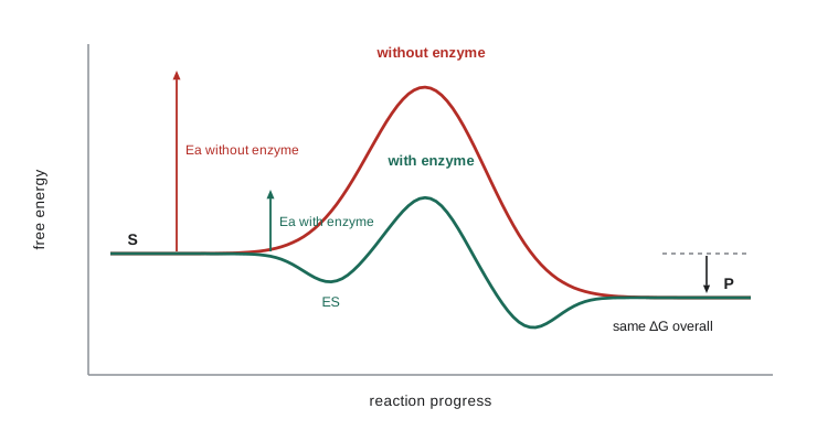
Enzymes lower barriers in several ways at once. They bind the substrate in the right orientation and bring reacting groups close together, removing the entropy cost of collision; this alone can speed a reaction by a factor of thousands. Their side chains act as acids and bases, donating and accepting protons at just the right moment, a mechanism called general acid-base catalysis. Some side chains, such as the serine of serine proteases, form a temporary covalent bond with the substrate, which is covalent catalysis. Metal ions stabilize negative charges and can activate water molecules. Above all, the active site binds the transition state, the fleeting, high-energy arrangement of atoms at the top of the barrier, more tightly than it binds the substrate itself. Linus Pauling proposed this idea in 1946, and it explains why molecules designed to resemble transition states make potent inhibitors.
Specificity was first pictured by Emil Fischer in 1894 as a lock and a key: the active site has a shape that fits only its substrate. Daniel Koshland's induced-fit model of 1958 refined the picture: the enzyme changes shape when the substrate binds, closing around it. Hexokinase, the first enzyme of glycolysis, is a classic example. Its two lobes swing together when glucose binds, shutting out water so that the phosphate of ATP is transferred to glucose rather than wasted on water.
A case study: chymotrypsin
Chymotrypsin, a digestive enzyme made by the pancreas, cuts peptide bonds on the carboxyl side of large hydrophobic residues such as phenylalanine, tyrosine and tryptophan. Its active site contains three residues that work as a team, called the catalytic triad: serine 195, histidine 57 and aspartate 102. Aspartate holds histidine in the right orientation and charge state; histidine pulls a proton off serine, making its oxygen a strong nucleophile that attacks the carbonyl carbon of the peptide bond. A tetrahedral intermediate forms, its negative oxygen stabilized by hydrogen bonds in a pocket called the oxyanion hole. The bond breaks, the first piece of the substrate leaves, and a water molecule, activated by the same histidine, then frees the second piece. A deep hydrophobic pocket next to the active site holds the aromatic side chain and explains the enzyme's specificity. The same triad occurs in hundreds of other enzymes, including trypsin, elastase, thrombin and many lipases, a striking example of evolution arriving at one good solution repeatedly.
Measuring enzyme activity
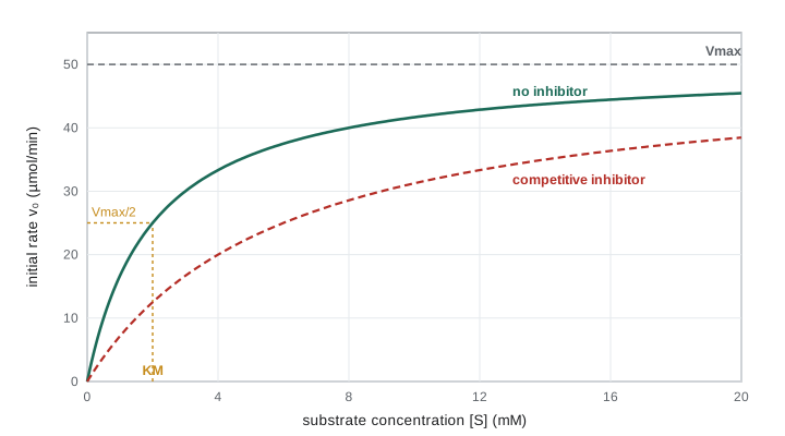
Enzyme rates are measured as initial velocities: the rate of product formation at the very start of a reaction, before much substrate is used up or product accumulates. Measured at a series of substrate concentrations, the initial velocity v₀ shows a characteristic pattern. At low substrate concentration the rate rises in proportion to [S]; at high concentration it levels off at a maximum, Vmax, because every enzyme molecule is busy. This saturation behaviour shows that the enzyme and substrate form a complex before the product is made.
In 1913 Leonor Michaelis and Maud Menten described this behaviour with a simple scheme. The enzyme E binds the substrate S to form a complex ES, which either falls apart again or goes on to form product P and free enzyme. Assuming that the concentration of ES quickly reaches a steady state, as George Briggs and J. B. S. Haldane showed in 1925, leads to the Michaelis–Menten equation:
The Michaelis constant KM is the substrate concentration at which the rate is half of Vmax. It has units of concentration and, roughly speaking, measures how much substrate the enzyme needs to work well: a low KM means the enzyme reaches half its maximum rate at low substrate concentrations. Typical values lie between about 1 µM and 10 mM, and many enzymes have KM values close to the normal concentration of their substrate in the cell, so their rates respond to changes in supply.
Vmax depends on how much enzyme is present. Dividing it by the total enzyme concentration gives the turnover number, kcat, the number of substrate molecules each active site converts per second when saturated. Turnover numbers range from less than one per second for some enzymes to about 600,000 per second for carbonic anhydrase, which converts carbon dioxide to bicarbonate in red blood cells. The ratio kcat/KM, called the specificity constant, measures how efficiently an enzyme works at low substrate concentrations. Its upper limit, about 10⁸ to 10⁹ per molar per second, is set by how fast substrate molecules can diffuse to the enzyme. A few enzymes, among them triose phosphate isomerase, fumarase and superoxide dismutase, operate close to that limit: they convert nearly every substrate molecule that bumps into them, and they have been called catalytically perfect.
Before computers were common, biochemists found KM and Vmax by turning the curve into a straight line. Taking the reciprocal of both sides gives the Lineweaver–Burk equation, 1/v₀ = (KM/Vmax)(1/[S]) + 1/Vmax, and a plot of 1/v₀ against 1/[S] is a line whose intercept on the y-axis is 1/Vmax and whose slope is KM/Vmax. The plot is still useful for seeing how inhibitors act, but because the reciprocals exaggerate errors in the slowest measurements, the parameters are now usually found by fitting the curve directly.
Temperature and pH
Enzyme rates rise with temperature, roughly doubling for each 10 °C rise, until the enzyme starts to denature and activity falls sharply. Human enzymes usually work best near 37 °C; enzymes from bacteria in hot springs may work best above 80 °C. Each enzyme also has an optimum pH, set by the ionization of side chains it needs for binding and catalysis. Pepsin, which works in the acid of the stomach, has its optimum near pH 2; trypsin, which works in the small intestine, near pH 8; most enzymes of the cytosol near pH 7.
Inhibition
Enzyme inhibitors are molecules that slow enzyme reactions, and many medicines and poisons work this way. Reversible inhibitors bind with weak interactions and can leave the enzyme again. A competitive inhibitor resembles the substrate and competes for the active site. It raises the apparent KM, because more substrate is needed to outcompete it, but it leaves Vmax unchanged, because enough substrate will always displace it. Statins, which lower blood cholesterol, are competitive inhibitors of HMG-CoA reductase; methotrexate, a cancer drug, competes with the substrate of dihydrofolate reductase.
A noncompetitive inhibitor binds elsewhere on the enzyme and reduces its ability to convert substrate, lowering Vmax without changing KM. An uncompetitive inhibitor binds only to the enzyme-substrate complex and lowers both. These patterns can be distinguished by measuring rates at several substrate and inhibitor concentrations: competitive inhibition gives Lineweaver–Burk lines that meet on the y-axis, noncompetitive inhibition gives lines that meet on the x-axis, and uncompetitive inhibition gives parallel lines.
Irreversible inhibitors form covalent bonds with the enzyme and inactivate it permanently. Aspirin acetylates a serine in the active site of cyclooxygenase, blocking the synthesis of prostaglandins that cause pain, fever and inflammation. Penicillin binds covalently to the enzyme that builds the cross-links of bacterial cell walls. Nerve agents and some insecticides phosphorylate the active-site serine of acetylcholinesterase, which is why they are so dangerous to the nervous system.
Regulating enzymes
A cell must control its enzymes, switching pathways on when their products are needed and off when they are not. It does so in several ways. It can change the amount of an enzyme by making more or by breaking it down, which takes minutes to hours. It can activate inactive precursors called zymogens by cutting them; the digestive proteases are made as zymogens such as trypsinogen so that they do not digest the pancreas that makes them. It can attach and remove chemical groups, most often phosphate: protein kinases add phosphate to serine, threonine or tyrosine side chains and protein phosphatases remove it, switching enzymes on or off within seconds.
The fastest control is allosteric regulation. Allosteric enzymes have regulatory sites, separate from the active site, where molecules called effectors bind and change the enzyme's shape and activity. Like hemoglobin, many allosteric enzymes have several subunits and show sigmoid rather than hyperbolic kinetics. A common pattern is feedback inhibition: the final product of a pathway inhibits the enzyme of the first committed step, so the pathway slows down when its product accumulates. In bacteria, the amino acid isoleucine inhibits threonine deaminase, the first enzyme of the five-step pathway that makes it, a case discovered in 1956 and one of the first examples of feedback control. Lesson 10 shows how allosteric control of phosphofructokinase governs the speed of glycolysis.
A worked example: Michaelis–Menten calculations
Worked problem
Using KM and Vmax
An enzyme has KM = 2.0 mM and Vmax = 50 µmol/min with a given amount of enzyme. (a) What is the rate at [S] = 0.5 mM, 2.0 mM and 20 mM? (b) A competitive inhibitor raises the apparent KM to 6.0 mM. What is the rate at [S] = 2.0 mM with the inhibitor present?
Show solution
- At 0.5 mM: v₀ = 50 × 0.5/(2.0 + 0.5) = 25/2.5 = 10 µmol/min, a fifth of Vmax.
- At 2.0 mM, where [S] = KM: v₀ = 50 × 2.0/4.0 = 25 µmol/min, exactly half of Vmax, as the definition of KM requires.
- At 20 mM: v₀ = 50 × 20/22 = 45.5 µmol/min, 91% of Vmax. Raising [S] tenfold above KM brings the rate close to, but never quite to, the maximum.
- With the inhibitor, Vmax is unchanged but KM(apparent) = 6.0 mM: v₀ = 50 × 2.0/(6.0 + 2.0) = 12.5 µmol/min, half the uninhibited rate.
- Raising [S] to 20 mM with inhibitor gives 50 × 20/26 = 38.5 µmol/min, showing that a high substrate concentration overcomes much of the competitive inhibition.
Answer: (a) 10, 25 and 45.5 µmol/min. (b) 12.5 µmol/min, half the uninhibited rate at 2.0 mM.
A program: fitting enzyme kinetics
Biochemists measure initial velocities and then find KM and Vmax that best fit them. The program generates measurements from an enzyme with KM = 2.0 mM and Vmax = 50 µmol/min, adding small fixed measurement errors, and recovers the parameters two ways: from a Lineweaver–Burk line fitted by least squares, and by a direct search for the KM and Vmax that minimize the squared error of the Michaelis–Menten curve itself. It then compares the rates with and without a competitive inhibitor.
def mm(s, vmax, km):
    return vmax * s / (km + s)

S = [0.25, 0.5, 1.0, 2.0, 4.0, 8.0, 16.0]          # substrate, mM
noise = [1.06, 0.97, 1.03, 0.98, 1.02, 0.99, 1.01]  # fixed "measurement errors"
V = [mm(s, 50.0, 2.0) * f for s, f in zip(S, noise)]

# 1. Lineweaver-Burk: straight line through (1/S, 1/v) by least squares
x = [1 / s for s in S]
y = [1 / v for v in V]
n = len(x)
mx, my = sum(x) / n, sum(y) / n
slope = sum((a - mx) * (b - my) for a, b in zip(x, y)) / sum((a - mx) ** 2 for a in x)
intercept = my - slope * mx
vmax_lb = 1 / intercept
km_lb = slope * vmax_lb

# 2. Direct fit: search a grid of Vmax and KM for the smallest squared error
best = None
for i in range(300, 701):
    vmax = i / 10
    for j in range(50, 501):
        km = j / 100
        err = sum((mm(s, vmax, km) - v) ** 2 for s, v in zip(S, V))
        if best is None or err < best[0]:
            best = (err, vmax, km)

print("[S] (mM)   v0 (umol/min)")
for s, v in zip(S, V):
    print(f"{s:8.2f} {v:14.2f}")
print(f"Lineweaver-Burk fit: Vmax = {vmax_lb:.1f}, KM = {km_lb:.2f} mM")
print(f"Direct fit:          Vmax = {best[1]:.1f}, KM = {best[2]:.2f} mM")
print("True values:         Vmax = 50.0, KM = 2.00 mM")
print()
print("[S] (mM)   no inhibitor   competitive inhibitor (apparent KM 6.0 mM)")
for s in (0.5, 2.0, 8.0, 32.0):
print(f"{s:8.1f} {mm(s, 50, 2.0):14.1f} {mm(s, 50, 6.0):14.1f}")[S] (mM) v0 (umol/min)
0.25 5.89
0.50 9.70
1.00 17.17
2.00 24.50
4.00 34.00
8.00 39.60
16.00 44.89
Lineweaver-Burk fit: Vmax = 47.2, KM = 1.79 mM
Direct fit: Vmax = 50.2, KM = 2.00 mM
True values: Vmax = 50.0, KM = 2.00 mM
[S] (mM) no inhibitor competitive inhibitor (apparent KM 6.0 mM)
0.5 10.0 3.8
2.0 25.0 12.5
8.0 40.0 28.6
32.0 47.1 42.1Both fits come close to the true values, but the direct fit is better because it treats every measurement fairly. The Lineweaver–Burk line gives the most weight to the measurement at the lowest substrate concentration, whose reciprocal is largest and whose error is magnified the most. The second table shows the signature of competitive inhibition: the inhibitor cuts the rate sharply at low substrate concentrations, but at 32 mM the inhibited enzyme runs within about 10% of the uninhibited rate, and more substrate would close the gap further.
Common mistakes
MistakeEnzymes make unfavourable reactions favourable.
InsteadEnzymes speed reactions but do not change the equilibrium; an unfavourable reaction must be coupled to a favourable one, for example ATP hydrolysis.
MistakeKM is the substrate concentration at which the enzyme reaches its maximum rate.
InsteadKM is the concentration at which the rate is half of Vmax; the maximum is approached only at concentrations far above KM.
MistakeA competitive inhibitor lowers Vmax.
InsteadIt raises the apparent KM; enough substrate outcompetes it, so Vmax is unchanged.
MistakeRates should be measured after the reaction has run for some time.
InsteadKinetics uses initial rates, measured before substrate is depleted or product builds up.
MistakeEnzymes are always proteins.
InsteadMost are, but ribozymes such as the ribosomal RNA that forms peptide bonds are catalytic RNA molecules.
MistakeAllosteric effectors bind at the active site.
InsteadThey bind at separate regulatory sites and change the enzyme's shape and activity.
Summary and key terms
- Enzymes are catalysts, mostly proteins, that speed reactions enormously by lowering the activation energy without changing the equilibrium.
- They work by binding substrates in the right orientation, acid-base and covalent catalysis, metal ions and, above all, tight binding of the transition state.
- Michaelis–Menten kinetics describe saturation: v₀ = Vmax[S]/(KM + [S]); kcat is the turnover number and kcat/KM measures efficiency.
- Competitive inhibitors raise the apparent KM, noncompetitive inhibitors lower Vmax, and irreversible inhibitors such as aspirin inactivate enzymes covalently.
- Cells control enzymes by changing their amount, activating zymogens, phosphorylation and allosteric regulation, including feedback inhibition.
- Active site
- The region of an enzyme where the substrate binds and the reaction takes place.
- Coenzyme
- A small organic molecule, often made from a vitamin, that an enzyme needs for activity.
- Transition state
- The highest-energy arrangement of atoms along the path from reactant to product.
- Michaelis constant (KM)
- The substrate concentration at which the reaction rate is half of Vmax.
- Turnover number (kcat)
- The number of substrate molecules converted per active site per second at saturation.
- Competitive inhibitor
- An inhibitor that competes with the substrate for the active site.
- Allosteric regulation
- Control of an enzyme by an effector binding at a site other than the active site.
Practice problems
Work each problem on paper first, then reveal the answer to check.
An enzyme has KM = 0.5 mM. At what substrate concentration does it run at 90% of Vmax?
Show answer
0.9 = [S]/(0.5 + [S]) gives [S] = 4.5 mM, nine times KM.
A solution contains 2 nM of an enzyme with Vmax = 1.2 µM per second. What is kcat?
Show answer
kcat = 1.2 µM/s ÷ 0.002 µM = 600 per second.
A Lineweaver–Burk plot has a y-intercept of 0.02 min/µM and a slope of 0.05 min. Find Vmax and KM.
Show answer
Vmax = 1/0.02 = 50 µM/min; KM = slope × Vmax = 0.05 × 50 = 2.5 µM.
Why does aspirin keep working after the drug has left the blood?
Show answer
It acetylates cyclooxygenase irreversibly, so the inactivated enzyme molecules stay inactive until new ones are made.
Why are digestive proteases made as zymogens?
Show answer
So they cannot digest the cells that make them; they are activated only once they reach the intestine.
How much faster is a reaction at 37 °C if an enzyme lowers its activation energy by 17.8 kJ/mol?
Show answer
The factor is e^(17,800/(8.314 × 310)) = e^6.9 ≈ 1,000.
Lesson quiz · 5 questions
Score 60% or more to complete this lesson.
Create a free account to save your progress, take quizzes and earn a certificate.