
BIOC-200 · Lesson 12 of 12 · 24 min
Capstone: fats, amino acids and the integration of metabolism
▶ Video coming soon A video version of this lesson is on the way.
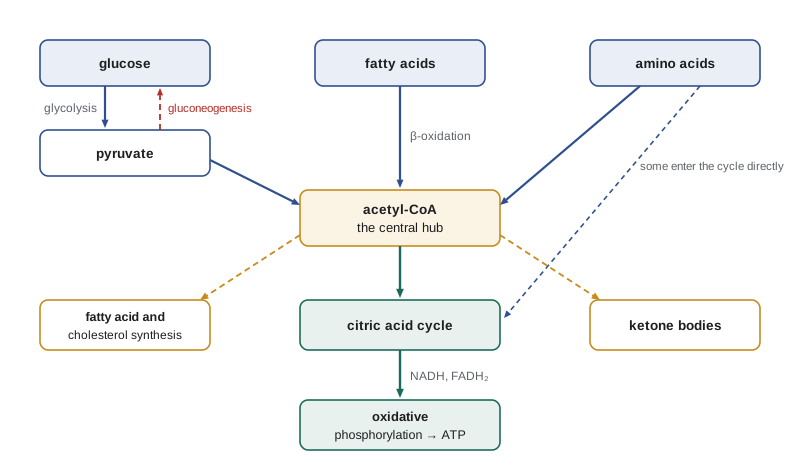
The last three lessons followed glucose from the cytosol to the mitochondria and to oxygen. But glucose is only one fuel. People also burn fat and protein, and they switch between fuels many times a day, after a meal, overnight, during a run, on a long fast. They also build: when food is plentiful, the body stores fat and glycogen, and every cell continually makes new proteins, membranes and nucleic acids. This capstone lesson surveys how fats and amino acids are broken down, how glucose can be made when it is scarce, and how the organs of the body, directed by hormones, share fuels among themselves. It ties together the molecules, enzymes and energy carriers of the whole course.
Burning fat: beta-oxidation
Stored triacylglycerols are first broken down by lipases in fat cells into glycerol and free fatty acids, which travel through the blood bound to the protein albumin. Glycerol goes to the liver, where it is converted to DHAP and enters glycolysis or gluconeogenesis. Fatty acids are taken up by muscle, heart, liver and other tissues. Inside a cell, a fatty acid is activated by attaching it to coenzyme A, at the cost of two phosphoanhydride bonds of ATP, and carried into the mitochondrial matrix by a shuttle that uses the small molecule carnitine.
In the matrix, the fatty acyl-CoA is broken down by beta-oxidation, a repeating sequence of four reactions that removes two carbons at a time as acetyl-CoA. The sequence is oxidation by FAD, addition of water, oxidation by NAD⁺ and cleavage by coenzyme A, and its chemistry closely mirrors the last steps of the citric acid cycle, from succinate to oxaloacetate. Each round produces one FADH₂, one NADH and one acetyl-CoA and leaves a fatty acyl-CoA two carbons shorter, which goes round again. Palmitate, with 16 carbons, goes through seven rounds to give eight acetyl-CoA, seven FADH₂ and seven NADH. The acetyl-CoA enters the citric acid cycle, and the reduced carriers feed the respiratory chain.
The payoff is large. Palmitate yields about 106 ATP, compared with about 32 for glucose, even though it has only 16 carbons to glucose's 6. That is because its carbons are almost fully reduced, as Lesson 7 explained. The program below works through the arithmetic.
Ketone bodies
When fatty acids are broken down faster than the citric acid cycle can use the acetyl-CoA, as happens in the liver during fasting, the liver turns the excess into ketone bodies: acetoacetate and beta-hydroxybutyrate, with a little acetone. They are water-soluble, travel freely in the blood and are taken up by heart, muscle and, after a few days of fasting, the brain, which converts them back to acetyl-CoA and burns it. During a long fast ketone bodies can supply about two thirds of the brain's energy, sparing glucose and therefore sparing the body's protein, which would otherwise be broken down to make glucose. In untreated type 1 diabetes, the lack of insulin lets fat breakdown run unchecked, and ketone bodies, which are acids, can accumulate to dangerous levels in diabetic ketoacidosis; the acetone gives the breath a fruity smell.
Amino acids: carbon and nitrogen
The body has no store of amino acids comparable with glycogen or fat. Amino acids from food or from the turnover of the body's own proteins that are not needed to make new proteins are broken down. Their carbon skeletons are burned for energy or converted into glucose or fat, and their nitrogen must be disposed of, because ammonia is toxic, especially to the brain.
The first step for most amino acids is transamination: an enzyme called an aminotransferase moves the amino group onto alpha-ketoglutarate, making glutamate and leaving the carbon skeleton as a keto acid. Aminotransferases use pyridoxal phosphate, from vitamin B6, as their coenzyme. Two of them, alanine aminotransferase and aspartate aminotransferase, are measured in blood tests, because they leak into the blood when liver cells are damaged. Glutamate then releases its amino group as ammonia in the liver mitochondria. The liver converts the ammonia, together with carbon dioxide and the nitrogen of aspartate, into urea in the urea cycle, which Hans Krebs and Kurt Henseleit described in 1932, five years before the citric acid cycle. Urea is carried in the blood to the kidneys and excreted. Fish excrete ammonia directly into the water, and birds and reptiles convert it to uric acid, which needs little water to excrete.
The carbon skeletons of the twenty amino acids are converted into seven molecules: pyruvate, acetyl-CoA, acetoacetyl-CoA, alpha-ketoglutarate, succinyl-CoA, fumarate and oxaloacetate. Amino acids whose skeletons become pyruvate or citric acid cycle intermediates can be used to make glucose and are called glucogenic. Those that become acetyl-CoA or acetoacetyl-CoA can make only ketone bodies or fat and are called ketogenic; leucine and lysine are purely ketogenic. Inherited defects in these pathways cause disease. In phenylketonuria, a deficiency of the enzyme that converts phenylalanine to tyrosine lets phenylalanine build up and damage the developing brain. Newborn babies in Canada are screened for it with a drop of blood, and a diet low in phenylalanine, started early, prevents the damage.
Making glucose: gluconeogenesis
The brain uses about 120 grams of glucose a day, and red blood cells, which have no mitochondria, can use nothing else. The liver's glycogen lasts only about a day without food, so the body must be able to make new glucose. This is gluconeogenesis, which takes place mainly in the liver, with some in the kidneys. Its starting materials are lactate from muscle and red cells, glycerol from fat, and the glucogenic amino acids, mainly alanine from muscle. Fatty acids cannot be used, because, as Lesson 11 explained, the pyruvate dehydrogenase reaction cannot be reversed.
Gluconeogenesis is not simply glycolysis run backward. Seven of the ten steps of glycolysis are near equilibrium and are shared, running in reverse. The three irreversible steps must be bypassed by different reactions. Pyruvate kinase is bypassed in two steps: pyruvate carboxylase makes oxaloacetate from pyruvate in the mitochondria, and PEP carboxykinase converts oxaloacetate into PEP, using GTP. PFK-1 is bypassed by fructose 1,6-bisphosphatase, which simply removes the phosphate by hydrolysis. Hexokinase is bypassed by glucose 6-phosphatase, found in liver and kidney but not muscle, which is why muscle glycogen fuels only muscle while liver glycogen can supply the blood. Making one glucose from two pyruvate costs six phosphoanhydride bonds, four ATP and two GTP, compared with the two ATP gained when glycolysis runs the other way. The extra cost is the price of making both directions favourable, so that each can be controlled independently, as Lesson 9 described.
The two pathways are controlled reciprocally, so that one runs when the other is off. Fructose 2,6-bisphosphate, which activates PFK-1, inhibits fructose 1,6-bisphosphatase; AMP does the same. Acetyl-CoA, a sign that fatty acids are being burned, activates pyruvate carboxylase and so diverts pyruvate toward glucose. Hormones set the overall direction, as the next section explains.
Building: storage and synthesis
When food supplies more energy than the body needs, the excess is stored. Glucose is built into glycogen by glycogen synthase, which adds glucose units from an activated form, UDP-glucose, to the ends of the chains, while a branching enzyme creates the α(1→6) branches. Once glycogen stores are full, extra glucose, and also extra amino acids, are converted to fat. Acetyl-CoA, exported from the mitochondria as citrate, is built into fatty acids in the cytosol by fatty acid synthase, using ATP and NADPH; much of the NADPH comes from the pentose phosphate pathway, a branch of glucose metabolism that also makes ribose 5-phosphate for nucleotides. The fatty acids are attached to glycerol to make triacylglycerols, which the liver packages into lipoproteins and sends to fat tissue for storage. Cholesterol is also made from acetyl-CoA, in a pathway whose controlling enzyme, HMG-CoA reductase, is the target of the statin drugs mentioned in Lesson 5.
Organs and hormones
Each organ has its own metabolic personality. The brain burns glucose, or ketone bodies during a long fast, and uses about a fifth of the body's resting energy. Skeletal muscle burns glucose, fatty acids and ketone bodies and keeps glycogen for its own use; during intense exercise it relies on its glycogen and phosphocreatine and makes lactate. Heart muscle burns mostly fatty acids and is rich in mitochondria. Fat tissue stores triacylglycerols after meals and releases fatty acids between them. The liver is the body's metabolic manager: it stores and releases glucose, makes ketone bodies, makes urea and processes almost everything absorbed from the gut.
Hormones coordinate the organs. Insulin, made by the beta cells of the pancreas when blood glucose rises after a meal, signals plenty. It speeds glucose uptake into muscle and fat by moving GLUT4 transporters to the cell surface, activates glycogen synthesis and fat synthesis, and inhibits gluconeogenesis and fat breakdown. Glucagon, made by the alpha cells of the pancreas when blood glucose falls, signals scarcity to the liver: it stimulates glycogen breakdown and gluconeogenesis and inhibits glycolysis by lowering fructose 2,6-bisphosphate. Adrenaline, released in stress and exercise, mobilizes glycogen in muscle and liver and fat in fat tissue within seconds. Cortisol, over hours, promotes the breakdown of muscle protein to supply amino acids for gluconeogenesis.
A day and a fast
Following one person through time shows these pieces at work. In the hours after a meal, insulin is high, the liver and muscle fill their glycogen stores, and the liver makes fat from excess carbohydrate. Overnight, insulin falls and glucagon rises; the liver releases glucose from its glycogen to keep blood glucose near 5 mM, and fat tissue begins to release fatty acids, which most tissues burn instead of glucose. By the next morning, after about 12 to 16 hours, gluconeogenesis is supplying a growing share of blood glucose. After two or three days without food, liver glycogen is gone, gluconeogenesis from amino acids and glycerol supplies the glucose, and the liver makes ketone bodies at a high rate. After a week or more, the brain gets most of its energy from ketone bodies, gluconeogenesis slows, and protein breakdown falls, preserving muscle. A person of normal weight carries enough fat to survive for one to two months in this state; how long depends mainly on how much fat they carry.
Type 2 diabetes, the most common form of the disease, shows what happens when this coordination breaks down. Tissues respond less to insulin, a state called insulin resistance, and the pancreas eventually cannot make enough extra insulin to compensate. Muscle and fat take up less glucose, the liver keeps making glucose as if the body were fasting, and blood glucose stays high. Treatments work at many of the points described in this course: metformin reduces gluconeogenesis in the liver, other drugs increase insulin release or block glucose reabsorption in the kidneys, and diet and exercise improve the response to insulin.
A worked example: the energy of palmitate
Worked problem
ATP from one molecule of palmitate
Calculate the ATP yield from the complete oxidation of palmitate (16 carbons), using 2.5 ATP per NADH, 1.5 per FADH₂ and 10 per acetyl-CoA oxidized in the citric acid cycle, and remembering that activation costs the equivalent of 2 ATP.
Show solution
- Rounds of beta-oxidation: a 16-carbon chain needs 16/2 − 1 = 7 rounds and gives 8 acetyl-CoA.
- From beta-oxidation: 7 FADH₂ × 1.5 = 10.5 ATP and 7 NADH × 2.5 = 17.5 ATP, total 28 ATP.
- From the citric acid cycle: 8 acetyl-CoA × 10 = 80 ATP.
- Gross yield = 28 + 80 = 108 ATP. Subtract 2 for activation (ATP → AMP + 2 Pi uses two phosphoanhydride bonds): net 106 ATP.
- Per carbon, palmitate gives 106/16 = 6.6 ATP, compared with 32/6 = 5.3 for glucose.
Answer: About 106 ATP per palmitate, roughly 6.6 ATP per carbon against 5.3 for glucose.
A program: fuels and a fast
The program first generalizes the worked example to saturated fatty acids of any even length and compares their ATP yield per carbon with glucose. It then runs a simple model of a long fast, estimating how many days a person's fat could cover their energy needs.
def fatty_acid_atp(carbons, nadh=2.5, fadh2=1.5, per_acetyl=10, activation=2):
    rounds = carbons // 2 - 1
    acetyl = carbons // 2
    return rounds * (fadh2 + nadh) + acetyl * per_acetyl - activation

print("fuel                  carbons   ATP   ATP per carbon")
print(f"{'glucose':20s} {6:8d} {32:5.0f} {32 / 6:15.2f}")
for name, c in (("lauric acid", 12), ("palmitic acid", 16), ("stearic acid", 18), ("arachidic acid", 20)):
    atp = fatty_acid_atp(c)
    print(f"{name:20s} {c:8d} {atp:5.0f} {atp / c:15.2f}")

# A long fast: how long would stored fat last?
energy_per_day = 7500.0      # kJ per day, a reduced need during fasting
fat_kg = 12.0                # stored triacylglycerol
fat_energy = fat_kg * 1000 * 37.0          # kJ, at 37 kJ per gram
glycogen_energy = 500 * 17.0               # kJ, about 500 g of glycogen
print()
print(f"glycogen lasts about {glycogen_energy / energy_per_day:.1f} days")
days = 0
reserve = fat_energy * 0.8                 # keep 20% of fat as a minimum
while reserve > 0:
    reserve -= energy_per_day * 0.9        # fat covers about 90% of needs
    days += 1
print(f"{fat_kg:.0f} kg of fat covers about {days} days of fasting")fuel carbons ATP ATP per carbon glucose 6 32 5.33 lauric acid 12 78 6.50 palmitic acid 16 106 6.62 stearic acid 18 120 6.67 arachidic acid 20 134 6.70 glycogen lasts about 1.1 days 12 kg of fat covers about 53 days of fasting
The ATP per carbon rises slightly with chain length because the activation cost is spread over more carbons, and all fatty acids yield more ATP per carbon than glucose. The fasting model is rough, since energy needs fall and the mix of fuels shifts over the course of a fast, but its answer, about seven weeks for 12 kg of fat, is in line with recorded cases of people surviving long fasts under medical supervision. Glycogen, by contrast, lasts about a day. The comparison sums up why animals store most of their energy as fat.
Looking back
This course began with water and the elements of life and ended with the coordinated metabolism of a whole person. Along the way the same ideas returned again and again. Weak interactions and the hydrophobic effect fold proteins, build membranes and stack the bases of DNA. Enzymes speed reactions by binding transition states and are controlled by allosteric effectors, phosphorylation and hormones. Free energy decides which way reactions go, ATP and the electron carriers move energy from where it is released to where it is needed, and irreversible steps are where pathways are controlled. The later biochemistry courses build on each of these foundations.
Common mistakes
MistakeFat can be converted into glucose when glucose is scarce.
InsteadFatty acids give acetyl-CoA, which cannot be converted to pyruvate; only the glycerol part of fat can make glucose.
MistakeGluconeogenesis is glycolysis run in reverse.
InsteadIt shares seven reversible steps but bypasses the three irreversible ones with different enzymes.
MistakeMuscle glycogen can raise blood glucose.
InsteadMuscle lacks glucose 6-phosphatase, so its glycogen fuels only the muscle itself.
MistakeKetone bodies are always a sign of disease.
InsteadThey are a normal fuel during fasting; only the uncontrolled excess of diabetic ketoacidosis is dangerous.
MistakeThe body stores surplus amino acids as protein for later use.
InsteadThere is no protein store; surplus amino acids are broken down, with their nitrogen excreted as urea.
MistakeInsulin and glucagon have the same effects on the liver.
InsteadInsulin signals plenty and promotes storage; glucagon signals scarcity and promotes glucose release and gluconeogenesis.
Summary and key terms
- Fatty acids are broken down by beta-oxidation to acetyl-CoA, NADH and FADH₂; palmitate yields about 106 ATP. The liver turns excess acetyl-CoA into ketone bodies during fasting.
- Amino acids lose their nitrogen by transamination and the urea cycle; their carbon skeletons become glucogenic or ketogenic intermediates.
- Gluconeogenesis makes glucose from lactate, glycerol and amino acids, bypassing the three irreversible steps of glycolysis, and is controlled reciprocally with glycolysis.
- Surplus fuel is stored as glycogen and, through acetyl-CoA, NADPH and fatty acid synthase, as fat.
- Insulin, glucagon, adrenaline and cortisol coordinate the organs, shifting the body between storage after meals and mobilization during fasting and exercise.
- Beta-oxidation
- The repeating four-step pathway that removes two carbons at a time from fatty acids as acetyl-CoA.
- Ketone bodies
- Acetoacetate and beta-hydroxybutyrate, water-soluble fuels made by the liver from acetyl-CoA.
- Transamination
- The transfer of an amino group from an amino acid to a keto acid.
- Urea cycle
- The liver pathway that converts ammonia into urea for excretion.
- Gluconeogenesis
- The synthesis of glucose from non-carbohydrate precursors such as lactate, glycerol and amino acids.
- Glucogenic amino acid
- An amino acid whose carbon skeleton can be converted into glucose.
- Insulin
- The pancreatic hormone that signals high blood glucose and promotes uptake and storage of fuels.
Practice problems
Work each problem on paper first, then reveal the answer to check.
How many rounds of beta-oxidation are needed for stearic acid (18 carbons), and how many acetyl-CoA result?
Show answer
8 rounds and 9 acetyl-CoA.
Why can the brain use ketone bodies but not fatty acids directly during a fast?
Show answer
Fatty acids travel bound to albumin and cross into the brain poorly; ketone bodies are small and water-soluble and cross readily.
Classify leucine and alanine as glucogenic or ketogenic.
Show answer
Leucine is purely ketogenic; alanine is glucogenic, since it is converted to pyruvate.
Which three enzymes bypass the irreversible steps of glycolysis in gluconeogenesis?
Show answer
Pyruvate carboxylase with PEP carboxykinase, fructose 1,6-bisphosphatase and glucose 6-phosphatase.
A person has eaten nothing for 24 hours. Where is most of their blood glucose coming from?
Show answer
Increasingly from gluconeogenesis in the liver, as liver glycogen is nearly used up.
What does a high blood level of alanine aminotransferase suggest?
Show answer
Damage to liver cells, which releases the enzyme into the blood.
Lesson quiz · 5 questions
Score 60% or more to complete this lesson.
Create a free account to save your progress, take quizzes and earn a certificate.