
BIOL-450 · Lesson 9 of 12 · 30 min
Epigenetics, cancer viruses and cancer stem cells
The first eight lessons treated cancer mainly as a disease of mutated genes. That picture is incomplete in three important ways. First, a gene can be switched off without any change to its DNA sequence, through chemical marks on DNA and on the histone proteins that package it; these epigenetic changes are as common in tumors as mutations and are often just as consequential. Second, about one cancer in eight worldwide is caused by an infection, mostly by viruses whose proteins disable the same tumor suppressors that mutations hit. Third, the cells within a tumor are not all equal: some can regenerate the whole tumor and others cannot, and that hierarchy, together with the genetic diversity of subclones, shapes how tumors respond to treatment. This lesson takes each of these in turn.
The epigenome in normal cells
Every cell in the body carries essentially the same genome, yet a liver cell and a neuron express very different genes. The difference is maintained by the epigenome: methylation of cytosine bases, chemical modifications of histone tails, the positioning of nucleosomes and the three-dimensional folding of chromatin. In mammals, DNA methylation occurs mostly at cytosines followed by guanines, called CpG sites. Most CpG sites in the genome are methylated, but short CpG-rich stretches called CpG islands, found at the promoters of about 60 percent of human genes, are usually unmethylated, leaving those genes available for transcription. DNA methyltransferases (DNMT3A and DNMT3B) lay down new methylation, DNMT1 copies the pattern to the new strand after each round of replication, and TET enzymes oxidize methylcytosine as the first step in removing it.
Histone marks add another layer. Acetylation of lysines on histone tails, added by histone acetyltransferases and removed by histone deacetylases (HDACs), loosens chromatin and accompanies active genes. Methylation of histone H3 at lysine 4 marks active promoters, whereas trimethylation at lysine 27, laid down by the Polycomb complex PRC2 and its catalytic subunit EZH2, and at lysine 9 marks repressed regions. Chromatin remodeling complexes such as SWI/SNF use ATP to slide or evict nucleosomes, opening DNA to transcription factors. Proteins that write, erase and read these marks keep cell identity stable through many divisions, which is exactly why their disruption can lock cells into an undifferentiated, proliferative state.
How the cancer epigenome differs
Tumors show two contrasting changes in DNA methylation. Across the bulk of the genome, methylation falls, a global hypomethylation first described in the early 1980s that can destabilize chromosomes and reactivate repetitive elements. At the same time, CpG islands at particular promoters become heavily methylated, silencing the genes they control. When the silenced gene is a tumor suppressor, promoter hypermethylation serves as one of Knudson's two hits, as lesson 3 described. Examples include CDKN2A (p16) in many tumor types, MLH1 in sporadic microsatellite-unstable colorectal and endometrial cancers, BRCA1 in a subset of breast and ovarian cancers, and VHL in some kidney cancers. A subset of colorectal cancers shows methylation of many CpG islands at once, a pattern called the CpG island methylator phenotype (CIMP), closely associated with BRAF V600E mutation and with the serrated route to colorectal cancer.
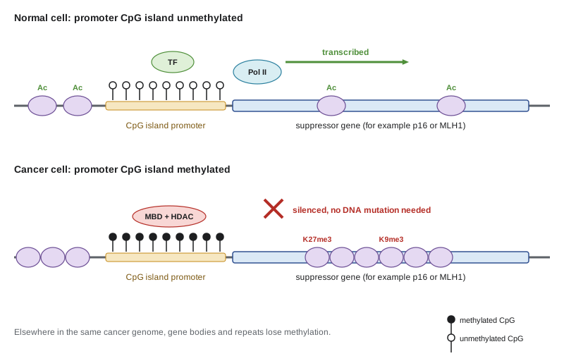
Methylation has practical value. In glioblastoma, methylation of the promoter of MGMT, the gene encoding a DNA repair enzyme that removes the damage caused by the alkylating drug temozolomide, predicts a better response to that drug and is tested routinely. Because tumor DNA circulates in blood, methylation patterns, which are tissue-specific, can reveal both the presence of cancer and its tissue of origin; some multi-cancer blood tests discussed in lesson 10 rely on this. Methylation-based classification of brain tumors, in which a tumor's methylation profile is compared with a reference library, has changed diagnosis in neuropathology and is part of the World Health Organization classification of central nervous system tumors.
Mutations in the epigenetic machinery
Genome sequencing revealed that genes encoding chromatin regulators are among the most frequently mutated in cancer, which links genetics and epigenetics directly. Mutations in the SWI/SNF chromatin remodeling complex occur in about a fifth of human cancers: ARID1A is lost in about half of ovarian clear cell carcinomas, and loss of SMARCB1 is essentially the only recurrent change in malignant rhabdoid tumors of young children, showing that a single epigenetic lesion can be enough to drive a lethal cancer. DNMT3A and TET2 mutations are common in acute myeloid leukemia and also appear, years earlier, in the blood stem cells of otherwise healthy older people, a condition called clonal hematopoiesis that raises the risk of blood cancers and, unexpectedly, of heart disease.
Lesson 6 introduced mutant IDH1 and IDH2, which produce the oncometabolite 2-hydroxyglutarate. 2-HG inhibits TET enzymes and histone demethylases, producing widespread hypermethylation that blocks differentiation. Mutations in histone genes themselves, the so-called oncohistones, define some childhood cancers: a lysine-to-methionine change at position 27 of histone H3 (H3 K27M) inhibits PRC2 throughout the genome and characterizes diffuse midline glioma, a brain tumor of children with a very poor prognosis. Rearrangements of the KMT2A gene (formerly MLL), which encodes a histone methyltransferase, cause aggressive infant and adult leukemias by recruiting partner proteins that keep developmental genes switched on.
Epigenetic drugs
Unlike mutations, epigenetic marks are reversible, which makes the enzymes that maintain them attractive drug targets. Several drug classes have reached the clinic.
| Drug class | Examples | Main approved uses | How it acts |
|---|---|---|---|
| DNMT inhibitors (hypomethylating agents) | Azacitidine, decitabine | Myelodysplastic syndromes, acute myeloid leukemia | Incorporated into DNA, they trap and deplete DNMT1, so methylation is lost with each division |
| HDAC inhibitors | Vorinostat, romidepsin | Cutaneous T-cell lymphoma | Increase histone acetylation and reopen chromatin |
| EZH2 inhibitor | Tazemetostat | Epithelioid sarcoma, follicular lymphoma | Blocks PRC2 methylation of H3 lysine 27; especially effective where SWI/SNF is lost |
| Mutant IDH inhibitors | Ivosidenib, enasidenib, vorasidenib | IDH-mutant acute myeloid leukemia, cholangiocarcinoma, low-grade glioma | Lower 2-HG and allow differentiation |
| Menin inhibitors | Revumenib | Acute leukemia with KMT2A rearrangement | Disrupt the menin–KMT2A interaction that keeps leukemia genes switched on |
| Differentiation therapy | All-trans retinoic acid with arsenic trioxide | Acute promyelocytic leukemia | Degrade the PML–RARA fusion and release the differentiation block |
Acute promyelocytic leukemia is the outstanding success. It is driven by a translocation fusing PML to the retinoic acid receptor gene RARA, whose product recruits repressive chromatin complexes and freezes cells at the promyelocyte stage. High doses of all-trans retinoic acid overcome the repression and force the leukemic cells to differentiate; combined with arsenic trioxide, which targets the fusion protein for degradation, the treatment cures most patients without conventional chemotherapy. The lesson that cancer cells can be pushed to mature, rather than killed, continues to guide drug development, and the hypomethylating agents may act partly in the same way, as well as by reactivating silenced genes and viral-like repeats that make tumor cells more visible to the immune system.
Infections and cancer
The idea that viruses cause cancer began in 1911, when Peyton Rous showed that a cell-free filtrate from a chicken sarcoma could transmit the tumor. The Rous sarcoma virus carries the src oncogene, and in 1976 J. Michael Bishop and Harold Varmus showed that src was a captured version of a normal cellular gene, the discovery that founded the proto-oncogene concept of lesson 2. In humans, infections cause roughly 13 percent of cancers worldwide, a share that is much higher in sub-Saharan Africa and parts of Asia than in Canada or the United States. The International Agency for Research on Cancer classifies the following agents as carcinogenic to humans.
| Agent | Main cancers | Key mechanism |
|---|---|---|
| High-risk human papillomaviruses (especially HPV16 and HPV18) | Cervix, anus, oropharynx, vulva, vagina, penis | E6 degrades p53; E7 inactivates RB |
| Hepatitis B virus | Hepatocellular carcinoma | Chronic inflammation and liver regeneration; viral DNA integration |
| Hepatitis C virus | Hepatocellular carcinoma, some lymphomas | Chronic inflammation and cirrhosis |
| Epstein–Barr virus | Burkitt lymphoma, Hodgkin lymphoma, nasopharyngeal carcinoma, some gastric cancers | Viral proteins such as LMP1 mimic growth and survival signals |
| Kaposi sarcoma herpesvirus (HHV-8) | Kaposi sarcoma, primary effusion lymphoma | Viral homologues of cyclin D and signaling receptors |
| Human T-cell lymphotropic virus type 1 | Adult T-cell leukemia/lymphoma | Tax protein activates proliferation pathways |
| Merkel cell polyomavirus | Merkel cell carcinoma | Integrated viral large T antigen binds RB |
| Helicobacter pylori (a bacterium) | Stomach cancer, gastric MALT lymphoma | Chronic gastritis; bacterial CagA protein disrupts cell signaling |
| HIV (indirectly) | Kaposi sarcoma, lymphomas, cervical cancer | Immune suppression lets other oncogenic viruses escape control |
Human papillomaviruses show the mechanism most clearly. Harald zur Hausen, who received the 2008 Nobel Prize for the work, identified HPV16 and HPV18 DNA in cervical cancers in the early 1980s. In cancers, the viral genome is often integrated into a host chromosome in a way that disrupts the viral E2 gene, which normally restrains expression of the two viral oncogenes. The E6 protein recruits a cellular ubiquitin ligase to tag p53 for destruction, and E7 binds RB and releases E2F transcription factors, driving cells into S phase. The virus thereby disables both of the tumor suppressor pathways that lesson 5 placed at the center of the cell cycle. This explains why HPV-positive cancers seldom carry TP53 mutations, and why staining for p16, which accumulates when RB is inactivated, is used as a surrogate marker for HPV in oropharyngeal cancer. HPV-positive oropharyngeal cancers respond better to treatment than smoking-related ones, and their incidence has risen in men in both Canada and the United States.
Most infections never lead to cancer. Persistent HPV infection lasting years, chronic hepatitis lasting decades, or a co-factor such as immune suppression, smoking or, for Burkitt lymphoma in equatorial Africa, malaria, is usually needed. The long interval is an opportunity. Vaccination against hepatitis B, introduced for infants in the 1980s and 1990s, has measurably reduced liver cancer in young adults in Taiwan, and HPV vaccination has sharply reduced cervical precancers and cervical cancer in vaccinated cohorts. Hepatitis C can now be cured with direct-acting antiviral drugs, and eradication of H. pylori lowers stomach cancer risk.
In the United States, the Advisory Committee on Immunization Practices recommends routine HPV vaccination at age 11 or 12, with vaccination possible from age 9 and catch-up through age 26. Uptake has lagged behind other adolescent vaccines, and coverage varies widely between states. The World Health Organization's global cervical cancer elimination strategy, launched in 2020, sets 2030 targets of 90 percent of girls fully vaccinated by age 15, 70 percent of women screened with a high-performance test by ages 35 and 45, and 90 percent of women with cervical disease treated, with elimination defined as fewer than four cases per 100,000 women.
Heterogeneity within tumors
A tumor biopsy samples one small region, yet a tumor can contain many genetically distinct subclones. Multiregion sequencing makes this visible. In a 2012 study of kidney cancers, Marco Gerlinger, Charles Swanton and colleagues sequenced several regions of each tumor and its metastases and found that about two thirds of mutations were not present in every region; a single biopsy would have suggested a misleadingly simple tumor. The TRACERx study of lung cancer, which follows patients like Maya from diagnosis, reported in 2017 that a high proportion of subclonal copy number changes was associated with a higher risk of recurrence or death. The usual reconstruction is a phylogenetic tree: truncal mutations, present in every cancer cell, sit on the trunk; branch mutations are shared by some subclones; and private mutations sit at the tips. Truncal drivers, such as KRAS and TP53 in Maya's tumor, make better therapeutic targets than branch drivers, because every cancer cell carries them.
Cancer stem cells and plasticity
A second kind of heterogeneity is not genetic. In 1997 John Dick and Dominique Bonnet sorted cells from patients with acute myeloid leukemia and transplanted them into immunodeficient mice. Only a rare population, carrying the surface marker CD34 but lacking CD38, could regenerate the leukemia, and the leukemias they produced recreated the full mix of cell types seen in the patient. This established the cancer stem cell model: a tumor can be organized like a normal tissue, with self-renewing cells at the top of a hierarchy producing progeny that divide for a while and then lose the ability to sustain the tumor. In 2003 Muhammad Al-Hajj and colleagues reported a similar population in breast cancers, marked as CD44 high and CD24 low, and lineage tracing in mouse intestinal adenomas later showed cells expressing the stem cell marker Lgr5 feeding the growth of the tumor in place, without transplantation.
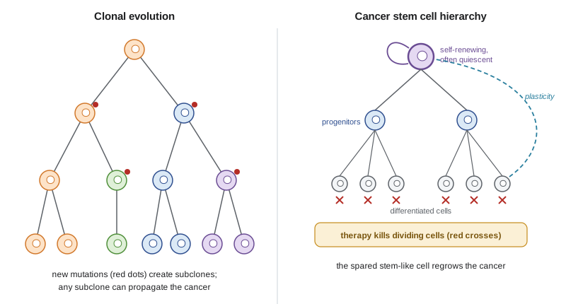
The model has been refined since. The frequency of tumor-propagating cells depends heavily on how well the recipient mice support human cells: when Elsa Quintana and Sean Morrison used more severely immunodeficient mice in 2008, about one in four unselected human melanoma cells could form tumors, far from rare. In many carcinomas, cells move between stem-like and more differentiated states depending on signals from their environment, a plasticity that links to the EMT programs of lesson 8 and that Hanahan's 2022 update proposed as an emerging hallmark, unlocking phenotypic plasticity. The clonal evolution model, in which genetically different subclones compete, and the stem cell model are best seen as complementary: each subclone may have its own hierarchy, and plasticity blurs the boundary between them.
The clinical relevance lies in treatment failure. Stem-like cells are often slowly dividing, express drug efflux pumps and repair DNA efficiently, so therapies that kill rapidly dividing cells can shrink a tumor dramatically while sparing the cells that will regrow it. The simulation below makes the point with a simple hierarchy.
# A two-compartment tumour hierarchy under a therapy that kills dividing differentiated cells far better than stem-like cells.
import numpy as np
days = np.arange(0, 241)
stem, diff = 1e7, 9e7                       # 10 per cent stem-like cells at the start
self_renew, diff_rate, prolif, death = 0.035, 0.03, 0.10, 0.06   # per-day rates
kill_stem, kill_diff = 0.02, 0.35          # extra daily kill during therapy
on = (days >= 30) & (days < 90)            # 60 days of treatment
S, D = [], []
for d in days:
    ks, kd = (kill_stem, kill_diff) if on[d] else (0, 0)
    new_s = stem * (1 + self_renew - diff_rate - ks)
    new_d = diff * (1 + prolif - death - kd) + stem * 2 * diff_rate
    stem, diff = new_s, new_d
    S.append(stem); D.append(diff)
S, D = np.array(S), np.array(D)
for d in (0, 30, 60, 90, 120, 180, 240):
    tot = S[d] + D[d]
    print(f"day {d:3d}: total {tot:9.2e} cells, stem-like fraction {S[d] / tot:5.1%}")
nadir = int(np.argmin(S + D))
print(f"smallest tumour on day {nadir}: {(S + D)[nadir] / (S + D)[0]:.1%} of starting size")
print(f"tumour back to starting size by day {int(np.argmax((days > nadir) & (S + D >= (S + D)[0])))}")day 0: total 1.04e+08 cells, stem-like fraction 9.6% day 30: total 2.38e+08 cells, stem-like fraction 4.8% day 60: total 8.75e+06 cells, stem-like fraction 83.1% day 90: total 5.99e+06 cells, stem-like fraction 78.7% day 120: total 2.64e+07 cells, stem-like fraction 20.7% day 180: total 3.14e+08 cells, stem-like fraction 2.4% day 240: total 3.35e+09 cells, stem-like fraction 0.3% smallest tumor on day 89: 5.4% of starting size tumor back to starting size by day 153
The numbers are invented, but the qualitative result is robust. During treatment the tumor shrinks to a small fraction of its starting size, which would look like an excellent response on a scan, but the surviving tumor is enriched for stem-like cells, and once treatment stops it regrows. Strategies proposed to deal with this include targeting stem cell signaling pathways such as Wnt, Notch and Hedgehog, forcing differentiation as in acute promyelocytic leukemia, and combining cytotoxic drugs with agents that exploit the stem-like state. The Hedgehog pathway inhibitors vismodegib and sonidegib, approved for advanced basal cell carcinoma, show that a developmental pathway can be targeted, although basal cell carcinoma is driven by Hedgehog mutations in essentially all of its cells rather than in a stem-like minority.
For Maya, two of these themes matter. Her tumor contains p16 loss, a change that in lung cancer can arise either from deletion of CDKN2A or from methylation of its promoter, and whichever route it took, the effect on RB control is the same. More importantly, her tumor is unlikely to be uniform: any biopsy samples only part of it, and subclones or drug-tolerant states may be present that the biopsy does not reveal. Lesson 10 shows how her tumor was profiled, and lesson 11 returns to resistance.
Worked problem
Reading a methylation result
A pathologist reports that a glioblastoma has a methylated MGMT promoter, and a separate colorectal cancer report states that the tumor is MSI-H with loss of MLH1 protein, MLH1 promoter methylation and a BRAF V600E mutation. What does each finding imply?
Show solution
- MGMT encodes a repair enzyme that removes the alkyl groups temozolomide adds to guanine.
- Promoter methylation silences MGMT, so tumor cells cannot repair temozolomide damage; the patient is more likely to benefit from the drug.
- For the colorectal cancer, loss of MLH1 explains the microsatellite instability.
- MLH1 promoter methylation with BRAF V600E points to a sporadic, CIMP-associated cancer rather than Lynch syndrome, so germline testing for MLH1 is less urgent, though a family history would still prompt referral.
Answer: Methylated MGMT predicts benefit from temozolomide; MLH1 methylation with BRAF V600E suggests a sporadic rather than inherited MSI-H colorectal cancer.
Common mistakes
MistakeThinking epigenetic changes are rare compared with mutations.
InsteadPromoter hypermethylation and global hypomethylation occur in most tumors, and chromatin regulators are among the most mutated genes.
MistakeAssuming all methylation in cancer increases.
InsteadCpG islands at certain promoters gain methylation, but the genome as a whole loses it.
MistakeBelieving infection alone causes virus-associated cancers.
InsteadPersistence over years and co-factors such as immune suppression are usually needed; most infections never cause cancer.
MistakeTreating the cancer stem cell model as fixed and universal.
InsteadThe frequency of tumor-propagating cells depends on the assay, and plasticity lets cells move between states.
MistakeJudging a treatment only by how much a tumor shrinks.
InsteadA large response can coexist with survival of the cells that regrow the tumor.
Summary and key terms
- DNA methylation, histone modifications and chromatin remodeling maintain cell identity without changing DNA sequence.
- Cancers combine global DNA hypomethylation with hypermethylation of CpG island promoters, which can silence tumor suppressors such as CDKN2A and MLH1.
- Chromatin regulators such as ARID1A, SMARCB1, DNMT3A, TET2, EZH2 and KMT2A are frequently mutated, linking genetic and epigenetic changes.
- Epigenetic drugs include DNMT, HDAC, EZH2, mutant IDH and menin inhibitors; differentiation therapy cures most acute promyelocytic leukemia.
- About 13 percent of cancers worldwide are caused by infections; HPV E6 and E7 inactivate p53 and RB.
- Vaccination against HPV and hepatitis B, antiviral cure of hepatitis C and eradication of H. pylori prevent cancer.
- Tumors contain genetically distinct subclones and, in many cases, hierarchies of stem-like and differentiated cells, with plasticity between states.
- epigenetics
- Heritable changes in gene activity that do not change the DNA sequence.
- CpG island
- A CpG-rich region, often at a promoter, that is usually unmethylated in normal cells.
- promoter hypermethylation
- Gain of DNA methylation at a CpG island promoter that silences the gene.
- CpG island methylator phenotype
- A tumor pattern in which many CpG islands are methylated together.
- oncohistone
- A mutated histone that drives cancer, such as H3 K27M.
- clonal hematopoiesis
- Expansion of a blood stem cell clone carrying a somatic mutation in a person without blood cancer.
- oncogenic virus
- A virus that contributes to cancer development, such as HPV or hepatitis B virus.
- intratumour heterogeneity
- Genetic or non-genetic differences among cells within one tumor.
- truncal mutation
- A mutation present in all cancer cells of a tumor, acquired early.
- cancer stem cell
- A tumor cell able to self-renew and regenerate the full tumor.
- cellular plasticity
- The ability of cells to change state, for example between stem-like and differentiated.
Practice problems
Work each problem on paper first, then reveal the answer to check.
What enzymes copy DNA methylation patterns after replication, and what enzymes begin their removal?
Show answer
DNMT1 maintains methylation; TET enzymes oxidize methylcytosine to start removal.
Give two tumor suppressors commonly silenced by promoter methylation.
Show answer
CDKN2A (p16), MLH1, BRCA1 or VHL.
Why is MGMT methylation tested in glioblastoma?
Show answer
Silencing of MGMT predicts benefit from temozolomide.
How does H3 K27M drive diffuse midline glioma?
Show answer
It inhibits PRC2 and loss of H3 lysine 27 trimethylation reprograms gene expression.
How do HPV E6 and E7 transform cells?
Show answer
E6 targets p53 for degradation and E7 inactivates RB, releasing E2F.
Why is p16 staining used as a marker of HPV in oropharyngeal cancer?
Show answer
RB inactivation by E7 causes p16 to accumulate.
What did the 1997 AML transplantation experiments show?
Show answer
Only a rare CD34-positive, CD38-negative population could regenerate the leukemia in mice.
Why are truncal mutations preferred as drug targets?
Show answer
They are present in every cancer cell, so targeting them can affect the whole tumor.
Lesson quiz · 5 questions
Score 60% or more to complete this lesson.
Create a free account to save your progress, take quizzes and earn a certificate.