
BIOL-450 · Lesson 6 of 12 · 23 min
Cancer metabolism
Dividing cells need more than energy. To make a daughter cell they must double their proteins, lipids and nucleic acids, which requires carbon skeletons, nitrogen and reducing power in the form of NADPH. Cancer cells rewire their metabolism to supply these building blocks and to survive in tissues where oxygen and nutrients run short. Reprogramming of metabolism became a hallmark of cancer in 2011. This lesson explains the Warburg effect and its modern interpretation, the role of glutamine and other nutrients, how mutations in metabolic enzymes can themselves drive cancer, and how cancer metabolism is used in diagnosis and treatment.
The Warburg effect
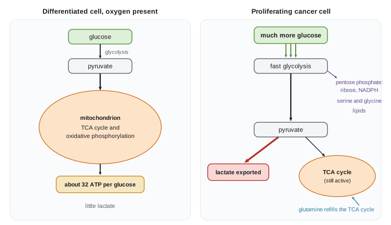
In the 1920s, Otto Warburg observed that tumor slices took up far more glucose than normal tissue and converted much of it to lactate even when oxygen was plentiful. Normal differentiated cells, in the presence of oxygen, oxidize most of the pyruvate from glycolysis in the mitochondria, yielding roughly 30 to 32 molecules of ATP per glucose, and switch to lactate production only when oxygen is short. Tumor cells used aerobic glycolysis, producing just 2 ATP per glucose from glycolysis while secreting lactate. Warburg concluded that cancer was caused by damaged mitochondria. That explanation turned out to be wrong for most tumors: their mitochondria work, and many cancers depend on oxidative phosphorylation. But the observation was right, and it has been confirmed in countless tumors and in rapidly dividing normal cells such as activated lymphocytes.
Why would a cell choose such an inefficient route? The modern answer, set out by Matthew Vander Heiden, Lewis Cantley and Craig Thompson in 2009, is that ATP is not the limiting resource for proliferation. A dividing cell has plenty of ATP; what it needs is carbon for biomass and NADPH for biosynthesis. High flux through glycolysis supplies intermediates that branch off into the pentose phosphate pathway (making ribose for nucleotides and NADPH), the serine synthesis pathway (feeding one-carbon metabolism for nucleotides and methylation) and glycerol for lipids. Secreting lactate regenerates NAD+ so glycolysis can keep running fast. The trade-off favors speed and building blocks over yield.
How oncogenes rewire metabolism
Metabolic rewiring is not separate from the oncogenic changes of lessons 2 and 3; it is driven by them. PI3K–AKT signaling increases expression and membrane localization of the glucose transporter GLUT1, activates hexokinase and phosphofructokinase, and through mTOR drives protein and lipid synthesis. MYC induces genes for glucose and glutamine uptake, glycolytic enzymes, ribosome biogenesis and nucleotide synthesis. Under hypoxia, and in tumors that have lost the VHL tumor suppressor, the transcription factor HIF-1 switches on glycolytic genes and pyruvate dehydrogenase kinase, which diverts pyruvate away from mitochondria. Loss of p53 removes a restraint on glycolysis and on the pentose phosphate pathway. Tumor cells also often express the M2 isoform of pyruvate kinase, whose regulated low activity can let glycolytic intermediates accumulate for biosynthesis.
Glutamine and anaplerosis
Many cancer cells consume glutamine avidly, a phenomenon called glutamine addiction. Glutamine supplies nitrogen for nucleotides and non-essential amino acids, and its carbon, as α-ketoglutarate, replenishes the TCA cycle as intermediates are drawn off for biosynthesis, a process called anaplerosis. Citrate leaves the mitochondrion to supply acetyl-CoA for fatty acid synthesis, and oxaloacetate feeds aspartate synthesis. Inhibitors of glutaminase have been tested in trials with limited success, partly because tumors in the body use nutrients differently from cells in culture, where glutamine is abundant. Tracer studies in patients, in which labeled glucose is infused before surgery, have shown that some human lung tumors oxidize glucose in their mitochondria more than expected and also use lactate as a fuel, a reminder that culture conditions can mislead.
Oncometabolites: when metabolic enzymes are the drivers
In some cancers, mutations in metabolic enzymes are themselves the oncogenic event. In 2008 and 2009, sequencing of gliomas and acute myeloid leukemia found recurrent point mutations in isocitrate dehydrogenase genes IDH1 and IDH2, at specific arginine residues (IDH1 R132, IDH2 R140 and R172). The mutant enzymes gain a new activity: instead of converting isocitrate to α-ketoglutarate, they reduce α-ketoglutarate to D-2-hydroxyglutarate (2-HG), which accumulates to millimolar levels. 2-HG competitively inhibits α-ketoglutarate-dependent dioxygenases, including the TET enzymes that demethylate DNA and the JmjC histone demethylases. The result is widespread hypermethylation of DNA and histones that blocks differentiation, a link to the epigenetics of lesson 9. Ivosidenib and enasidenib, inhibitors of mutant IDH1 and IDH2, are approved for IDH-mutant acute myeloid leukemia, and vorasidenib was approved in 2024 in the United States for low-grade IDH-mutant gliomas, with Canadian authorization following (approval status as of 2026; check current labels).
Mutations in succinate dehydrogenase (SDH) and fumarate hydratase (FH), enzymes of the TCA cycle, act as tumor suppressor losses. Germline variants cause hereditary paragangliomas and pheochromocytomas (SDH) and hereditary leiomyomatosis and renal cell cancer (FH). The accumulated succinate and fumarate also inhibit α-ketoglutarate-dependent enzymes and stabilize HIF, mimicking hypoxia. These discoveries confirmed that metabolism can be a cause, not just a consequence, of cancer.
Imaging cancer metabolism: FDG-PET
The high glucose uptake of tumors is used every day in clinics. 18F-fluorodeoxyglucose (FDG) is a glucose analog labeled with a positron-emitting fluorine isotope. Cells take it up through glucose transporters and hexokinase phosphorylates it, but it cannot proceed further down glycolysis and is trapped. Positron emission tomography (PET), usually combined with computed tomography (CT), shows where FDG accumulates. Uptake is often summarized as a standardized uptake value (SUV), the tissue activity concentration divided by the injected activity per kilogram of body weight. FDG-PET is used to stage cancers such as lung cancer and lymphoma, to look for distant spread and to monitor response. It has limits: the brain, inflamed tissue and healing wounds also take up glucose avidly, and some slow-growing tumors, such as many prostate cancers, take up little FDG. Other tracers, such as PSMA-targeted agents for prostate cancer, fill those gaps.
# ATP per glucose, lactate output and the FDG standardized uptake value (illustrative numbers).
glucose_uptake = {'differentiated cell': 1.0, 'proliferating tumour cell': 10.0}   # relative uptake
frac_to_lactate = {'differentiated cell': 0.05, 'proliferating tumour cell': 0.85}
ATP_glycolysis, ATP_oxidative = 2, 30          # per glucose, approximate
for cell in glucose_uptake:
    g = glucose_uptake[cell]; f = frac_to_lactate[cell]
    atp = g * (ATP_glycolysis + (1 - f) * ATP_oxidative)
    lactate = g * f * 2                        # 2 lactate per glucose fermented
    print(f"{cell:26s} ATP {atp:5.1f}  lactate {lactate:5.1f}  ATP per glucose {atp / g:4.1f}")
# SUV = tissue activity (kBq/mL) / (injected activity (kBq) / body weight (g)), taking 1 mL of tissue as about 1 g
injected_MBq, weight_kg = 300.0, 75.0
for tissue, kBq_per_mL in [('lung tumour', 32.0), ('normal lung', 2.0), ('liver', 9.6)]:
    suv = kBq_per_mL / (injected_MBq * 1000 / (weight_kg * 1000))
print(f"SUV {tissue:12s} = {suv:4.1f}")differentiated cell ATP 30.5 lactate 0.1 ATP per glucose 30.5 proliferating tumor cell ATP 65.0 lactate 17.0 ATP per glucose 6.5 SUV lung tumor = 8.0 SUV normal lung = 0.5 SUV liver = 2.4
The numbers make Warburg's paradox concrete. Per molecule of glucose, the tumor cell makes much less ATP, yet with ten times the uptake it still makes more ATP overall, while exporting large amounts of lactate and keeping glycolytic intermediates available for biosynthesis. An SUV of about 8 in a lung nodule, against about 0.5 in normal lung, is the kind of contrast that makes FDG-PET useful for staging, though thresholds depend on the scanner and protocol.
The metabolic microenvironment
Metabolism also shapes the space around tumor cells. Lactate and protons exported by glycolytic cells acidify the extracellular space, which favors invasion by activating proteases and harms immune cells; T cells, which also rely on glycolysis when activated, compete with tumor cells for glucose and are suppressed by lactate. Poorly perfused regions are short of oxygen, glucose and amino acids, and cells there may scavenge nutrients by macropinocytosis, engulfing extracellular proteins, a behavior seen in KRAS-mutant pancreatic cancer. Neighboring stromal cells can feed tumor cells with lactate, ketone bodies or amino acids. These interactions link this lesson to the tumor microenvironment of lesson 7 and to tumor immunology in BIOL-380.
Measuring metabolism in tumors
How do researchers know what tumors actually do with nutrients? Steady-state metabolomics measures the levels of hundreds of metabolites by mass spectrometry, but levels alone do not reveal fluxes, because a metabolite can be abundant because it is made quickly or because it is used slowly. Stable isotope tracing solves this. Cells, mice or patients are given glucose, glutamine or lactate labeled with carbon-13, and the pattern of label appearing in downstream metabolites shows which pathways carried it. Such studies, including infusions of labeled glucose into patients shortly before surgery, revealed that tumors in the body use mixtures of fuels that differ from cells in culture, that lactate can be a major fuel for the TCA cycle, and that the same oncogene can produce different metabolic programs in different tissues. Hyperpolarized carbon-13 magnetic resonance imaging, which follows the conversion of labeled pyruvate to lactate in real time, is being tested as a way to measure tumor metabolism in patients without radiation.
Targeting metabolism
Antimetabolites were among the first chemotherapy drugs and are still central: methotrexate blocks dihydrofolate reductase and thus one-carbon metabolism, 5-fluorouracil blocks thymidylate synthase, and gemcitabine and cytarabine are nucleoside analogues that halt DNA synthesis. Sidney Farber's use of the folate antagonist aminopterin to induce remissions in children with acute leukemia in 1948 is often counted as the start of modern chemotherapy. Newer approaches target mutant IDH, nutrient transporters, glutaminase, fatty acid synthesis and the dependence of some tumors on particular amino acids, such as asparagine in acute lymphoblastic leukemia, which is treated with the enzyme asparaginase. Diet has also been studied: ketogenic diets, fasting-mimicking diets and restriction of specific amino acids show effects in mouse models, but evidence in patients is limited, and no diet has been shown to cure cancer.
| Metabolic change | Driver | Consequence | Clinical use |
|---|---|---|---|
| Aerobic glycolysis | PI3K–AKT, MYC, HIF-1, p53 loss | High glucose uptake, lactate export, biosynthetic intermediates | FDG-PET imaging |
| Glutamine addiction | MYC | Nitrogen supply and TCA anaplerosis | Glutaminase inhibitors (trials) |
| IDH1 or IDH2 mutation | Gain-of-function mutation | 2-HG, DNA and histone hypermethylation, blocked differentiation | Ivosidenib, enasidenib, vorasidenib |
| SDH or FH loss | Tumor suppressor loss | Succinate or fumarate accumulation, pseudohypoxia | Genetic testing of families |
| Folate and nucleotide synthesis | Proliferation demand | Dependence on one-carbon metabolism | Methotrexate, 5-fluorouracil |
| Asparagine dependence | Low asparagine synthetase | Needs external asparagine | Asparaginase in ALL |
Maya's staging illustrates the clinical side. Her FDG-PET-CT showed an avid primary tumor in the right upper lobe with an SUV of about 9 and an FDG-avid lymph node on the same side of the chest, but no distant uptake. That placed her in stage III, a decision that shaped her treatment as described in lesson 11.
Worked problem
Calculating an SUV
A patient weighing 80 kg receives 320 MBq of FDG. One hour later a lymph node has an activity concentration of 20 kBq/mL. Calculate the SUV, taking 1 mL of tissue as about 1 g.
Show solution
- Injected activity per gram of body weight = 320,000 kBq / 80,000 g = 4.0 kBq/g.
- SUV = tissue concentration / injected activity per gram = 20 / 4.0.
- SUV = 5.0.
- An SUV of 5 in a lymph node is suspicious, but inflammation can give similar values, so biopsy or follow-up may be needed.
Answer: An SUV of about 5.
Common mistakes
MistakeSaying the Warburg effect means tumor mitochondria are broken.
InsteadMost tumors have working mitochondria; aerobic glycolysis supports biosynthesis and fast flux.
MistakeThinking ATP is the main thing proliferating cells lack.
InsteadCarbon building blocks and NADPH usually limit growth more than ATP does.
MistakeAssuming every hot spot on FDG-PET is cancer.
InsteadInflammation, infection, healing tissue and brain also take up FDG.
MistakeTreating IDH mutations as loss-of-function.
InsteadMutant IDH gains a new activity, producing the oncometabolite 2-HG.
MistakeConcluding from mouse studies that a diet can treat cancer.
InsteadEvidence in patients is limited; diets are studied as adjuncts, not cures.
Summary and key terms
- Cancer cells reprogram metabolism to supply carbon, nitrogen and NADPH for biomass, not just ATP.
- The Warburg effect, aerobic glycolysis, supports fast flux and biosynthesis; most tumor mitochondria still work.
- Oncogenes and tumor suppressors, including PI3K–AKT, MYC, HIF and p53, drive metabolic rewiring.
- Glutamine supplies nitrogen and replenishes the TCA cycle in many cancers.
- Mutant IDH1 and IDH2 produce 2-HG, an oncometabolite that alters DNA and histone methylation; SDH and FH losses act similarly.
- FDG-PET exploits high glucose uptake for staging and monitoring, with known pitfalls.
- Antimetabolites, IDH inhibitors and asparaginase show that cancer metabolism can be targeted.
- Warburg effect
- Aerobic glycolysis: high glucose uptake and lactate production by cancer cells even when oxygen is present.
- aerobic glycolysis
- Conversion of glucose to lactate despite adequate oxygen.
- pentose phosphate pathway
- A branch of glucose metabolism producing ribose and NADPH.
- anaplerosis
- Replenishment of TCA cycle intermediates drawn off for biosynthesis.
- glutamine addiction
- Dependence of cancer cells on glutamine for nitrogen and TCA anaplerosis.
- oncometabolite
- A metabolite that accumulates in cancer and drives tumor formation, such as 2-HG.
- 2-hydroxyglutarate
- The oncometabolite produced by mutant IDH enzymes that inhibits dioxygenases.
- FDG-PET
- Positron emission tomography using a radiolabelled glucose analog to image glucose uptake.
- standardized uptake value
- PET tracer concentration in tissue normalized to injected activity per body weight.
- antimetabolite
- A drug that blocks a metabolic pathway needed for DNA synthesis.
Practice problems
Work each problem on paper first, then reveal the answer to check.
Why does aerobic glycolysis suit a proliferating cell despite its low ATP yield?
Show answer
It supplies carbon intermediates and NADPH for biosynthesis and regenerates NAD+ for fast flux.
Name two oncogenic changes that increase glucose uptake.
Show answer
PI3K–AKT activation and MYC activation (also HIF stabilization).
What is anaplerosis and how does glutamine contribute?
Show answer
Replenishing TCA intermediates; glutamine becomes α-ketoglutarate.
What new activity does mutant IDH1 R132 have?
Show answer
It converts α-ketoglutarate to 2-hydroxyglutarate.
Why can inflammation give a false positive on FDG-PET?
Show answer
Activated immune cells also take up glucose avidly.
A tumor has SUV 12 and normal tissue SUV 1.5. What is the tumor-to-background ratio?
Show answer
8.
Which enzyme is used to treat acute lymphoblastic leukemia, and why does it work?
Show answer
Asparaginase; leukemic cells cannot make enough asparagine.
Name an inherited syndrome caused by a TCA cycle enzyme defect.
Show answer
Hereditary paraganglioma–pheochromocytoma (SDH) or hereditary leiomyomatosis and renal cell cancer (FH).
Lesson quiz · 5 questions
Score 60% or more to complete this lesson.
Create a free account to save your progress, take quizzes and earn a certificate.