
BIOL-450 · Lesson 5 of 12 · 24 min
Cell cycle control, apoptosis and senescence
A cell that has acquired a growth signal and lost a brake still faces internal safeguards. Checkpoints halt the cell cycle when DNA is damaged or chromosomes are misaligned. Apoptosis removes cells that are damaged or behaving abnormally. Senescence permanently withdraws cells from the cycle. Each of these is a barrier that a developing cancer must overcome, and each has become a target for therapy. BIOL-214 covered the machinery of the cell cycle and the basic pathways of cell death; here the focus is on how cancer cells disable them and how drugs can restore or exploit them.
Checkpoints and cancer
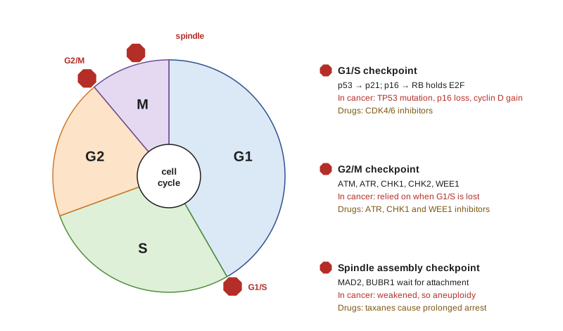
Three checkpoints matter most. The G1/S checkpoint, governed by p53 and the RB pathway, stops cells with damaged DNA from replicating it. The intra-S and G2/M checkpoints, driven by ATR and CHK1 and by ATM and CHK2, slow replication and block entry into mitosis until damage is repaired, largely by inhibiting the phosphatase CDC25 so that CDK1 stays switched off. The spindle assembly checkpoint holds cells in metaphase until every kinetochore is attached to the spindle, preventing chromosome missegregation.
Cancer cells commonly lose the G1/S checkpoint, through TP53 mutation, loss of p16INK4a, or amplification of cyclin D or CDK4. That leaves them dependent on the remaining G2/M checkpoint to survive damage, a weakness that inhibitors of ATR, CHK1 and WEE1 aim to exploit: knock out the last checkpoint in a cell that has already lost the first, and DNA damage drives the cell into a catastrophic mitosis. Normal cells, with intact G1/S control, can stop and repair. This is another form of synthetic lethality, related to the logic met in lesson 4.
CDK4/6 inhibitors: restoring a brake
Lesson 3 explained that the RB pathway is disrupted upstream in many cancers, leaving RB intact but constantly phosphorylated by cyclin D–CDK4/6. Palbociclib, ribociclib and abemaciclib block CDK4 and CDK6 and hold RB in its active, growth-restraining state. In hormone receptor-positive, HER2-negative breast cancer, where estrogen drives cyclin D expression, adding a CDK4/6 inhibitor to endocrine therapy roughly doubled progression-free survival in pivotal trials and, for some combinations, improved overall survival. The drugs mainly cause arrest rather than death, so they work best in combination, and tumors that lose RB, amplify cyclin E or activate CDK2 become resistant.
p53 dynamics: pulses as signals
The p53–MDM2 feedback loop, introduced in lesson 3, has a striking consequence. Because p53 induces its own inhibitor after a delay, its levels after DNA damage do not simply rise and stay high. In single cells imaged over time, Galit Lahav and colleagues found that p53 rises and falls in a series of pulses of fixed size and duration, with the number of pulses increasing with the amount of damage. Sustained p53, produced artificially, pushed cells toward senescence rather than recovery. The temporal pattern of a transcription factor can therefore carry information, and delayed negative feedback is enough to generate pulses.
# Delayed negative feedback between p53 and MDM2 produces pulses (illustrative parameters).
import numpy as np
dt, T, tau = 0.01, 40.0, 1.0          # hours; tau = delay for MDM2 to be made from p53
n = int(T / dt); lag = int(tau / dt)
p = np.zeros(n); m = np.zeros(n)
p[0], m[0] = 0.1, 0.1
damage = 1.0                          # constant damage signal keeps p53 production high
for i in range(1, n):
    p_delayed = p[i - lag] if i >= lag else p[0]
    dp = damage * 1.5 - 4.0 * m[i - 1] * p[i - 1] / (p[i - 1] + 0.1)   # production minus MDM2-driven loss
    dm = 1.0 * p_delayed ** 4 / (0.5 ** 4 + p_delayed ** 4) - 0.8 * m[i - 1]
    p[i] = max(p[i - 1] + dt * dp, 0); m[i] = max(m[i - 1] + dt * dm, 0)
peaks = [i for i in range(1, n - 1) if p[i] > p[i - 1] and p[i] >= p[i + 1] and p[i] > 0.4]
times = np.array(peaks) * dt
print("p53 peak times (h):", ", ".join(f"{t:.1f}" for t in times[:8]))
if len(times) > 2:
    print(f"mean interval between pulses: {np.diff(times).mean():.1f} h")
print(f"p53 range after the first 10 h: {p[int(10 / dt):].min():.2f} to {p[int(10 / dt):].max():.2f}")p53 peak times (h): 1.8, 7.3, 12.4, 17.6, 22.7, 27.8, 33.0, 38.1 mean interval between pulses: 5.2 h p53 range after the first 10 h: 0.06 to 1.39
The model is minimal, two variables and a delay, and the parameters are illustrative, but it reproduces the qualitative behavior seen in living cells: repeated pulses of p53 under continuing damage rather than a single sustained rise. Real cells add further feedback loops through WIP1 and ATM that shape the pulses.
Apoptosis: two pathways to the same end
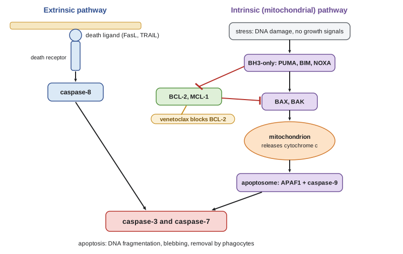
Apoptosis is a tidy form of cell death carried out by caspases, proteases that cleave hundreds of cellular proteins and dismantle the cell into membrane-bound fragments that neighbors engulf without inflammation. Two pathways activate the executioner caspases, caspase-3 and caspase-7. The extrinsic pathway begins at the cell surface, where death ligands such as FAS ligand or TRAIL bind death receptors, recruiting the adaptor FADD and activating caspase-8. The intrinsic, or mitochondrial, pathway begins inside the cell in response to DNA damage, oncogene stress, loss of survival signals or other insults. It is controlled by the BCL-2 family.
The BCL-2 family has three groups. Pro-survival members, BCL-2, BCL-XL and MCL-1, restrain the pore-forming effectors BAX and BAK. BH3-only proteins, such as BIM, PUMA, NOXA and BID, are the sensors: activated by stress, they neutralize the pro-survival proteins or directly activate BAX and BAK. When BAX and BAK oligomerize in the outer mitochondrial membrane, cytochrome c is released into the cytoplasm, binds APAF1 to form the apoptosome, and activates caspase-9, which activates the executioners. This step, mitochondrial outer membrane permeabilization, is usually the point of no return. p53 feeds into the intrinsic pathway by inducing PUMA and NOXA, which is one reason TP53 loss makes cells resistant to DNA-damaging chemotherapy.
How cancers evade apoptosis
BCL-2 was discovered at the breakpoint of the t(14;18) translocation in follicular lymphoma, which places BCL2 next to the immunoglobulin heavy chain enhancer and drives its overexpression. It was the first oncogene shown to work not by promoting division but by preventing death, a finding by David Vaux, Suzanne Cory and Jerry Adams that broadened the concept of oncogenes. Other tumors amplify MCL1, lose BAX or APAF1, or lose p53 so that stress no longer induces PUMA and NOXA. Many solid tumors rely on survival signals from the PI3K–AKT pathway, which inactivates pro-apoptotic proteins and keeps MCL-1 high.
Venetoclax, a BH3 mimetic that binds BCL-2 and frees the effectors, was approved for chronic lymphocytic leukemia in 2016 and later for acute myeloid leukemia in older adults when combined with hypomethylating agents. Its development rested on decades of structural biology of BCL-2 family interactions. Tumors that rely on BCL-2 are said to be primed for death, a state that can be measured by BH3 profiling, in which isolated mitochondria are exposed to BH3 peptides to see which pro-survival protein the cell depends on. MCL-1 inhibitors have been more difficult because of heart toxicity in trials.
Apoptosis also helps explain why some cancers respond to chemotherapy and others do not. Testicular germ cell tumors and many childhood leukemias are highly curable with chemotherapy, and their cells are strongly primed for death, with pro-survival BCL-2 family proteins already near capacity. Many solid tumors of adults are poorly primed and tolerate the same damage. Anthony Letai and colleagues showed that mitochondrial priming measured in patient samples correlated with clinical response across several cancer types, a reminder that the decision to die after damage is as important as the damage itself.
Other forms of regulated cell death
Apoptosis is not the only regulated death. Necroptosis, triggered by the kinases RIPK1 and RIPK3 and the pore-forming protein MLKL, is an inflammatory backup when caspase-8 is blocked. Ferroptosis, an iron-dependent death driven by lipid peroxidation and prevented by the enzyme GPX4, is of interest because some therapy-resistant, mesenchymal-like cancer cells are especially vulnerable to it. Autophagy, the self-digestion of cellular components, usually helps stressed cancer cells survive by recycling nutrients, although in some contexts it suppresses tumor formation. These pathways matter because a cancer cell that blocks apoptosis may still die by another route.
Senescence: a permanent arrest
Senescence is a stable arrest of proliferation in which cells remain alive and metabolically active. It is triggered by critically short telomeres (lesson 4), by DNA damage, and by strong oncogene signaling. Oncogene-induced senescence was discovered when Manuel Serrano, Scott Lowe and colleagues found in 1997 that introducing activated RAS into normal human fibroblasts did not transform them but instead arrested them permanently, through p16INK4a and p53. In people, benign naevi (moles), most of which carry BRAF V600E, contain senescent melanocytes that may stay arrested for decades. Senescence is therefore a barrier to cancer, and escaping it, usually by losing p16 or p53, is a step on the road to malignancy.
Senescent cells are not inert. They secrete inflammatory cytokines, growth factors and proteases, the senescence-associated secretory phenotype (SASP). In the short term the SASP recruits immune cells that clear the senescent cells; in the long term, persistent senescent cells can promote inflammation, tissue dysfunction and even the growth of neighboring tumor cells. Chemotherapy and radiation induce senescence in tumor cells, which can contribute both to response and, through the SASP, to later relapse. Drugs that selectively kill senescent cells, called senolytics, are being studied, as are approaches that combine induction of senescence with immune clearance.
| Barrier | What triggers it | How cancer escapes | Therapeutic approach |
|---|---|---|---|
| G1/S checkpoint | DNA damage, oncogene stress | TP53 mutation, p16 loss, cyclin D/CDK4 gain | CDK4/6 inhibitors where RB is intact |
| G2/M checkpoint | DNA damage, replication stress | Rarely lost; tumors depend on it | ATR, CHK1, WEE1 inhibitors |
| Intrinsic apoptosis | Damage, oncogene stress, loss of survival signals | BCL-2 or MCL-1 overexpression, p53 loss, BAX loss | BH3 mimetics such as venetoclax |
| Extrinsic apoptosis | Death ligands from immune cells | Decoy receptors, FLIP overexpression | Death receptor agonists (research) |
| Senescence | Short telomeres, damage, oncogene signaling | p16 and p53 loss | Senolytics, senescence plus immune clearance |
For Maya, these barriers explain both her tumor's biology and her treatment. Her tumor's TP53 mutation removed the G1/S checkpoint and blunted apoptosis after DNA damage, which may reduce the benefit of platinum chemotherapy. Her KRAS G12C mutation would normally risk oncogene-induced senescence, a barrier her tumor escaped through loss of p16, a common partner of KRAS in lung adenocarcinoma.
Worked problem
Choosing a drug from a tumor's profile
Three tumors are profiled. Tumor A: hormone receptor-positive breast cancer with intact RB and amplified cyclin D1. Tumor B: chronic lymphocytic leukemia with high BCL-2 dependence on BH3 profiling. Tumor C: RB-null small cell lung cancer. Which tumors are reasonable candidates for a CDK4/6 inhibitor or venetoclax?
Show solution
- Tumor A has intact RB driven hard by cyclin D1, the setting in which CDK4/6 inhibitors work.
- Tumor B depends on BCL-2, the target of venetoclax.
- Tumor C lacks RB, so blocking CDK4/6 cannot restore the brake; it is a poor candidate for CDK4/6 inhibition.
- Each choice follows from which barrier the tumor has lost and which dependency remains.
Answer: A is a candidate for a CDK4/6 inhibitor, B for venetoclax; C would not benefit from a CDK4/6 inhibitor.
Common mistakes
MistakeAssuming cancer cells have lost all checkpoints.
InsteadMany lose G1/S control but depend on G2/M, which is why ATR, CHK1 and WEE1 inhibitors are being tested.
MistakeTreating apoptosis and necrosis as the same.
InsteadApoptosis is a regulated, non-inflammatory death executed by caspases; necrosis is typically uncontrolled and inflammatory.
MistakeThinking BCL-2 promotes division.
InsteadBCL-2 blocks death; it was the first oncogene shown to work by preventing apoptosis.
MistakeCalling senescent cells harmless because they do not divide.
InsteadTheir secretory phenotype can promote inflammation and tumor growth.
MistakeExpecting CDK4/6 inhibitors to kill tumor cells directly.
InsteadThey mainly cause arrest, so they are combined with other treatments such as endocrine therapy.
Summary and key terms
- Checkpoints, apoptosis and senescence are intrinsic barriers that cancer cells must overcome.
- Cancers usually lose G1/S control but depend on G2/M checkpoints, a vulnerability for ATR, CHK1 and WEE1 inhibitors.
- CDK4/6 inhibitors restore the RB brake in tumors with intact RB, such as hormone receptor-positive breast cancer.
- Delayed negative feedback makes p53 pulse after damage; the temporal pattern influences cell fate.
- The BCL-2 family controls intrinsic apoptosis at the mitochondrion; BH3 mimetics such as venetoclax target dependence on BCL-2.
- Necroptosis, ferroptosis and autophagy offer other routes to death or survival.
- Oncogene-induced senescence is a barrier to cancer; the senescence-associated secretory phenotype can help or harm.
- checkpoint
- A control mechanism that halts the cell cycle until a condition, such as repaired DNA, is met.
- CDK4/6 inhibitor
- A drug that blocks cyclin D-dependent kinases and keeps RB active.
- apoptosis
- Regulated, non-inflammatory cell death carried out by caspases.
- caspase
- A cysteine protease that executes apoptosis by cleaving cellular proteins.
- BCL-2 family
- Proteins that control mitochondrial outer membrane permeabilization and intrinsic apoptosis.
- BH3 mimetic
- A drug that mimics BH3-only proteins to neutralize pro-survival BCL-2 family members.
- apoptosome
- A complex of cytochrome c and APAF1 that activates caspase-9.
- ferroptosis
- Iron-dependent cell death driven by lipid peroxidation.
- senescence
- A stable arrest of proliferation in living cells.
- oncogene-induced senescence
- Permanent arrest triggered by strong oncogene signaling, a barrier to transformation.
- SASP
- The senescence-associated secretory phenotype: cytokines, growth factors and proteases released by senescent cells.
Practice problems
Work each problem on paper first, then reveal the answer to check.
Why might a WEE1 inhibitor kill TP53-mutant cells more than normal cells?
Show answer
Without G1/S control, TP53-mutant cells depend on the G2/M checkpoint that WEE1 enforces.
What do CDK4/6 inhibitors need in a tumor to work?
Show answer
Functional RB.
What causes p53 to pulse rather than rise steadily after damage?
Show answer
Delayed negative feedback through MDM2, which p53 induces.
Name the step usually considered the point of no return in intrinsic apoptosis.
Show answer
Mitochondrial outer membrane permeabilization by BAX and BAK.
How was BCL-2 first linked to cancer?
Show answer
Through the t(14;18) translocation in follicular lymphoma, which overexpresses BCL-2.
What does BH3 profiling measure?
Show answer
Which pro-survival BCL-2 family protein a cell depends on, by exposing mitochondria to BH3 peptides.
Why are most moles not cancer despite carrying BRAF V600E?
Show answer
Their melanocytes undergo oncogene-induced senescence.
Give one way the SASP can promote cancer.
Show answer
Inflammatory cytokines and growth factors can stimulate neighboring tumor cells and remodel tissue.
Lesson quiz · 5 questions
Score 60% or more to complete this lesson.
Create a free account to save your progress, take quizzes and earn a certificate.