
PSYC-104 · Lesson 5 of 12 · 20 min
The brain and nervous system
▶ Video coming soon A video version of this lesson is on the way.
Navarro arrives for the fifth week with a plastic model of a human brain that comes apart in pieces. She passes the halves around the room. Fatima is surprised by how small it is, about the size of two fists pressed together and weighing roughly 1.4 kilograms. 'That is about one-fiftieth of your body weight,' Navarro says, 'and it uses about one-fifth of the energy your body burns at rest. It is the most expensive organ you own.' This week the class moves from single neurons to the systems they form.
The organization of the nervous system
The nervous system has two main divisions. The central nervous system consists of the brain and spinal cord. The peripheral nervous system consists of all the nerves that connect the central nervous system to the rest of the body. Nerves are bundles of axons, like cables, carrying information in both directions.
The peripheral nervous system has two parts. The somatic nervous system controls voluntary movements of the skeletal muscles and carries sensations from the skin, muscles and joints. When Owen taps a key to start recording, his somatic nervous system carries the command. The autonomic nervous system controls glands and the muscles of internal organs, such as the heart, lungs and digestive tract, mostly without conscious control.
The autonomic system itself has two branches that usually work in opposition. The sympathetic nervous system arouses the body for action in an emergency, the response often called fight or flight: it speeds the heart, raises blood pressure, dilates the pupils, slows digestion and releases sugar into the blood. The parasympathetic nervous system calms the body and conserves energy, slowing the heart, lowering blood pressure and restarting digestion. Navarro asks the class to remember a near miss on the road: the pounding heart is sympathetic arousal, and the slow return to calm over the next half hour is the parasympathetic branch taking over.
| Division | Part of | Main job |
|---|---|---|
| Central nervous system | Whole nervous system | Brain and spinal cord; integrates information and makes decisions |
| Somatic nervous system | Peripheral nervous system | Voluntary movement; sensations from skin, muscles and joints |
| Autonomic nervous system | Peripheral nervous system | Controls glands and internal organs |
| Sympathetic branch | Autonomic nervous system | Arouses the body for action |
| Parasympathetic branch | Autonomic nervous system | Calms the body and conserves energy |
The spinal cord and reflexes
The spinal cord is an information highway connecting the brain to the peripheral nervous system. It also handles some responses on its own. A reflex is a simple, automatic response to a stimulus. When a hand touches a hot stove, sensory neurons carry the signal to the spinal cord, where interneurons pass it directly to motor neurons that pull the hand away. The pain message also travels up to the brain, but the hand has often moved before the person feels anything. Injuries that sever the spinal cord cut communication between the brain and the body below the injury, which is why a person with such an injury may lose sensation and voluntary movement but still show reflexes.
Tools for studying the brain
For most of history, the only way to learn what a brain region did was to study people whose brains had been damaged by injury or disease. The case of Phineas Gage, a railway construction foreman injured in 1848 when an iron rod passed through the front of his skull, is the most famous. Gage survived, but people who knew him reported that he had become impulsive and unreliable, a change that suggested the frontal lobes were involved in planning and self-control. Historians have since found that accounts of Gage were exaggerated over time, and that he later worked for years as a stagecoach driver, a reminder that even famous case studies need careful checking.
Modern tools allow researchers to study healthy brains. Electroencephalography, or EEG, records the brain's electrical activity through electrodes on the scalp; it shows changes from millisecond to millisecond but cannot pinpoint exactly where activity comes from. Computed tomography and magnetic resonance imaging, CT and MRI, produce detailed pictures of brain structure. Positron emission tomography, PET, tracks where a mildly radioactive form of glucose or another substance is used. Functional MRI, fMRI, measures changes in blood oxygen that follow neural activity, showing which regions are more active during a task, with a delay of a few seconds. Researchers can also temporarily alter brain activity with transcranial magnetic stimulation, which uses magnetic pulses through the scalp.
Navarro warns that brain images are easily over-interpreted. A region 'lighting up' on fMRI means that blood flow there changed relative to some comparison task, not that the region is the seat of an emotion or ability. Most functions depend on networks of regions working together, and most regions take part in many functions.
The brainstem and cerebellum
The oldest and innermost parts of the brain manage basic life functions. The brainstem begins where the spinal cord enters the skull. Its lowest part, the medulla, controls heartbeat and breathing. Just above it, the pons helps coordinate movement and plays a role in sleep. Running through the brainstem is the reticular formation, a network of neurons that helps control arousal and alertness; damage to it can cause coma.
At the back of the brainstem is the cerebellum, or 'little brain', which contains more than half of the brain's neurons. It coordinates voluntary movement and balance, and it helps with timing, with learning motor skills and with some kinds of memory that operate without awareness, such as the conditioned eye-blink studied in lesson 10. Alcohol impairs the cerebellum early, which is why roadside sobriety tests ask people to walk a straight line or touch their nose.
The limbic system
Above the brainstem sits a set of structures, sometimes called the limbic system, involved in emotion, motivation and memory. The thalamus acts as the brain's sensory relay station: it receives information from all the senses except smell and routes it to the appropriate areas of the cortex. The hypothalamus, below the thalamus, regulates hunger, thirst, body temperature and sexual behaviour, and it controls the pituitary gland, linking the nervous and endocrine systems.
The amygdala, two almond-shaped clusters of neurons, is involved in detecting threats and in emotional learning, especially fear. People with damage to both amygdalae may fail to recognize fear in other people's faces and may approach situations that others find threatening. The hippocampus, curved like a seahorse, helps form new explicit memories of facts and events and supports spatial navigation. London taxi drivers, who spend years memorizing the city's streets, have been found to have a larger rear portion of the hippocampus than comparison groups, and the size grows with experience, an example of the brain changing with use.
The cerebral cortex
The cerebral cortex is the thin, wrinkled outer layer of the brain's two cerebral hemispheres, only a few millimetres thick but containing billions of neurons. Its folds pack a large surface area into the skull. Each hemisphere is divided into four lobes, named after the skull bones that cover them.
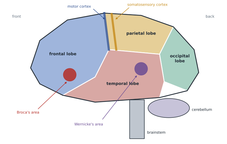
The frontal lobes, behind the forehead, are involved in planning, judgment, decision-making, working memory and controlling impulses, as well as producing speech and controlling movement. The parietal lobes, at the top and rear, receive sensory input for touch and body position and help with spatial reasoning and attention. The occipital lobes, at the back of the head, process vision. The temporal lobes, above the ears, process sound and play important roles in language, memory and recognizing faces and objects.
At the rear of the frontal lobe runs the motor cortex, which controls voluntary movements. Just behind it, at the front of the parietal lobe, lies the somatosensory cortex, which registers touch, temperature and pain. In the 1930s and 1940s, Wilder Penfield and his colleagues at the Montreal Neurological Institute mapped these strips by stimulating the exposed cortex of patients who were awake during surgery for epilepsy and asking them what they felt. Their maps showed that body parts are represented in proportion to their sensitivity or the precision of their movements, not their size: the hands, lips and tongue occupy far more cortex than the back or legs. The resulting distorted figure, drawn on the cortex, is called the homunculus, or 'little person'.
Most of the cortex is not devoted to simple sensation or movement. Association areas integrate information, link it with memories and support higher mental functions such as reasoning, language and planning. They are found in all four lobes. The prefrontal cortex, at the very front, is the area most associated with executive functions: holding goals in mind, switching between tasks and inhibiting responses that would be inappropriate.
Language and the two hemispheres
Two areas of the left hemisphere played a historic part in showing that functions can be localized. In 1861, the French physician Paul Broca described a patient who could understand speech but could say little more than one syllable. After the patient died, Broca found damage in the lower left frontal lobe, now called Broca's area. In 1874, Carl Wernicke described patients with damage in the left temporal lobe who spoke fluently but made little sense and had trouble understanding others; that region is now called Wernicke's area. Language is now known to depend on a wider network, but these findings established that the left hemisphere is dominant for language in most people.
The two hemispheres are connected by the corpus callosum, a thick band of axons. In the 1960s, some patients with severe epilepsy had the corpus callosum cut to stop seizures spreading. Roger Sperry and Michael Gazzaniga studied these split-brain patients. Because each hemisphere receives visual information from the opposite side of space, the researchers could flash a word to just one hemisphere. When a word appeared to the right hemisphere only, patients said they saw nothing, because the speaking left hemisphere had not received it, yet the left hand, controlled by the right hemisphere, could pick out the named object. Sperry shared the Nobel Prize in 1981 for this work.
Popular claims that people are 'left-brained' or 'right-brained' go far beyond this evidence. The hemispheres do specialize to some degree, with the left more involved in language and the right more involved in some aspects of spatial attention and face perception. But in intact brains the hemispheres share information constantly through the corpus callosum, and large studies of brain activity have found no evidence that individuals rely more on one hemisphere overall.
Plasticity
The brain is not fixed. Neuroplasticity is the brain's ability to change its structure and function in response to experience and injury. It is greatest in childhood. Children who have had an entire hemisphere removed to stop life-threatening seizures can, with intensive rehabilitation, develop surprisingly good language and movement, as the remaining hemisphere takes over. In people who are blind from early life, parts of the visual cortex become active when they read Braille, and in people who are deaf, auditory areas may respond to visual input.
Adults show plasticity too, though less dramatically. Learning a skill such as juggling has been found to increase grey matter in areas that process visual motion, with some of the change reversing when practice stops. After a stroke, rehabilitation helps undamaged areas take over some functions. Navarro tells the class that plasticity is one reason the brain they leave the course with will be physically different from the one they brought in.
Worked problem
Locating a problem
A 64-year-old patient has a stroke. Afterward, he understands what people say and knows what he wants to say, but his speech is slow, effortful and missing small words. He also has weakness in his right arm. Where is the damage most likely to be, and why?
Show solution
- Speech that is effortful but with good comprehension suggests damage to Broca's area.
- Broca's area is in the lower part of the left frontal lobe.
- Weakness in the right arm suggests damage to the motor cortex of the left hemisphere, because each hemisphere controls the opposite side of the body.
- The motor cortex is at the rear of the frontal lobe, close to Broca's area.
- Both findings point to the left frontal lobe.
Answer: The damage is most likely in the left frontal lobe, affecting Broca's area and the nearby motor cortex that controls the right arm.
Common mistakes
MistakeWe use only a small fraction of our brains.
InsteadImaging shows activity throughout the brain, and damage to almost any region causes problems; the brain is too expensive to carry unused tissue.
MistakeA region lighting up on a scan is where an ability lives.
InsteadImaging shows relative changes in activity; most functions depend on networks of regions.
MistakePeople are either left-brained or right-brained.
InsteadThe hemispheres specialize somewhat but work together; individuals do not rely more on one overall.
MistakeThe adult brain cannot change.
InsteadThe brain remains plastic throughout life, though most plastic in childhood.
Summary and key terms
- The central nervous system is the brain and spinal cord; the peripheral nervous system connects it to the body.
- The somatic system controls voluntary movement; the autonomic system controls organs through sympathetic arousal and parasympathetic calming.
- EEG, MRI, PET, fMRI and brain stimulation reveal structure and activity, but images are easily over-interpreted.
- The brainstem controls vital functions; the cerebellum coordinates movement and timing.
- The thalamus relays sensation; the hypothalamus regulates basic drives; the amygdala and hippocampus support emotion and memory.
- The cortex has frontal, parietal, occipital and temporal lobes, with motor, sensory and association areas.
- Language depends mostly on left-hemisphere networks; split-brain studies show hemispheric specialization; the brain is plastic.
- Sympathetic nervous system
- The branch of the autonomic system that arouses the body for action.
- Cerebellum
- The structure at the back of the brain that coordinates movement, balance and timing.
- Hippocampus
- A limbic structure that helps form new explicit memories and supports navigation.
- Amygdala
- Limbic structures involved in detecting threats and in emotional learning.
- Corpus callosum
- The band of axons connecting the two cerebral hemispheres.
- Neuroplasticity
- The brain's ability to change its structure and function with experience and injury.
Practice problems
Work each problem on paper first, then reveal the answer to check.
Which branch of the nervous system is active when you calm down after a scare?
Show answer
The parasympathetic branch of the autonomic nervous system.
Which lobe processes vision?
Show answer
The occipital lobe, at the back of the brain.
Why are the hands larger than the back on the homunculus?
Show answer
Cortical area reflects sensitivity and precision of movement, and the hands are far more sensitive and precise.
In a split-brain patient, a word is flashed to the right hemisphere. Why can the patient not say it?
Show answer
Speech depends mainly on the left hemisphere, which did not receive the information because the corpus callosum is cut.
Lesson quiz · 5 questions
Score 60% or more to complete this lesson.
Create a free account to save your progress, take quizzes and earn a certificate.