
BIOL-380 · Lesson 8 of 12 · 26 min
Tolerance and immune regulation
Because B and T cell receptors are generated at random, every person makes lymphocytes capable of attacking their own tissues. That most people never develop autoimmune disease is due to tolerance, the specific unresponsiveness of the adaptive immune system to self antigens, and to regulation, the mechanisms that limit the size and duration of all immune responses. This lesson examines central tolerance in the thymus and bone marrow, peripheral tolerance in the rest of the body, the regulatory T cells whose discovery was recognized by the 2025 Nobel Prize in Physiology or Medicine, inhibitory receptors and checkpoints, and the special cases of tolerance to food, commensal microbes and the fetus. Lesson 10 then examines what happens when tolerance fails.
Central tolerance
Central tolerance is induced in the primary lymphoid organs while lymphocytes are developing. In the thymus, as lesson 6 described, thymocytes that bind self peptide–MHC strongly are deleted by apoptosis, a process called clonal deletion, or are diverted into the regulatory T cell lineage. The transcription factor AIRE, and a second factor called FEZF2, drive medullary thymic epithelial cells to express thousands of tissue-restricted proteins, so that T cells specific for insulin, thyroid proteins and many other organ-specific antigens can be removed before they leave. Remarkably, each AIRE-expressing cell makes only a fraction of these antigens, and the full set is displayed across the population, with dendritic cells picking up and re-presenting antigen from the epithelial cells.
B cells undergo similar tests in the bone marrow. An immature B cell whose receptor binds a multivalent self antigen strongly does not die at once; it first tries receptor editing, rearranging a new light chain to change its specificity. Studies in mice suggest that a quarter or more of B cells edit their receptors at least once. If editing fails, the cell dies. B cells that bind soluble self antigens more weakly may survive in an unresponsive, anergic state. Central tolerance is efficient but incomplete: some self-reactive cells escape because their antigen is not present in the thymus or bone marrow, because their affinity falls just below the deletion threshold, or because the antigen is modified in tissues after development. In healthy adults, a substantial minority of mature naive B cells still bind self antigens weakly.
Peripheral tolerance
Self-reactive lymphocytes that escape central tolerance are controlled by peripheral mechanisms. The first is ignorance: many self antigens are present in places naive T cells do not visit, such as behind the blood–brain barrier, or at concentrations too low to activate them. Ignorance can be broken, for example when injury releases sequestered antigens, as in sympathetic ophthalmia, a rare condition in which injury to one eye triggers autoimmune attack on both eyes.
The second mechanism is anergy. As lesson 6 explained, a T cell that recognizes its antigen without costimulation, as happens when resting dendritic cells present self antigens in the absence of infection, becomes functionally unresponsive. Anergic T cells have reduced signalling through the TCR and fail to make interleukin-2. The third mechanism is deletion of mature T cells by apoptosis after repeated stimulation with abundant self antigen, which involves the proteins Bim and Fas. The fourth, and perhaps most important, is suppression by regulatory T cells.
| Mechanism | Where it acts | How it works | What happens when it fails |
|---|---|---|---|
| Clonal deletion | Thymus, bone marrow | Strongly self-reactive developing lymphocytes die | AIRE deficiency: multi-organ autoimmunity (APS-1) |
| Receptor editing | Bone marrow | Self-reactive B cells rearrange new light chains | Possibly more autoreactive B cells |
| Ignorance | Periphery | Antigen sequestered or too scarce to activate T cells | Release of hidden antigen after injury |
| Anergy | Periphery | Antigen without costimulation inactivates T and B cells | Inflammation can provide costimulation to autoreactive cells |
| Deletion of mature cells | Periphery | Repeated stimulation triggers apoptosis via Bim and Fas | Fas defects: autoimmune lymphoproliferative syndrome |
| Regulatory T cells | Lymphoid organs and tissues | FOXP3 Tregs suppress effector cells | FOXP3 mutations: IPEX syndrome |
| Inhibitory receptors | Periphery | CTLA-4, PD-1 and others limit activation | CTLA-4 haploinsufficiency; checkpoint inhibitor side effects |
Regulatory T cells: a history of discovery
The idea of suppressor T cells was popular in the 1970s, but the cells could not be reliably identified, the proposed mechanisms proved illusory and the field fell into disrepute. Shimon Sakaguchi, working in Japan, persisted. Earlier experiments had shown that removing the thymus from mice three days after birth led to autoimmune disease of several organs. In 1995 Sakaguchi showed that a small population of CD4 T cells expressing high levels of CD25, the α chain of the interleukin-2 receptor, prevented autoimmune disease when transferred into mice that lacked them, and that removing these cells from normal mice caused autoimmunity. He called them regulatory T cells.
The molecular key came from a strain of mice called scurfy, discovered at Oak Ridge National Laboratory in the 1940s, in which males developed fatal multi-organ autoimmune inflammation within weeks of birth. In 2001 Mary Brunkow, Fred Ramsdell and colleagues at the biotechnology company Celltech Chiroscience in Bothell, Washington, identified the mutated gene, which they named Foxp3, and showed with collaborators that mutations in the human equivalent, FOXP3, cause IPEX syndrome (immune dysregulation, polyendocrinopathy, enteropathy, X-linked), a devastating disease of infant boys. In 2003 Sakaguchi and two other groups showed that FOXP3 is the master transcription factor that controls regulatory T cell development. For these discoveries concerning peripheral immune tolerance, Brunkow, Ramsdell and Sakaguchi received the 2025 Nobel Prize in Physiology or Medicine, announced on 6 October 2025.
Regulatory T cells (Tregs) make up about 5 to 10 per cent of CD4 T cells in the blood. Most are thymic Tregs, generated in the thymus from thymocytes with relatively high-affinity TCRs for self antigens; others are peripheral Tregs, induced from conventional CD4 T cells in the periphery, especially in the gut, by TGF-β, retinoic acid and microbial metabolites. Tregs depend on interleukin-2, which they cannot make themselves but consume from activated effector cells, an arrangement that ties the number of Tregs to the intensity of a response. Their stability depends on demethylation of a region of the FOXP3 gene, which locks in its expression.
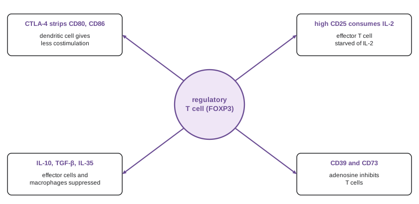
Tregs suppress through several mechanisms. They express high levels of CTLA-4, which binds CD80 and CD86 on dendritic cells and strips them from the cell surface by a process called trans-endocytosis, depriving naive T cells of costimulation. They consume interleukin-2, starving effector cells. They secrete the inhibitory cytokines interleukin-10, TGF-β and interleukin-35, which suppress dendritic cells, macrophages and effector T cells; interleukin-10 is particularly important at mucosal surfaces, and mice lacking it develop colitis. They can also produce adenosine, which inhibits T cells, through the surface enzymes CD39 and CD73. Tregs in tissues have further roles in repair, such as Tregs in muscle that produce the growth factor amphiregulin after injury.
# Toy model: effector T cell expansion with and without regulatory T cells.
# Effectors grow when stimulated; Tregs grow by consuming IL-2 made by effectors and suppress effectors.
def run(treg0, days=14, dt=0.001):
    e, r, t = 1.0, treg0, 0.0
    out = []
    while t <= days + 1e-9:
        if abs(t - round(t)) < dt / 2 and round(t) in (0, 3, 6, 9, 12, 14):
            out.append((round(t), e, r))
        il2 = e                                   # IL-2 made in proportion to effectors
        de = 1.2 * e * (1 - e / 1000) - 0.8 * r * e / (1 + 0.01 * e)   # growth minus suppression
        dr = 0.6 * r * il2 / (50 + il2) - 0.05 * r                     # Tregs need IL-2
        e = max(e + de * dt, 0.0); r = max(r + dr * dt, 0.0); t += dt
    return out
for label_, treg0 in (("no Tregs", 0.0), ("Tregs present", 1.0)):
    print(label_)
    for day, e, r in run(treg0):
print(f" day {day:2d}: effectors {e:8.1f} Tregs {r:6.2f}")no Tregs day 0: effectors 1.0 Tregs 0.00 day 3: effectors 35.3 Tregs 0.00 day 6: effectors 572.0 Tregs 0.00 day 9: effectors 980.0 Tregs 0.00 day 12: effectors 999.4 Tregs 0.00 day 14: effectors 999.9 Tregs 0.00 Tregs present day 0: effectors 1.0 Tregs 1.00 day 3: effectors 3.8 Tregs 0.92 day 6: effectors 16.7 Tregs 1.03 day 9: effectors 50.3 Tregs 1.78 day 12: effectors 17.5 Tregs 3.50 day 14: effectors 0.8 Tregs 3.56
The model shows a core feature of Treg biology: because Tregs depend on interleukin-2 produced by effector cells, they expand when a response grows and then restrain it, a negative feedback loop. Without Tregs, effector cells grow until they reach the limit imposed by the model's carrying capacity, a stand-in for the uncontrolled expansion seen in scurfy mice and IPEX patients. With Tregs, expansion is limited to a small peak, after which the accumulated Tregs drive the response back down. The parameters are illustrative, but models of this kind have helped explain why low doses of interleukin-2, which preferentially expand Tregs because of their high-affinity receptor, are being tested as treatments for autoimmune diseases such as lupus and type 1 diabetes.
Inhibitory receptors and checkpoints
Activated T cells express inhibitory receptors that act as brakes. CTLA-4 (cytotoxic T-lymphocyte–associated protein 4), discovered by Pierre Golstein's group in 1987 and shown to be inhibitory in the mid-1990s by James Allison and Jeffrey Bluestone, competes with CD28 for CD80 and CD86, binding them with higher affinity. Mice lacking CTLA-4 die of massive lymphoproliferation within weeks, and people with one defective copy of CTLA4 develop autoimmune disease and immunodeficiency. CTLA-4 acts mainly early, during priming in lymphoid organs, and on Tregs.
PD-1 (programmed cell death protein 1), discovered by Tasuku Honjo's group in 1992, binds the ligands PD-L1 and PD-L2. PD-L1 is expressed on many cells, especially in inflamed tissues, where interferon-γ induces it. PD-1 signalling recruits the phosphatase SHP-2, which dephosphorylates the TCR and CD28 pathways. PD-1 acts mainly in tissues, damping effector T cells during chronic stimulation; mice lacking PD-1 develop autoimmune disease more slowly and in a strain-dependent manner. Other inhibitory receptors include LAG-3, TIM-3 and TIGIT. Allison and Honjo shared the 2018 Nobel Prize for showing that blocking these checkpoints unleashes T cells against cancer (lesson 12), and the autoimmune side effects of such treatment, which can affect the colon, skin, liver, thyroid and other organs, reveal how much these brakes normally restrain self-reactive cells.
Exhaustion
During chronic infections such as HIV, hepatitis B and hepatitis C, and in many tumours, persistent antigen drives T cells into a distinct state called exhaustion, first characterized in mice infected with chronic strains of lymphocytic choriomeningitis virus. Exhausted T cells express multiple inhibitory receptors, especially PD-1, progressively lose the ability to produce interleukin-2, TNF and interferon-γ and to proliferate, and have a distinctive epigenetic landscape controlled by the transcription factor TOX. Exhaustion is a form of regulation that limits immunopathology when a pathogen cannot be cleared, at the cost of allowing it to persist. A subset of progenitor exhausted cells, expressing the transcription factor TCF-1, retains the ability to proliferate and is the population that expands after PD-1 blockade.
Tolerance at special sites
The gut presents a particular challenge. It contains the largest population of immune cells in the body, faces tonnes of food antigens over a lifetime and is home to trillions of commensal bacteria. Feeding an antigen typically induces oral tolerance, a reduced systemic response to later exposure, mediated by peripheral Tregs induced in the gut-associated lymphoid tissue by dendritic cells that make retinoic acid and TGF-β. Commensal bacteria actively promote this: certain Clostridia species produce short-chain fatty acids such as butyrate, which promote Treg differentiation by inhibiting histone deacetylases, and the human commensal Bacteroides fragilis makes a polysaccharide that induces interleukin-10-producing Tregs. Germ-free mice have fewer colonic Tregs and are prone to inappropriate inflammation.
Oral tolerance has practical implications for food allergy. For many years parents were advised to delay giving infants peanuts to prevent allergy. The LEAP trial, published in 2015 by Gideon Lack and colleagues, randomized infants at high risk of peanut allergy to eat or avoid peanut from 4 to 11 months of age; at five years, peanut allergy was present in about 3 per cent of those who ate peanut versus 17 per cent of those who avoided it, a relative reduction of over 80 per cent. Guidelines in Canada and the United States now recommend introducing peanut and other common allergens early, at around six months but not before four months, especially for infants at high risk.
The fetus poses a different problem: it carries paternal antigens foreign to the mother, yet is not rejected. Several mechanisms contribute. The placental trophoblast cells that contact maternal blood do not express classical HLA-A or HLA-B but express HLA-G and HLA-C, which engage inhibitory receptors on maternal NK cells; the enzyme indoleamine 2,3-dioxygenase depletes tryptophan, inhibiting T cells; and pregnancy expands maternal Tregs specific for fetal antigens, which persist after delivery and protect subsequent pregnancies with the same father. Peter Medawar posed the puzzle of fetal tolerance in 1953; it remains incompletely solved.
Some sites, including the eye, brain and testis, were long called immune privileged, because foreign grafts placed there survive longer than elsewhere. Privilege is now understood as an active state maintained by barriers, local expression of Fas ligand and inhibitory cytokines such as TGF-β, and the absence of conventional lymphatics, rather than complete isolation. Corneal transplants, among the most common transplants performed, often succeed without HLA matching for this reason.
Therapeutic tolerance
Inducing antigen-specific tolerance, switching off only the harmful response while leaving the rest of the immune system intact, is a long-standing goal. Allergen immunotherapy, in which gradually increasing doses of an allergen are given under the skin or tongue, induces Tregs and IgG4 antibodies that block IgE, and oral immunotherapy for peanut allergy, offered in allergy clinics in both countries, received its first US approval for a standardized peanut product in 2020. Treg cell therapy, in which a patient's own Tregs are expanded or engineered and reinfused, is in clinical trials for type 1 diabetes, transplantation and graft-versus-host disease; engineered Tregs carrying chimeric antigen receptors that target the transplanted organ are an active area of research. Teplizumab, an anti-CD3 antibody approved by the US Food and Drug Administration in 2022, delays the onset of clinical type 1 diabetes in at-risk people by about two to three years on average, probably by inducing partial exhaustion of autoreactive T cells.
Worked problem
Reasoning about a tolerance defect
An infant boy develops severe watery diarrhoea, eczema and diabetes in the first months of life. Blood tests show autoantibodies against islet cells and the gut. His maternal uncle died in infancy with similar symptoms. Which gene is most likely mutated, and why does the defect produce this pattern?
Show solution
- Onset in infancy, X-linked inheritance through the maternal line and autoimmunity of the gut, skin and endocrine organs together suggest IPEX syndrome.
- IPEX is caused by mutations in FOXP3, the master regulator of regulatory T cells.
- Without functional Tregs, self-reactive T cells that escaped central tolerance are not suppressed, and inflammation targets organs with high antigen exposure, such as the gut and skin, and endocrine tissues such as the pancreas.
- Treatment involves immunosuppression and, ideally, hematopoietic stem cell transplantation to supply normal Tregs; gene-corrected Treg therapies are being studied.
Answer: A FOXP3 mutation causing IPEX syndrome; loss of regulatory T cells allows multi-organ autoimmunity to develop early in life.
Common mistakes
MistakeAssuming central tolerance removes all self-reactive lymphocytes.
InsteadCentral tolerance is incomplete; peripheral mechanisms control escapees.
MistakeTreating tolerance as the inability to respond.
InsteadTolerance is an active, antigen-specific state maintained by deletion, anergy and regulation.
MistakeEquating Tregs with the discredited suppressor cells of the 1970s.
InsteadTregs are defined by FOXP3 and have well-characterized mechanisms.
MistakeThinking exhaustion is a defect with no purpose.
InsteadIt limits immunopathology during chronic stimulation, at the cost of pathogen persistence.
MistakeAdvising delayed introduction of peanut to prevent allergy.
InsteadTrials such as LEAP showed early introduction greatly reduces peanut allergy in high-risk infants.
Summary and key terms
- Central tolerance deletes or edits strongly self-reactive lymphocytes; AIRE and FEZF2 expose T cells to tissue antigens in the thymus.
- Peripheral tolerance includes ignorance, anergy, deletion of mature cells and suppression by regulatory T cells.
- Tregs, identified by CD25 and FOXP3, suppress through CTLA-4, IL-2 consumption, IL-10, TGF-β and adenosine; FOXP3 mutations cause IPEX.
- Brunkow, Ramsdell and Sakaguchi received the 2025 Nobel Prize for discoveries concerning peripheral immune tolerance.
- CTLA-4 and PD-1 are inhibitory checkpoints acting in lymphoid organs and tissues respectively.
- Chronic antigen drives T cell exhaustion, marked by PD-1 and controlled by TOX.
- Oral tolerance, fetal tolerance and immune privilege are maintained by active mechanisms; early allergen introduction reduces food allergy.
- tolerance
- Specific unresponsiveness of the adaptive immune system to an antigen, especially self.
- central tolerance
- Tolerance induced in the thymus and bone marrow during lymphocyte development.
- peripheral tolerance
- Mechanisms that control mature self-reactive lymphocytes outside the primary organs.
- receptor editing
- Rearrangement of a new light chain by a self-reactive immature B cell.
- regulatory T cell
- A FOXP3-expressing CD4 T cell that suppresses immune responses.
- FOXP3
- The master transcription factor of regulatory T cells.
- IPEX syndrome
- Lethal X-linked autoimmunity caused by FOXP3 mutations.
- CTLA-4
- An inhibitory receptor that competes with CD28 for CD80 and CD86.
- PD-1
- An inhibitory receptor that dampens T cell signalling when bound by PD-L1 or PD-L2.
- T cell exhaustion
- A dysfunctional state of T cells under chronic antigen stimulation.
- oral tolerance
- Reduced systemic responsiveness to an antigen after it is eaten.
Practice problems
Work each problem on paper first, then reveal the answer to check.
What does receptor editing achieve?
Show answer
It replaces a self-reactive B cell receptor by rearranging a new light chain.
Name four peripheral tolerance mechanisms.
Show answer
Ignorance, anergy, deletion of mature cells and suppression by Tregs.
What experiment led Sakaguchi to identify Tregs?
Show answer
Transfer of CD4 CD25-high T cells prevented autoimmunity; their removal caused it.
What gene is mutated in scurfy mice and IPEX patients?
Show answer
FOXP3.
How does CTLA-4 on Tregs suppress responses?
Show answer
It removes CD80 and CD86 from dendritic cells by trans-endocytosis, reducing costimulation.
Why do Tregs depend on effector T cells?
Show answer
They need interleukin-2, which they cannot make but effector cells produce.
What did the LEAP trial show?
Show answer
Early peanut introduction reduced peanut allergy in high-risk infants by over 80 per cent.
Give two mechanisms of fetal tolerance.
Show answer
HLA-G on trophoblast engaging NK inhibitory receptors; expansion of fetal-specific maternal Tregs.
Lesson quiz · 5 questions
Score 60% or more to complete this lesson.
Create a free account to save your progress, take quizzes and earn a certificate.