
BIOL-380 · Lesson 7 of 12 · 26 min
Effector responses and immunological memory
Activation in a lymph node produces an army of effector lymphocytes. This lesson follows them into action: how cytotoxic T cells find and kill infected cells, how helper T cells arm macrophages and other cells, how antibodies and cells cooperate, and how the response is shut down once the infection is controlled. It then examines the most practically important feature of adaptive immunity, memory: what memory cells are, where they live, how long protection lasts and why some infections and vaccines give lifelong immunity while others do not.
Cytotoxic T lymphocytes
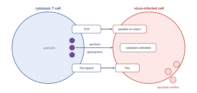
Effector CD8 T cells leave the lymph node and enter infected tissues, where they scan cells for their specific peptide on MHC class I. When a cytotoxic T lymphocyte (CTL) recognizes a target, it forms an immunological synapse and within minutes reorients its microtubule-organizing centre and secretory apparatus toward the target, delivering lytic granules precisely into the narrow space between the cells, so that neighbouring uninfected cells are spared. The granules contain perforin, which inserts into the target membrane and forms pores, and granzymes, serine proteases that pass through the pores or are taken up by endocytosis and trigger apoptosis by cleaving caspases and other substrates. The CTL then detaches and can kill again; a single CTL can kill several targets in succession, and live imaging shows that several CTLs often cooperate to kill a single virus-infected cell.
CTLs can also kill through death receptors: Fas ligand on the CTL binds Fas (CD95) on the target, activating caspase-8 and apoptosis. Killing by apoptosis rather than lysis is important, because the infected cell's contents, including virus particles, are packaged into apoptotic bodies and engulfed by phagocytes without spilling into the tissue. CTLs also release interferon-γ and TNF, which inhibit viral replication and increase MHC class I expression on surrounding cells. The importance of perforin is shown by familial hemophagocytic lymphohistiocytosis, in which inherited perforin deficiency prevents CTLs and NK cells from killing infected cells; T cells and macrophages remain activated, producing a devastating cytokine storm unless treated by stem cell transplantation.
Helper T cell effector functions
Effector CD4 T cells act mainly by recognizing antigen on other cells and delivering signals to them. Th1 cells recognize peptides from engulfed microbes on the MHC class II of macrophages and activate them through interferon-γ and CD40 ligand. Activated macrophages fuse their phagosomes with lysosomes more efficiently, produce more reactive oxygen and nitrogen species and make more inflammatory cytokines, enabling them to kill microbes such as Mycobacterium tuberculosis and Leishmania that survive inside resting macrophages. If the microbe still persists, chronic Th1 activation leads to the formation of a granuloma, a ball of activated macrophages, some fused into giant cells, surrounded by T cells, which walls off the infection at the cost of tissue damage. Roughly a quarter of the world's population is estimated to carry latent tuberculosis contained in this way.
Th2 cells act at barrier surfaces against worms. Their interleukin-4 and interleukin-13 induce alternatively activated macrophages, goblet cell hyperplasia and mucus production, increased smooth muscle contraction and faster turnover of the gut epithelium, a weep-and-sweep response that dislodges worms, while interleukin-5 recruits eosinophils, which release toxic granule proteins onto larvae coated with IgE or IgG. Th17 cells recruit neutrophils by inducing epithelial cells to make chemokines, and induce antimicrobial peptides. Tfh cells, as described in lesson 5, help B cells in germinal centres. The type of helper response thus determines which effector mechanisms dominate, and a mismatch, such as a Th2 response to an intracellular bacterium, can be harmful, as in lepromatous leprosy, where a weak Th1 and stronger Th2 response allows Mycobacterium leprae to multiply unchecked.
| Type of response | Typical threat | Main effector mechanisms |
|---|---|---|
| Type 1 (Th1, CTL, NK, IgG1/IgG3) | Viruses, intracellular bacteria and protozoa | Macrophage activation by IFN-γ, CTL killing, opsonizing antibodies |
| Type 2 (Th2, ILC2, IgE, eosinophils, mast cells) | Helminths, also venoms and allergens | Mucus, smooth muscle contraction, eosinophils, IgE-armed mast cells, tissue repair |
| Type 3 (Th17, ILC3, neutrophils) | Extracellular bacteria and fungi | Neutrophil recruitment, antimicrobial peptides, barrier maintenance |
| Antibody-dominated (Tfh, plasma cells) | Toxins, extracellular microbes, viruses before cell entry | Neutralization, opsonization, complement |
Cooperation between antibodies and cells
Antibodies bridge the specificity of adaptive immunity and the killing power of innate cells. Fc receptors are expressed on many cell types and recognize the Fc portions of different antibody classes. Activating Fcγ receptors on macrophages and neutrophils trigger phagocytosis of IgG-coated microbes, CD16 on NK cells triggers ADCC, and the high-affinity IgE receptor FcεRI arms mast cells and basophils with IgE. An inhibitory Fcγ receptor, FcγRIIB, on B cells and myeloid cells dampens responses when immune complexes accumulate, providing negative feedback. Different IgG subclasses bind these receptors differently, which is why therapeutic antibodies are engineered for particular Fc functions: an antibody meant to kill tumour cells is designed to engage activating receptors strongly, while one meant only to block a cytokine is designed to engage them weakly.
Contraction: ending the response
At the peak of a strong response, the expanded effector population may make up a large share of the lymphocytes in the body. Once antigen is cleared, 90 to 95 per cent of these cells die over the following weeks, a phase called contraction. Contraction occurs largely by apoptosis as the survival signals that sustained the cells, antigen, costimulation and cytokines such as interleukin-2, disappear. The proteins Bim and other members of the Bcl-2 family regulate this death, and repeatedly activated T cells can also kill one another through Fas. Inhibitory receptors such as CTLA-4 and PD-1 and regulatory T cells further damp the response. People with defects in Fas or Fas ligand develop autoimmune lymphoproliferative syndrome, with enlarged lymph nodes and spleen and autoimmune destruction of blood cells, because activated lymphocytes fail to die.
import math
# Toy model of a T cell response: expansion while antigen is present, contraction after clearance,
# with a fraction of cells surviving as long-lived memory. Rates are illustrative, per day.
naive = 100.0          # epitope-specific precursors
growth = 1.4           # net expansion rate while antigen persists (about 12 h doubling)
death_eff = 0.45       # effector death rate after antigen is cleared
mem_frac = 0.05        # fraction of the peak that becomes memory
clear_day = 8          # antigen cleared at day 8
dt = 0.01
eff, mem, t = naive, 0.0, 0.0
peak = 0.0
print("day   effector cells   memory cells")
records = {0, 4, 8, 10, 14, 21, 30, 60}
while t <= 60 + 1e-9:
    d = round(t, 2)
    if d in records:
        print(f"{int(d):3d}   {eff:14,.0f}   {mem:12,.0f}")
    if t < clear_day:
        eff += growth * eff * dt
        peak = max(peak, eff)
    else:
        if mem == 0.0:
            mem = mem_frac * peak
            eff -= mem
        eff -= death_eff * eff * dt
        mem -= 0.0005 * mem * dt          # memory decays very slowly (half-life years)
    t += dt
print(f"peak expansion: {peak / naive:,.0f}-fold; memory pool is {mem / naive:,.0f} times the naive precursor pool")day effector cells memory cells 0 100 0 4 26,012 0 8 6,766,499 0 10 2,656,667 342,720 14 437,364 342,036 21 18,609 340,841 30 321 339,310 60 0 334,258 peak expansion: 68,612-fold; memory pool is 3,343 times the naive precursor pool
The model reproduces the familiar shape of a T cell response: exponential expansion to a peak around day 8, rapid contraction as effector cells die, and a stable memory population that is much smaller than the peak but thousands of times larger than the original pool of naive precursors. This larger pool, together with qualitative changes in the cells themselves, explains why a second response is faster. The exact numbers depend on the chosen rates; real responses vary with the pathogen, the dose and the host.
Memory T cells
Memory T cells differ from naive cells in number, location and function. They are present at frequencies tens to hundreds of times higher; they respond to lower doses of antigen and are less dependent on costimulation; they rapidly produce effector cytokines and, for CD8 cells, cytotoxic molecules; and they have altered homing receptors that give them access to tissues. In 1999 Federica Sallusto and Antonio Lanzavecchia divided human memory T cells into central memory cells, which express CCR7 and L-selectin, circulate through lymph nodes and proliferate vigorously on re-exposure, and effector memory cells, which lack these molecules, patrol the blood and non-lymphoid tissues and act immediately.
A third population, discovered in the 2000s, has transformed thinking about protection. Tissue-resident memory T cells (TRM) settle permanently in tissues such as the skin, lungs, gut and reproductive tract after an infection there, without recirculating. Marked by CD69 and, for many CD8 TRM, the integrin CD103, they act as sentinels: on detecting their antigen, they kill infected cells and release interferon-γ, which puts the surrounding tissue on alert and recruits circulating cells within hours. TRM explain why immunity at a site of previous infection is often stronger than elsewhere, and they have encouraged development of vaccines delivered to mucosal surfaces, such as nasal influenza vaccines, that can establish them. Memory T cells are maintained for years by slow homeostatic proliferation driven by interleukin-7 and interleukin-15, rather than by persistent antigen.
Memory B cells and long-lived plasma cells
Humoral memory has two components. Long-lived plasma cells, generated in germinal centres, migrate to niches in the bone marrow where they receive survival signals from stromal cells and cytokines such as APRIL and interleukin-6, and continue to secrete antibody without needing antigen. These cells provide the circulating antibody that can prevent infection outright. Remarkable studies show how long they can last: people vaccinated against smallpox decades earlier still have neutralizing antibodies, and a 2008 study of survivors of the 1918 influenza pandemic, then aged over 90, found B cells making antibodies that neutralized the 1918 virus. Memory B cells, the second component, are class-switched, have mutated high-affinity receptors and circulate or reside in lymphoid tissue; they do not secrete antibody until re-exposed, when they rapidly become plasma cells, giving the fast secondary response seen in lesson 5.
Antibody levels after an infection or vaccine typically fall in two phases: a rapid early decline as short-lived plasma cells die, then a much slower decline sustained by long-lived plasma cells. The half-life of the slow phase differs strikingly between antigens. A study by Ian Amanna, Nichole Carlson and Mark Slifka in 2007 estimated antibody half-lives of more than 200 years for measles and mumps after infection, around 50 years for rubella, but only about 11 years for tetanus and 19 years for diphtheria after vaccination, explaining why tetanus boosters are recommended every ten years in adults while one or two measles doses give lifelong protection.
import math
# Antibody decline after vaccination: two-phase model, and when a booster might be needed.
# Concentrations in arbitrary units; protective threshold illustrative.
def level(t_years, peak, fast_frac, fast_half, slow_half):
    fast = peak * fast_frac * 0.5 ** (t_years / fast_half)
    slow = peak * (1 - fast_frac) * 0.5 ** (t_years / slow_half)
    return fast + slow
threshold = 0.6
for name, slow_half in (("tetanus-like (slow half-life 11 y)", 11.0), ("measles-like (slow half-life 200 y)", 200.0)):
    print(name)
    for t in (0, 1, 5, 10, 20, 40):
        print(f"   year {t:2d}: antibody {level(t, 10.0, 0.8, 0.3, slow_half):6.2f}")
    # find the year the level falls below threshold
    t = 0.0
    while level(t, 10.0, 0.8, 0.3, slow_half) > threshold and t < 300:
        t += 0.1
print(f" falls below threshold {threshold} after about {t:.0f} years" if t < 300 else " stays above threshold for over 300 years")tetanus-like (slow half-life 11 y) year 0: antibody 10.00 year 1: antibody 2.67 year 5: antibody 1.46 year 10: antibody 1.07 year 20: antibody 0.57 year 40: antibody 0.16 falls below threshold 0.6 after about 19 years measles-like (slow half-life 200 y) year 0: antibody 10.00 year 1: antibody 2.79 year 5: antibody 1.97 year 10: antibody 1.93 year 20: antibody 1.87 year 40: antibody 1.74 stays above threshold for over 300 years
Real decisions about boosters depend on more than antibody levels. Memory B and T cells can respond quickly even after antibodies have fallen, which is enough for diseases with long incubation periods, such as hepatitis B, where a memory response can control infection before disease develops; that is why routine hepatitis B boosters are not recommended for healthy people who completed the vaccine series. For toxin-mediated diseases such as tetanus, with incubation periods of days, protection depends on antibody already present, so boosters matter more.
Why memory sometimes fails
Immunity is not always lasting. Some pathogens change their antigens faster than memory can keep up: influenza viruses accumulate mutations in their surface proteins (antigenic drift, described in BIOL-330), so antibodies against last year's strain bind this year's poorly, and seasonal vaccines must be updated. Rhinoviruses circulate as more than 150 serotypes. Some pathogens, such as respiratory syncytial virus and the seasonal coronaviruses, cause infections of the airway surface where circulating IgG has limited access, and mucosal IgA responses wane within months, so reinfection is common. Others, such as Plasmodium and HIV, actively subvert immune responses. And some vaccines, especially inactivated and subunit vaccines without strong adjuvants, induce weaker germinal centre responses and shorter-lived antibodies than live infections.
A further complication is original antigenic sin, a term coined by Thomas Francis Jr. in 1960 and now often called immune imprinting: the first influenza strains a person meets in childhood shape responses to later strains, because memory B cells recognizing conserved epitopes are recalled preferentially over naive cells that could target new ones. Imprinting can be helpful, as when childhood exposure to H1 or H2 viruses appears to protect against later infection with avian H5N1, which belongs to the same group, or unhelpful, when recall of old memory limits responses to new variants. It is one reason researchers are developing universal influenza vaccines that target conserved parts of the virus.
Worked problem
Interpreting antibody kinetics
Two years after receiving a vaccine, a person's antibody level has fallen to one quarter of its value at six months. Assuming the decline over this period is in the slow phase and exponential, estimate the half-life, and predict the level at ten years.
Show solution
- From six months to two years is 1.5 years, during which the level falls by a factor of four, which is two half-lives.
- The half-life is therefore 1.5 / 2 = 0.75 years, about nine months.
- From two years to ten years is 8 years, or 8 / 0.75 ≈ 10.7 half-lives, so the level falls by a further factor of about \(2^{10.7} \approx 1{,}600\).
- Such a short half-life suggests the response was sustained mainly by short-lived plasma cells; whether protection is lost also depends on memory B and T cells, which antibody measurements do not capture.
Answer: The half-life is about nine months, and the level at ten years would be roughly 1/1,600 of the two-year value, though memory cells may still protect.
Common mistakes
MistakeThinking cytotoxic T cells lyse targets indiscriminately.
InsteadThey deliver granules directionally across a synapse and induce apoptosis, sparing neighbours.
MistakeBelieving memory depends on continuous exposure to antigen.
InsteadMemory T cells are maintained by IL-7 and IL-15 and long-lived plasma cells by bone marrow niches.
MistakeEquating falling antibody levels with loss of immunity.
InsteadMemory B and T cells can still mount rapid responses, enough for some diseases.
MistakeAssuming all memory cells circulate.
InsteadTissue-resident memory T cells stay in tissues as local sentinels.
MistakeExpecting every vaccine to give lifelong protection like measles.
InsteadAntibody half-lives vary greatly; antigen variation and mucosal sites also limit durability.
Summary and key terms
- CTLs kill infected cells by directional release of perforin and granzymes and through Fas ligand, inducing apoptosis.
- Th1 cells activate macrophages; Th2 cells drive anti-worm responses; Th17 cells recruit neutrophils; Tfh cells help B cells.
- Fc receptors link antibodies to phagocytes, NK cells and mast cells, with inhibitory FcγRIIB providing feedback.
- Most effector cells die during contraction; defects in Fas cause autoimmune lymphoproliferative syndrome.
- Memory T cells include central, effector and tissue-resident subsets maintained by IL-7 and IL-15.
- Long-lived plasma cells in bone marrow sustain antibody for years to decades; memory B cells give rapid recall responses.
- Durability varies with antigen; antigen variation, mucosal sites and imprinting limit protection.
- cytotoxic T lymphocyte
- An effector CD8 T cell that kills target cells.
- granzyme
- A serine protease delivered by cytotoxic cells that triggers apoptosis.
- Fas ligand
- A death receptor ligand that induces apoptosis in Fas-bearing cells.
- granuloma
- An organized collection of activated macrophages and T cells around persistent antigen.
- Fc receptor
- A cell receptor that binds the Fc region of antibodies.
- contraction
- The death of most effector cells after antigen is cleared.
- central memory T cell
- A memory T cell that circulates through lymph nodes and proliferates on re-exposure.
- effector memory T cell
- A memory T cell that patrols blood and tissues and acts rapidly.
- tissue-resident memory T cell
- A memory T cell that remains in a tissue as a local sentinel.
- long-lived plasma cell
- An antibody-secreting cell that survives for years in bone marrow niches.
- immune imprinting
- The lasting influence of first exposures on later responses to related antigens.
Practice problems
Work each problem on paper first, then reveal the answer to check.
Why do CTLs kill by apoptosis rather than lysis?
Show answer
Apoptosis packages cell contents, including virus, for clean removal by phagocytes.
How do Th1 cells help control tuberculosis?
Show answer
Interferon-γ and CD40 ligand activate macrophages to kill engulfed mycobacteria.
What is the weep-and-sweep response?
Show answer
Th2-driven mucus secretion and gut contraction that expel intestinal worms.
What proportion of effector T cells typically die during contraction?
Show answer
About 90 to 95 per cent.
Distinguish central memory from effector memory T cells.
Show answer
Central memory cells express CCR7 and home to lymph nodes; effector memory cells lack CCR7 and patrol tissues.
Where do long-lived plasma cells live?
Show answer
In survival niches in the bone marrow.
Why are tetanus boosters given every ten years but measles vaccine is not boosted in adults?
Show answer
Tetanus antibody has a half-life of about 11 years; measles antibody lasts much longer.
What is immune imprinting?
Show answer
Preferential recall of memory from first exposures when related antigens are met later.
Lesson quiz · 5 questions
Score 60% or more to complete this lesson.
Create a free account to save your progress, take quizzes and earn a certificate.