
BIOL-380 · Lesson 10 of 12 · 28 min
Hypersensitivity and autoimmunity
The immune system can cause disease in two broad ways besides failing to protect. It can respond excessively or inappropriately to harmless environmental antigens, such as pollen, peanuts or nickel, causing hypersensitivity; or it can attack the body's own tissues, causing autoimmunity. Together these conditions affect a large share of the population: allergic diseases affect perhaps a quarter or more of people in high-income countries, and autoimmune diseases together affect roughly 5 to 8 per cent. This lesson classifies hypersensitivity reactions by mechanism, examines allergy in detail, and then explores how tolerance breaks down to produce autoimmune disease and how such diseases are treated.
Classifying hypersensitivity
In 1963 Philip Gell and Robin Coombs classified hypersensitivity reactions into four types according to the immune mechanism. Types I to III are mediated by antibodies and type IV by T cells. The classification simplifies reality, since many diseases involve more than one mechanism, but it remains a useful framework for linking symptoms to their causes.
| Type | Mechanism | Timing | Examples |
|---|---|---|---|
| I (immediate) | IgE on mast cells and basophils cross-linked by allergen; release of histamine and other mediators | Minutes | Hay fever, allergic asthma, food allergy, anaphylaxis |
| II (antibody against cells or matrix) | IgG or IgM against cell-surface or matrix antigens; complement, phagocytosis or altered receptor function | Hours to days | Transfusion reactions, hemolytic disease of the newborn, Graves disease, myasthenia gravis |
| III (immune complex) | Soluble antigen–antibody complexes deposit in vessels and tissues and activate complement | Hours to weeks | Serum sickness, lupus nephritis, post-streptococcal glomerulonephritis |
| IV (delayed-type) | T cells (Th1, Th17, CD8) activate macrophages or kill cells | 24–72 hours or longer | Contact dermatitis, tuberculin skin test, type 1 diabetes |
Type I hypersensitivity: allergy
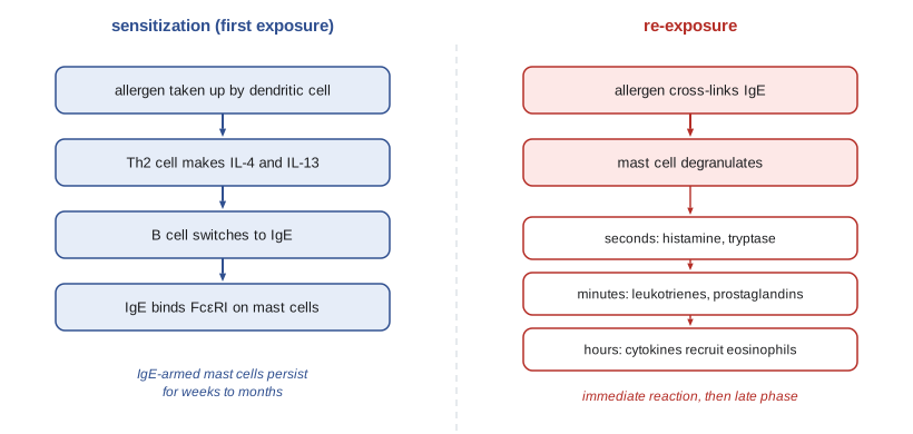
Allergy begins with sensitization. On first exposure to an allergen, a person with a genetic predisposition, called atopy, mounts a Th2 response: dendritic cells present allergen peptides to naive CD4 T cells in an environment rich in epithelial cytokines such as thymic stromal lymphopoietin, interleukin-25 and interleukin-33, which favour Th2 and type 2 innate lymphoid cells. Interleukin-4 and interleukin-13 from Th2 and Tfh cells induce B cells to switch to IgE. IgE then binds the high-affinity receptor FcεRI on mast cells in tissues and on basophils in blood, where it persists for weeks to months, arming them. Most allergens are small soluble proteins that enter across mucosal surfaces at low doses, and many have enzymatic activity, such as the cysteine protease Der p 1 of house dust mites, which disrupts epithelial tight junctions.
On re-exposure, allergen cross-links IgE on mast cells, triggering degranulation within minutes. Preformed mediators, histamine, the protease tryptase and heparin, are released at once; histamine dilates and leaks blood vessels, causes itching and contracts bronchial smooth muscle. Within minutes the mast cell also makes lipid mediators, prostaglandin D2 and the leukotrienes C4, D4 and E4, which are far more potent bronchoconstrictors than histamine and are the target of leukotriene-receptor antagonists used in asthma. Over hours, mast cells produce cytokines such as TNF and interleukin-5, recruiting eosinophils, basophils and Th2 cells, which cause a late-phase reaction four to eight hours later. In chronic allergic diseases such as asthma and atopic dermatitis, repeated cycles of these reactions remodel the tissue, thickening the airway wall and increasing mucus production.
Where the allergen enters determines the disease. Inhaled pollen causes allergic rhinitis and conjunctivitis; inhaled dust mite and animal allergens contribute to allergic asthma; food allergens cause hives, vomiting and, in severe cases, systemic reactions; insect venoms cause local or systemic reactions. Anaphylaxis is a rapid, potentially fatal systemic reaction in which mediators released throughout the body cause widespread vasodilation and capillary leak, falling blood pressure, airway swelling and bronchoconstriction. The first-line treatment is intramuscular epinephrine, which constricts blood vessels, relaxes airway smooth muscle and stabilizes mast cells; antihistamines act too slowly to treat anaphylaxis. People at risk carry epinephrine auto-injectors, and a nasal epinephrine spray was approved in the United States in 2024. Anaphylaxis can also be triggered without IgE, for example by some drugs that activate mast cells directly through the receptor MRGPRX2.
The prevalence of allergic disease rose sharply in high-income countries during the second half of the twentieth century. In 1989 David Strachan observed that hay fever was less common in children with many older siblings and proposed the hygiene hypothesis: reduced exposure to infections in early life might predispose to allergy. The idea has been refined into the old friends or microbial diversity hypothesis, which emphasizes contact with diverse environmental microbes, helminths and a rich gut microbiota rather than infections as such. Supporting evidence includes lower rates of asthma and allergy in children raised on traditional farms, such as Amish children in the United States compared with Hutterite children who farm with modern methods, and associations between early antibiotic use, caesarean birth and allergy. The CHILD Cohort Study, which has followed more than 3,000 Canadian children from before birth, has linked differences in the infant gut microbiota at three months to later wheeze and allergic sensitization. Diet, vitamin D, air pollution and timing of food introduction (lesson 8) also contribute.
Treatment of allergy includes allergen avoidance, antihistamines, corticosteroids and leukotriene antagonists. Biological drugs target the type 2 pathway: omalizumab, an antibody against IgE, reduces asthma attacks and, since its 2024 US approval for food allergy, the severity of reactions to accidental exposure; dupilumab, which blocks the shared receptor for interleukin-4 and interleukin-13, treats atopic dermatitis, asthma and several other type 2 diseases; and antibodies against interleukin-5 or its receptor reduce eosinophils in severe eosinophilic asthma. Allergen immunotherapy (lesson 8) is the only treatment that alters the underlying immune response.
Type II hypersensitivity
In type II reactions, antibodies bind antigens on cell surfaces or in the extracellular matrix. They can destroy cells by complement-mediated lysis or opsonization and phagocytosis, cause inflammation, or alter the function of receptors. Transfusion reactions are a classic example: people make natural IgM antibodies against the A and B blood group antigens they lack, probably induced by similar carbohydrates on gut bacteria, so a mismatched transfusion causes rapid intravascular hemolysis. Hemolytic disease of the fetus and newborn occurs when an RhD-negative mother, sensitized by a previous RhD-positive pregnancy, makes IgG anti-D that crosses the placenta and destroys fetal red cells. Since the late 1960s, giving RhD-negative mothers anti-D immunoglobulin during pregnancy and after delivery prevents sensitization, probably by clearing fetal cells before they can trigger a response, and has made the disease rare in Canada and the United States.
Some type II reactions alter function without destroying cells. In Graves disease, the most common cause of hyperthyroidism, autoantibodies stimulate the receptor for thyroid-stimulating hormone, driving excess hormone production; in myasthenia gravis, autoantibodies block and remove acetylcholine receptors at the neuromuscular junction, causing muscle weakness. Babies born to mothers with these conditions can show transient disease, because maternal IgG crosses the placenta, which demonstrates that the antibodies themselves cause the disease. Drug-induced hemolytic anaemia and the kidney and lung damage of Goodpasture syndrome, caused by antibodies against type IV collagen, are further examples.
Type III hypersensitivity
When antibodies form complexes with soluble antigens, the complexes are normally cleared by complement and phagocytes in the liver and spleen. If complexes form in large amounts, particularly with a slight excess of antigen, they can escape clearance and deposit in small blood vessels, the kidney glomeruli and joints, where they activate complement and Fc receptors and recruit neutrophils, causing vasculitis, nephritis and arthritis. Clemens von Pirquet described serum sickness in 1905 in patients treated with horse antitoxin serum, who developed fever, rash, joint pain and kidney inflammation about one to two weeks later, as antibodies against horse proteins appeared and formed complexes with the remaining serum. Serum sickness still occurs with some drugs and therapeutic antibodies. Post-streptococcal glomerulonephritis follows some throat or skin infections with Streptococcus pyogenes, and immune complex deposition is central to lupus nephritis.
Type IV hypersensitivity
Delayed-type reactions are mediated by T cells and take one to three days to develop, the time needed for memory T cells to be recruited and activated at the site. The tuberculin skin test, in which a small amount of mycobacterial protein is injected into the skin, causes a firm swelling at 48 to 72 hours in people with T cell memory from infection or BCG vaccination, as Th1 cells accumulate and activate macrophages. Contact dermatitis to nickel, rubber chemicals or urushiol, the oil of poison ivy, occurs when small reactive chemicals called haptens bind skin proteins, creating new epitopes recognized by T cells. Type IV mechanisms also cause tissue damage in many autoimmune diseases and in transplant rejection, and severe drug reactions such as Stevens–Johnson syndrome, which are linked to particular HLA alleles (lesson 4).
Autoimmunity: how tolerance breaks
More than 80 autoimmune diseases are recognized. Some are organ-specific, such as type 1 diabetes, in which T cells destroy the insulin-producing β cells of the pancreas, and Hashimoto thyroiditis; others are systemic, such as systemic lupus erythematosus, in which autoantibodies against nuclear antigens form immune complexes that damage skin, joints, kidneys and blood vessels. Most autoimmune diseases are more common in women; lupus affects about nine women for every man. The reasons include the immune-enhancing effects of estrogens, genes on the X chromosome such as TLR7 that can escape X inactivation, and, as reported in 2024, the RNA Xist itself, which coats the inactive X chromosome and forms complexes with proteins that can provoke autoantibodies.
Genetic susceptibility is substantial. Identical twins of people with type 1 diabetes have a lifetime risk of roughly 30 to 50 per cent, compared with about 0.4 per cent in the general population, but the fact that concordance is far from 100 per cent shows that environment and chance matter too. The strongest genetic associations are with HLA alleles (lesson 4), which determine which self peptides are presented. Genome-wide association studies have identified hundreds of other variants, many in genes controlling lymphocyte activation and tolerance, such as PTPN22, CTLA4, IL2RA and the interleukin-23 receptor; many are shared among several autoimmune diseases. Rare single-gene defects, such as in AIRE, FOXP3 and Fas (lessons 6 to 8), produce severe autoimmunity and reveal the essential tolerance mechanisms.
import math
# Strength of an HLA association: odds ratio and 95% CI from a case-control study (invented counts).
#                 carrier   non-carrier
cases     = (180, 20)      # people with the disease
controls  = (16, 184)      # people without it
a, b = cases; c, d = controls
odds_ratio = (a * d) / (b * c)
se = math.sqrt(1 / a + 1 / b + 1 / c + 1 / d)
lo, hi = math.exp(math.log(odds_ratio) - 1.96 * se), math.exp(math.log(odds_ratio) + 1.96 * se)
print(f"carriers among cases: {a / (a + b):.0%}; among controls: {c / (c + d):.0%}")
print(f"odds ratio = {odds_ratio:.0f} (95% CI {lo:.0f} to {hi:.0f})")
# Even a strong association gives a modest absolute risk when the disease is uncommon.
prevalence = 0.002              # disease prevalence in the population (illustrative)
carrier_freq = 0.08             # fraction of the population carrying the allele
p_carrier_given_disease = a / (a + b)
risk_carrier = p_carrier_given_disease * prevalence / carrier_freq
risk_non = (1 - p_carrier_given_disease) * prevalence / (1 - carrier_freq)
print(f"absolute risk in carriers: {risk_carrier:.1%}; in non-carriers: {risk_non:.3%}")carriers among cases: 90%; among controls: 8% odds ratio = 104 (95% CI 52 to 206) absolute risk in carriers: 2.3%; in non-carriers: 0.022%
The calculation shows the difference between relative and absolute risk. An allele carried by 90 per cent of patients but only 8 per cent of the population gives a very large odds ratio, yet because the disease is uncommon, only a few per cent of carriers develop it. This is why HLA typing is used to support a diagnosis in someone with symptoms, rather than to screen healthy people.
Environmental triggers include infections, drugs, diet, smoking and the microbiota. Molecular mimicry occurs when a microbial antigen resembles a self antigen, so that a response against the microbe cross-reacts with self: in rheumatic fever, antibodies and T cells against group A streptococcal M protein cross-react with heart proteins, damaging heart valves, a disease now uncommon in Canada and the United States but still a leading cause of acquired heart disease in young people in low-income countries and in some Indigenous communities in Australia and New Zealand. Multiple sclerosis, in which T cells and antibodies attack myelin in the central nervous system, has been strongly linked to Epstein–Barr virus: a 2022 study by Kjetil Bjornevik, Alberto Ascherio and colleagues, following more than ten million US military personnel, found that EBV infection increased the risk of later multiple sclerosis about 32-fold, and in the same year researchers found antibodies that cross-react between an EBV protein and a protein of the central nervous system. Canada has one of the highest rates of multiple sclerosis in the world, with an estimated 90,000 or more people living with the disease, and the United States has nearly a million. Smoking promotes the conversion of arginine to citrulline in proteins in the lungs, and antibodies against citrullinated proteins appear years before rheumatoid arthritis develops in genetically susceptible people.
Once a response to one self antigen has begun, tissue damage can release further antigens, and the response can broaden to new epitopes on the same or different proteins, a process called epitope spreading. Inflammation can also activate bystander autoreactive T cells by providing costimulation. Most autoimmune diseases have a long preclinical phase, sometimes many years, during which autoantibodies are detectable before symptoms; in type 1 diabetes, children with two or more islet autoantibodies have a lifetime risk of clinical disease approaching 100 per cent, which is why screening programs and the preventive drug teplizumab (lesson 8) focus on this stage.
Treating hypersensitivity and autoimmune disease
Treatment has moved from broad immunosuppression toward targeted biological drugs. Corticosteroids suppress many immune and inflammatory genes and remain widely used despite long-term side effects. Conventional drugs such as methotrexate inhibit lymphocyte proliferation. TNF inhibitors, introduced in the late 1990s, transformed rheumatoid arthritis, inflammatory bowel disease, psoriasis and ankylosing spondylitis. Rituximab and other anti-CD20 antibodies deplete B cells and are used in rheumatoid arthritis and are among the most effective treatments for multiple sclerosis, such as ocrelizumab and ofatumumab. Abatacept, a fusion of CTLA-4 with an IgG Fc region, blocks costimulation through CD28. Antibodies against interleukin-17 and interleukin-23 treat psoriasis. JAK inhibitors block signalling by many cytokines at once. Each of these agents increases the risk of certain infections, reflecting the role of the pathway it blocks.
A striking recent development is the use of CAR-T cells, engineered T cells that recognize CD19 on B cells (lesson 12), to treat severe autoimmune disease. In 2022 Andreas Mackensen, Georg Schett and colleagues in Erlangen reported that five patients with severe refractory lupus treated with anti-CD19 CAR-T cells went into drug-free remission, apparently because the treatment deeply depleted B cells, including those in tissues, allowing a new, non-autoreactive B cell population to emerge. Larger trials in lupus, systemic sclerosis, myositis and other diseases are under way, and the durability and safety of such treatment remain under study.
Worked problem
Classifying a reaction
A gardener develops an itchy, blistering rash on her forearms two days after clearing a patch of poison ivy. Another person develops hives, lip swelling and wheezing within ten minutes of eating a cashew. A third patient develops fever, rash, joint pain and protein in the urine ten days after receiving a therapeutic monoclonal antibody made in mice. Classify each reaction.
Show solution
- The poison ivy rash appears after two days and is caused by urushiol acting as a hapten that modifies skin proteins: type IV, T cell-mediated contact dermatitis.
- The cashew reaction occurs within minutes, with hives, swelling and bronchoconstriction: type I, IgE-mediated food allergy, potentially anaphylaxis requiring epinephrine.
- Fever, rash, arthritis and kidney involvement one to two weeks after a foreign protein indicate immune complexes formed by antibodies against the mouse protein: type III, serum sickness.
- Timing is one of the most useful clues: minutes suggests type I, days after a first dose suggests type III, and one to three days after re-exposure suggests type IV.
Answer: Poison ivy: type IV; cashew: type I; reaction to the mouse antibody: type III.
Common mistakes
MistakeTreating anaphylaxis with antihistamines first.
InsteadIntramuscular epinephrine is the first-line treatment; antihistamines act too slowly.
MistakeAssuming allergy is caused by a weak immune system.
InsteadAllergy is an excessive type 2 response to harmless antigens.
MistakeBelieving type IV reactions are antibody-mediated.
InsteadDelayed-type hypersensitivity is mediated by T cells and takes one to three days.
MistakeExpecting identical twins to share autoimmune diseases in every case.
InsteadConcordance is well below 100 per cent; environment and chance also matter.
MistakeInterpreting a large odds ratio as a high absolute risk.
InsteadFor uncommon diseases, most carriers of a strongly associated allele stay healthy.
Summary and key terms
- Gell and Coombs classified hypersensitivity into IgE-mediated (I), antibody-to-cell (II), immune complex (III) and T cell-mediated (IV) types.
- Allergy involves Th2-driven IgE sensitization and mast cell degranulation with immediate and late-phase reactions; anaphylaxis is treated with epinephrine.
- The hygiene and microbial diversity hypotheses link reduced early microbial exposure to rising allergy.
- Type II reactions include transfusion reactions, hemolytic disease of the newborn, Graves disease and myasthenia gravis.
- Type III reactions involve immune complex deposition, as in serum sickness and lupus nephritis; type IV includes contact dermatitis and the tuberculin test.
- Autoimmunity arises from genetic susceptibility, especially HLA, together with environmental triggers such as infection, mimicry and smoking.
- Treatments range from corticosteroids to biologics targeting TNF, CD20, costimulation and cytokines, and CAR-T cells are being tested.
- hypersensitivity
- An excessive or inappropriate immune response that causes tissue damage.
- atopy
- A genetic predisposition to IgE-mediated allergy.
- degranulation
- Release of preformed mediators from mast cell or basophil granules.
- anaphylaxis
- A rapid, severe, potentially fatal systemic allergic reaction.
- late-phase reaction
- Inflammation four to eight hours after allergen exposure, with eosinophils and Th2 cells.
- hygiene hypothesis
- The proposal that reduced early microbial exposure increases allergy risk.
- immune complex
- An aggregate of antigen and antibody that can deposit in tissues.
- hapten
- A small molecule that becomes immunogenic when bound to a protein.
- molecular mimicry
- Cross-reaction between microbial and self antigens that triggers autoimmunity.
- epitope spreading
- Broadening of an immune response to additional self epitopes after tissue damage.
Practice problems
Work each problem on paper first, then reveal the answer to check.
What makes a person sensitized to an allergen?
Show answer
Prior Th2 response leading to allergen-specific IgE bound to mast cells and basophils.
Why do leukotriene antagonists help asthma?
Show answer
Leukotrienes are potent bronchoconstrictors released by mast cells.
How does anti-D immunoglobulin prevent hemolytic disease of the newborn?
Show answer
It clears fetal RhD-positive cells before the mother becomes sensitized.
Why can babies of mothers with Graves disease have transient hyperthyroidism?
Show answer
Maternal stimulating IgG crosses the placenta.
Describe serum sickness.
Show answer
Immune complexes of antibody and foreign serum proteins cause fever, rash, arthritis and nephritis one to two weeks after exposure.
What is the mechanism of poison ivy dermatitis?
Show answer
Urushiol acts as a hapten, modifying skin proteins that T cells recognize: type IV hypersensitivity.
What did the 2022 military cohort study find about EBV and multiple sclerosis?
Show answer
EBV infection increased MS risk about 32-fold.
Why do autoimmune diseases show a female bias?
Show answer
Estrogen effects, X-linked immune genes escaping inactivation and Xist-protein complexes.
Lesson quiz · 5 questions
Score 60% or more to complete this lesson.
Create a free account to save your progress, take quizzes and earn a certificate.