
BIOL-380 · Lesson 6 of 12 · 27 min
T cell development and activation
T cells are the coordinators and the assassins of adaptive immunity. Helper T cells decide what kind of response the body makes and help B cells and macrophages do their jobs; cytotoxic T cells find and kill cells that harbour viruses or have become cancerous; regulatory T cells keep the whole system in check. This lesson follows T cells from their origin in the thymus, where they acquire their receptors and are tested against self, through their activation in lymph nodes by dendritic cells, to their differentiation into the subsets that shape each immune response. Lesson 7 then examines what effector T cells do and how memory forms.
The T cell receptor
The T cell receptor (TCR) resembles one arm of an antibody. Most T cells carry an αβ TCR, a heterodimer of an α and a β chain, each with a variable and a constant immunoglobulin domain; a minority carry a γδ TCR. Like antibody genes, TCR genes are assembled by V(D)J recombination using RAG1 and RAG2: the β chain from V, D and J segments, the α chain from V and J segments. Junctional diversity at the joints, again concentrated in CDR3, generates an enormous potential repertoire, estimated at more than 10^15 distinct αβ receptors, although an individual has perhaps 10^8 to 10^10 different TCRs at any time. Unlike B cells, T cells do not undergo somatic hypermutation or class switching, and the TCR is never secreted.
The TCR recognizes a composite surface formed by a peptide and the MHC molecule holding it. Structures solved since 1996 show that TCRs generally dock diagonally across the peptide–MHC surface, with their germline-encoded CDR1 and CDR2 loops contacting mainly the MHC helices and their hypervariable CDR3 loops sitting over the centre of the peptide. The affinity of a TCR for peptide–MHC is low, with dissociation constants typically in the micromolar range, a thousand times weaker than many antibodies. Sensitivity comes from other sources: the CD4 or CD8 coreceptor binds the MHC and brings the kinase Lck to the complex; adhesion molecules hold the cells together; and repeated brief binding by many TCRs over minutes allows a T cell to respond to as few as one to ten peptide–MHC complexes on a target cell.
The TCR itself has short cytoplasmic tails and cannot signal. It is associated with the CD3 complex, made of γ, δ and ε chains, and a ζ chain dimer, whose cytoplasmic tails carry immunoreceptor tyrosine-based activation motifs (ITAMs). When the TCR binds peptide–MHC, Lck phosphorylates the ITAMs, which recruit the kinase ZAP-70, which in turn phosphorylates adaptor proteins and sets off pathways that raise intracellular calcium, activate the transcription factors NFAT, NF-κB and AP-1 and reorganize the cytoskeleton. The immunosuppressant drugs cyclosporine and tacrolimus, essential in organ transplantation (lesson 11), block the phosphatase calcineurin, which is required to activate NFAT.
T cell development in the thymus
T cell progenitors leave the bone marrow and enter the thymus as cells lacking CD4, CD8 and a TCR, called double-negative cells. In the thymic cortex they proliferate and rearrange first the β chain. A functional β chain pairs with a surrogate pre-Tα chain to form a pre-TCR, which signals the cell to stop rearranging β, to proliferate and to express both CD4 and CD8, becoming a double-positive thymocyte. Double-positive cells then rearrange the α chain, and can try several successive α rearrangements on each chromosome, until a complete αβ TCR appears on the surface. Each day an adult mouse thymus produces tens of millions of double-positive cells, yet only about 2 to 5 per cent survive to leave the thymus. The rest die by apoptosis because they fail one of two tests.
The first test is positive selection. Cortical thymic epithelial cells display self peptides on MHC class I and class II. A double-positive thymocyte whose TCR binds weakly to one of these complexes receives a survival signal; one whose TCR cannot bind self MHC at all, as is the case for most random receptors, dies by neglect within a few days. Positive selection ensures that surviving T cells can recognize peptides on the person's own MHC molecules, which explains MHC restriction. It also determines lineage: thymocytes selected on class II become CD4 single-positive cells and those selected on class I become CD8 single-positive cells, a decision governed by the transcription factors ThPOK and Runx3. Cortical epithelial cells use a special thymoproteasome to generate the peptides used for positive selection.
The second test is negative selection. Thymocytes that bind self peptide–MHC strongly are dangerous, because they could attack the body. In the thymic medulla, dendritic cells and medullary thymic epithelial cells present a wide range of self peptides, and thymocytes that bind them with high avidity are deleted by apoptosis or diverted to become regulatory T cells. To expose developing T cells to proteins that are normally found only in particular organs, such as insulin in the pancreas or thyroglobulin in the thyroid, medullary epithelial cells express thousands of tissue-restricted antigens under the control of the transcription factor AIRE (autoimmune regulator). People with mutations in AIRE develop autoimmune polyendocrine syndrome type 1, with autoimmune attack on several endocrine organs and chronic candidiasis, a striking demonstration of the importance of central tolerance (lesson 8).
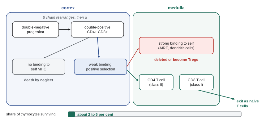
The selection model implies that the fate of a thymocyte depends on the strength of its TCR signal: too weak and the cell dies; moderate and it survives; too strong and it is deleted. This affinity model raises an apparent paradox, since the same receptor must be selected on self yet be able to respond to foreign peptides. The resolution is that a TCR that weakly binds some self peptide–MHC is likely to bind strongly to some of the vast number of possible foreign peptides on the same MHC, and that thresholds for activation differ between thymocytes and mature T cells. Selection is also leaky: some self-reactive T cells escape, and peripheral mechanisms must control them.
import random
# Toy model of thymic selection: each thymocyte gets a random TCR whose strongest
# binding to self peptide-MHC is drawn from a skewed distribution (arbitrary units).
random.seed(380)
N = 200_000
neglect, positive, negative, treg = 0, 0, 0, 0
for _ in range(N):
    s = random.expovariate(1.0) ** 2        # most receptors bind self MHC very weakly
    if s < 5.3:
        neglect += 1                         # too weak: death by neglect
    elif s < 7.9:
        positive += 1                        # positively selected, survives
    elif s < 9.0:
        treg += 1                            # strong but below deletion: some become Tregs
    else:
        negative += 1                        # deleted by negative selection
for name, k in (("death by neglect", neglect), ("survive as conventional T cells", positive),
                ("diverted to regulatory T cells", treg), ("deleted (negative selection)", negative)):
print(f"{name:34s} {k / N:6.1%}")death by neglect 90.1% survive as conventional T cells 4.0% diverted to regulatory T cells 1.0% deleted (negative selection) 4.9%
The thresholds in the code were chosen only to give proportions of the right order: most thymocytes die by neglect because random receptors rarely fit self MHC well, a few per cent survive as conventional T cells, a smaller fraction with relatively strong self-reactivity become regulatory T cells, and a similar fraction is deleted. Real estimates suggest that in mice roughly 5 per cent of double-positive cells mature, and that negative selection removes a substantial share of those that are positively selected, although the exact proportions remain debated.
The thymus shrinks with age, and by middle age much of it is replaced by fat. The output of new naive T cells falls, and in older adults the T cell pool is maintained largely by division of existing cells, reducing its diversity. This contributes to immunosenescence, the decline of immune function with age that makes older people more vulnerable to infections and less responsive to vaccines. Children born without a thymus, as in complete DiGeorge syndrome, have almost no T cells and can be treated by transplanting thymus tissue.
Activating naive T cells: three signals
Naive T cells circulate between the blood and secondary lymphoid organs, guided by the chemokine receptor CCR7 and the adhesion molecule L-selectin, and survive for months to years with the help of interleukin-7 and contact with self peptide–MHC. To become an effector, a naive T cell must meet a dendritic cell presenting its specific peptide. Activation requires three kinds of signal, a model that explains much about how responses are controlled.
Signal 1 is antigen: the TCR binds peptide–MHC on the dendritic cell, forming a structured contact zone called the immunological synapse, in which TCRs cluster at the centre surrounded by a ring of adhesion molecules. Signal 2 is costimulation: the molecule CD28 on the T cell binds CD80 (B7-1) or CD86 (B7-2) on the dendritic cell, which are increased when pattern recognition receptors detect microbes. Signal 1 without signal 2 tends to make T cells unresponsive, a state called anergy, or to kill them, which is one reason why self antigens presented by resting dendritic cells do not trigger responses. Signal 3 is cytokines, released by the dendritic cell and other cells, which direct the T cell toward a particular type of effector.
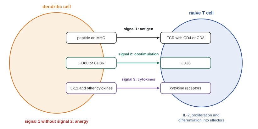
Once activated, a T cell produces the cytokine interleukin-2 and expresses the high-affinity IL-2 receptor, including the α chain CD25, which drives its own rapid proliferation and that of its neighbours, the clonal expansion described in lesson 1. Activation also changes the T cell's homing molecules, so that effector cells leave the lymph node and enter inflamed tissues. In parallel, activated T cells begin to express inhibitory receptors such as CTLA-4 and PD-1, which provide negative feedback; CTLA-4 binds CD80 and CD86 more strongly than CD28 does and competes with it. Lesson 8 shows how these checkpoints maintain tolerance, and lesson 12 how blocking them treats cancer.
Helper T cell subsets
Naive CD4 T cells can differentiate into several kinds of helper cell, each specialized for a different type of threat. The concept arose in 1986, when Tim Mosmann and Robert Coffman found that mouse helper T cell clones fell into two groups, Th1 and Th2, based on the cytokines they made. Since then further subsets have been added. Each subset is induced by particular cytokines present during activation, is controlled by a master transcription factor and produces characteristic cytokines that mobilize particular effector cells. The subsets are not rigidly fixed, and some cells change their profile, but the framework remains useful.
| Subset | Induced by | Master transcription factor | Signature cytokines | Main role | Disease when excessive |
|---|---|---|---|---|---|
| Th1 | IL-12, IFN-γ | T-bet | IFN-γ, TNF | Activate macrophages against intracellular microbes; help CD8 T cells | Some autoimmune diseases, granulomas |
| Th2 | IL-4 | GATA3 | IL-4, IL-5, IL-13 | Defence against worms; IgE class switching; eosinophils | Allergy and asthma |
| Th17 | IL-6, TGF-β, IL-23, IL-1β | RORγt | IL-17, IL-22 | Recruit neutrophils against extracellular bacteria and fungi; barrier repair | Psoriasis, some arthritis and bowel disease |
| Tfh (T follicular helper) | IL-6, IL-21, ICOS signalling | Bcl-6 | IL-21, IL-4 | Help B cells in germinal centres | Autoantibody diseases |
| Treg (regulatory T cell) | TGF-β, IL-2 | FOXP3 | IL-10, TGF-β, IL-35 | Suppress immune responses; maintain tolerance | Deficiency causes autoimmunity |
Th1 cells are induced when dendritic cells detect intracellular bacteria or viruses and make interleukin-12. Their interferon-γ activates macrophages to kill microbes they have engulfed, as in tuberculosis, and promotes switching to IgG subclasses that opsonize and fix complement. People with inherited defects in the IL-12–interferon-γ axis are highly susceptible to mycobacteria, including the weakened BCG vaccine strain, a condition called Mendelian susceptibility to mycobacterial disease. Th2 cells, induced by interleukin-4 in responses to helminths and allergens, produce interleukin-4, which drives IgE class switching, interleukin-5, which recruits eosinophils, and interleukin-13, which increases mucus production and gut contractility, together helping expel worms. Th17 cells, identified in 2005, recruit neutrophils and maintain barriers against extracellular bacteria and fungi; patients with defects in the IL-17 pathway suffer chronic mucocutaneous candidiasis, and antibodies blocking IL-17 or IL-23 are highly effective treatments for psoriasis.
Tfh cells, which express the chemokine receptor CXCR5 that draws them into B cell follicles, provide the CD40 ligand and interleukin-21 that germinal centre B cells need, as described in lesson 5, and they are the subset induced by most vaccines that work through antibodies. Regulatory T cells, characterized by FOXP3, suppress other immune cells and are the subject of lesson 8. Their discovery earned the 2025 Nobel Prize in Physiology or Medicine.
CD8 T cells
Naive CD8 T cells are activated by dendritic cells presenting peptides on MHC class I, often through cross-presentation. For many responses, CD4 help is required to generate effective and long-lived CD8 memory. Help is delivered indirectly: a CD4 T cell recognizing antigen on the same dendritic cell activates it through CD40 ligand, a process called licensing, so that the dendritic cell expresses more costimulatory molecules and cytokines and can fully activate CD8 T cells. Activated CD8 T cells proliferate and become cytotoxic T lymphocytes carrying granules of perforin and granzymes. Lesson 7 describes how they kill.
Unconventional T cells
Not all T cells recognize peptides on classical MHC molecules. γδ T cells, which make up 1 to 10 per cent of blood T cells but are far more common in the skin and gut epithelium, recognize a variety of antigens, including phosphorylated metabolites called phosphoantigens made by stressed cells and some bacteria, often without MHC. Natural killer T (NKT) cells use a semi-invariant TCR to recognize lipid antigens presented by the MHC-like molecule CD1d. Mucosal-associated invariant T (MAIT) cells, abundant in human blood and liver, recognize vitamin B metabolites made by bacteria and yeasts and presented by the molecule MR1. These cells respond quickly, more like innate cells, and bridge innate and adaptive immunity.
Worked problem
Diagnosing a T cell development defect
An infant has recurrent viral and fungal infections. Blood tests show normal numbers of B cells and NK cells but very few T cells. Chest imaging shows no thymic shadow. Newborn screening had flagged a low level of T cell receptor excision circles (TRECs). What do these findings mean?
Show solution
- TRECs are circles of DNA excised during TCR α chain rearrangement; they do not replicate, so their level in blood reflects the output of new T cells from the thymus.
- Low TRECs, absent thymic shadow and few T cells indicate a failure of T cell production.
- Normal B and NK cells point to a defect specific to T cell development or the thymus, such as complete DiGeorge syndrome or certain forms of severe combined immunodeficiency, rather than a RAG defect, which would also eliminate B cells.
- Early diagnosis allows treatment by hematopoietic stem cell transplantation or, if the thymus is absent, thymus transplantation, before live vaccines or severe infections occur.
Answer: The infant has a severe defect in T cell production; TREC screening detects such defects at birth so that curative treatment can begin early.
Common mistakes
MistakeThinking T cell receptors bind antigen as strongly as antibodies do.
InsteadTCR affinity is low (micromolar); sensitivity comes from coreceptors, adhesion and serial binding.
MistakeBelieving most thymocytes survive selection.
InsteadOnly a few per cent survive; most die by neglect or are deleted.
MistakeConfusing positive and negative selection.
InsteadPositive selection keeps cells that bind self MHC weakly; negative selection removes cells that bind self strongly.
MistakeAssuming antigen recognition alone activates a naive T cell.
InsteadCostimulation (signal 2) and cytokines (signal 3) are also needed; signal 1 alone can cause anergy.
MistakeTreating helper subsets as permanently fixed.
InsteadSubsets are defined by master regulators and cytokines but show some plasticity.
Summary and key terms
- The αβ TCR is assembled by V(D)J recombination, recognizes peptide–MHC with low affinity and signals through CD3 ITAMs, Lck and ZAP-70.
- In the thymus, positive selection keeps thymocytes that bind self MHC weakly and sets CD4 or CD8 lineage.
- Negative selection, aided by AIRE-driven expression of tissue antigens, deletes strongly self-reactive thymocytes or diverts them to Tregs.
- Naive T cells need antigen (signal 1), costimulation through CD28 (signal 2) and cytokines (signal 3); signal 1 alone can cause anergy.
- Th1, Th2, Th17, Tfh and Treg subsets are induced by cytokines, defined by master transcription factors and specialized for different threats.
- CD8 T cells are primed by dendritic cells, often licensed by CD4 help, and become cytotoxic lymphocytes.
- γδ T cells, NKT cells and MAIT cells recognize non-peptide antigens and respond rapidly.
- T cell receptor
- The antigen receptor of T cells, recognizing peptide bound to MHC.
- CD3 complex
- Signalling proteins associated with the TCR, carrying ITAMs.
- positive selection
- Survival of thymocytes whose TCRs bind self MHC weakly.
- negative selection
- Deletion of thymocytes whose TCRs bind self peptide–MHC strongly.
- AIRE
- A transcription factor that drives expression of tissue-restricted antigens in the thymus.
- costimulation
- A second activating signal, chiefly CD28 binding CD80 or CD86.
- anergy
- A state of unresponsiveness induced by antigen recognition without costimulation.
- immunological synapse
- The organized contact zone between a T cell and an antigen-presenting cell.
- Th17 cell
- A helper T cell subset making IL-17 that recruits neutrophils.
- T follicular helper cell
- A helper T cell that helps B cells in germinal centres.
- TREC
- T cell receptor excision circle, a marker of new T cell output used in newborn screening.
Practice problems
Work each problem on paper first, then reveal the answer to check.
How does the TCR signal despite its short cytoplasmic tails?
Show answer
Through the associated CD3 and ζ chains, whose ITAMs are phosphorylated by Lck and recruit ZAP-70.
What decides whether a thymocyte becomes CD4 or CD8?
Show answer
Whether its TCR is selected on MHC class II or class I, enforced by ThPOK and Runx3.
What does AIRE do, and what happens when it is defective?
Show answer
It drives expression of tissue antigens in the thymus; defects cause autoimmune polyendocrine syndrome type 1.
What happens to a naive T cell that receives signal 1 without signal 2?
Show answer
It becomes anergic or dies.
Which subset defends against worms, and which cytokines does it make?
Show answer
Th2, making IL-4, IL-5 and IL-13.
Why do patients with IL-17 pathway defects get chronic candidiasis?
Show answer
Th17 responses recruit neutrophils and maintain barriers against fungi.
What is CD8 T cell licensing?
Show answer
Activation of a dendritic cell by CD4 T cells through CD40 ligand so it can fully prime CD8 T cells.
What do MAIT cells recognize?
Show answer
Microbial vitamin B metabolites presented by MR1.
Lesson quiz · 5 questions
Score 60% or more to complete this lesson.
Create a free account to save your progress, take quizzes and earn a certificate.