
BIOL-380 · Lesson 3 of 12 · 28 min
Innate immunity II: inflammation, cytokines and innate cells
Recognition is only the start. Once pattern recognition receptors have detected a microbe or tissue damage, the innate system must summon reinforcements, kill the invader, contain the damage and then restore the tissue. This lesson follows that sequence: the inflammatory response and how leukocytes leave the blood; the cytokines and interferons that coordinate innate defence; the killing mechanisms of phagocytes; natural killer cells and innate lymphoid cells; the systemic effects of inflammation such as fever; and finally the resolution of inflammation and the newer concept of trained immunity. Throughout, a theme recurs: the same mechanisms that protect can cause damage when they are too strong, last too long or are triggered in the wrong place.
Inflammation
The Roman writer Celsus described the four cardinal signs of inflammation in the first century: rubor, calor, tumor and dolor, redness, heat, swelling and pain; a fifth, loss of function, was added later. Each has a mechanism. Mediators released by activated macrophages and mast cells, including histamine, prostaglandins, nitric oxide and the cytokines tumour necrosis factor (TNF) and interleukin-1, dilate local arterioles, increasing blood flow and producing redness and heat. They also make the endothelial cells lining postcapillary venules contract and pull apart, so that plasma proteins and fluid leak into the tissue, causing swelling. The leaked fluid brings antibodies, complement and clotting proteins to the site, and fibrin clots help wall off the infection. Pain arises from bradykinin and prostaglandins acting on sensory nerves; the anti-inflammatory drugs ibuprofen and acetylsalicylic acid relieve it by inhibiting the cyclooxygenase enzymes that make prostaglandins.
The most important consequence of inflammation is the recruitment of leukocytes. Neutrophils arrive first, within hours, followed by monocytes over the next day or two. Leaving the blood is a precise, multistep process called the leukocyte adhesion cascade. TNF and interleukin-1 induce endothelial cells to display selectins, adhesion molecules that bind carbohydrate ligands on passing leukocytes and make them roll along the vessel wall. Chemokines on the endothelial surface, such as CXCL8 (interleukin-8) for neutrophils, activate integrins on the rolling leukocyte, switching them to a high-affinity form that binds firmly to endothelial ICAM-1. The arrested leukocyte then crawls to a junction between endothelial cells and squeezes through, a step called transmigration or diapedesis, before following chemokine gradients and bacterial products such as formylated peptides through the tissue to the site of infection.
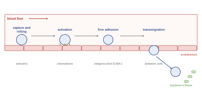
The importance of each step is shown by rare inherited diseases. In leukocyte adhesion deficiency type 1, mutations in the gene for the β2 integrin subunit CD18 prevent firm adhesion. Neutrophils accumulate in the blood, often at several times normal levels, but cannot enter tissues, so patients suffer severe bacterial infections without pus, and the umbilical cord often fails to separate normally. Drugs that block leukocyte migration exploit the same steps: natalizumab, an antibody against the α4 integrin, prevents lymphocytes from entering the brain and is used to treat multiple sclerosis, although by impairing immune surveillance of the brain it carries a small risk of a viral brain infection, progressive multifocal leukoencephalopathy.
Cytokines and chemokines
Cytokines are small secreted proteins that carry signals between cells. They act at very low concentrations, often picomolar, by binding high-affinity receptors, and most act locally on nearby cells or on the secreting cell itself, although some, such as interleukin-6, have systemic effects. Cytokines are typically pleiotropic, meaning one cytokine has many effects on different cells, and redundant, meaning several cytokines can have similar effects. They act in networks, inducing or inhibiting one another, which makes their effects hard to predict from any single molecule. Many cytokine receptors signal through the JAK–STAT pathway introduced in BIOL-214, which is the target of the JAK inhibitor drugs used in rheumatoid arthritis and other diseases.
| Cytokine | Main sources | Main effects in innate immunity |
|---|---|---|
| TNF | Macrophages, T cells, mast cells | Endothelial activation, fever, acute-phase response; in excess, shock |
| Interleukin-1β | Macrophages (requires inflammasome processing) | Fever, endothelial activation, acute-phase response |
| Interleukin-6 | Macrophages, endothelium, T cells | Acute-phase proteins such as CRP, fever, B cell growth |
| CXCL8 (IL-8) | Macrophages, epithelium, endothelium | Recruits and activates neutrophils |
| Interleukin-12 | Dendritic cells, macrophages | Activates NK cells; drives Th1 differentiation (lesson 6) |
| Type I interferons (IFN-α, IFN-β) | Almost any infected cell; plasmacytoid dendritic cells | Antiviral state in neighbouring cells; raise MHC class I; activate NK cells |
| Interferon-γ | NK cells, Th1 cells, CD8 T cells | Activates macrophages to kill intracellular microbes |
| Interleukin-10 | Macrophages, regulatory T cells | Suppresses macrophage and dendritic cell activation |
| TGF-β | Many cells, regulatory T cells | Suppression, tissue repair, fibrosis; Treg and Th17 differentiation |
Chemokines are a family of about 50 small cytokines that direct cell migration. They are classified by the arrangement of conserved cysteine residues near their amino terminus, into CC, CXC, CX3C and XC families, and their receptors are G protein-coupled receptors. Different cell types express different chemokine receptors, which is how each is guided to the right place: neutrophils respond to CXCL8 through CXCR1 and CXCR2; naive T cells home to lymph nodes through CCR7; and activated T cells enter inflamed tissues through receptors such as CXCR3. Some viruses exploit chemokine receptors: HIV uses CCR5 or CXCR4 as a coreceptor with CD4, and people homozygous for the CCR5-Δ32 deletion, about one per cent of people of northern European descent, are highly resistant to the common R5 strains of HIV, a fact used in the bone marrow transplants that cured a handful of patients described in lesson 11.
Type I interferons, IFN-α and IFN-β, were the first cytokines discovered, by Alick Isaacs and Jean Lindenmann in 1957, as a factor that interfered with viral replication. Almost any cell infected with a virus can make them after RIG-I, MDA5, cGAS or TLR3 detect viral nucleic acids, and plasmacytoid dendritic cells, using TLR7 and TLR9, can make enormous amounts. Interferons bind receptors on neighbouring cells and induce hundreds of interferon-stimulated genes, establishing an antiviral state: the protein kinase PKR halts protein synthesis when it detects double-stranded RNA, the enzyme OAS activates RNase L to degrade RNA, and proteins such as Mx and the IFITMs block viral entry and replication. Interferons also increase MHC class I expression and activate NK cells. Their importance was dramatically shown during the COVID-19 pandemic: work by the COVID Human Genetic Effort found that about 15 to 20 per cent of patients with critical COVID-19 pneumonia had either inborn errors of type I interferon immunity or, more commonly, autoantibodies that neutralized type I interferons, which were rare in the general population but became more common with age.
The danger of excessive cytokines is clearest in sepsis and cytokine storms. When bacteria or their products reach the bloodstream in large amounts, macrophages throughout the body release TNF and interleukin-1, causing widespread vasodilation, capillary leak, clotting in small vessels and falling blood pressure: septic shock. Sepsis remains a leading cause of death in hospitals worldwide. A similar storm of cytokines can follow some viral infections, CAR-T cell therapy (lesson 12) and certain drugs, and drugs blocking interleukin-6, such as tocilizumab, are used to treat it.
Phagocytes and how they kill
Neutrophils and macrophages are professional phagocytes. They recognize microbes directly through receptors such as the mannose receptor, scavenger receptors and Dectin-1, and much more efficiently when microbes are opsonized by antibodies, which bind Fc receptors, or complement fragments, which bind complement receptors such as CR3. Binding triggers actin polymerization that extends the membrane around the particle, enclosing it in a phagosome, which then fuses with lysosomes and, in neutrophils, with granules, forming a phagolysosome. Inside, microbes are attacked by acid, proteases, antimicrobial peptides, iron-sequestering proteins and reactive chemicals.
The most potent of these chemicals are reactive oxygen species. On activation, phagocytes assemble the NADPH oxidase complex in the phagosome membrane, which transfers electrons from NADPH to oxygen, producing superoxide; this consumes so much oxygen that the event is called the respiratory burst. Superoxide is converted into hydrogen peroxide and, in neutrophils, by the enzyme myeloperoxidase into hypochlorous acid, the active ingredient of bleach. Macrophages activated by interferon-γ also produce nitric oxide through inducible nitric oxide synthase. Children with chronic granulomatous disease, caused by mutations in subunits of NADPH oxidase, cannot mount a respiratory burst and suffer recurrent infections with catalase-positive bacteria such as Staphylococcus aureus and fungi such as Aspergillus, together with granulomas, clusters of macrophages that form around organisms they cannot kill.
Neutrophils have a further weapon. In 2004 Volker Brinkmann, Arturo Zychlinsky and colleagues showed that activated neutrophils can expel their chromatin, decorated with granule proteins, as web-like neutrophil extracellular traps (NETs) that ensnare and kill bacteria and fungi. NETs are double-edged: they promote blood clotting and have been implicated in thrombosis, lupus and the lung damage of severe COVID-19.
Macrophages are remarkably plastic. Exposure to interferon-γ and microbial products produces classically activated, or M1, macrophages that make inflammatory cytokines and reactive oxygen and nitrogen species and kill intracellular microbes. Exposure to interleukin-4 and interleukin-13 produces alternatively activated, or M2, macrophages that promote tissue repair, fibrosis and defence against worms. The M1 and M2 labels describe the ends of a continuum, and in tissues macrophages adopt many intermediate states. Macrophages also clear the billions of cells that die by apoptosis each day, a process called efferocytosis, recognizing phosphatidylserine on the outer face of the dying cells' membranes and responding with anti-inflammatory signals rather than inflammation.
Natural killer cells and innate lymphoid cells
Natural killer cells make up about 5 to 15 per cent of blood lymphocytes. They kill target cells by releasing granules containing perforin, which forms pores in the target membrane, and granzymes, proteases that enter the target and trigger apoptosis; they can also kill through death receptors such as Fas. NK cells are also a major early source of interferon-γ, which activates macrophages. As described in lesson 2, NK cells integrate signals from inhibitory receptors, which recognize MHC class I (killer cell immunoglobulin-like receptors, KIRs, and the CD94–NKG2A receptor in humans), and activating receptors, such as NKG2D, which binds stress-induced proteins, and CD16, an Fc receptor that allows NK cells to kill antibody-coated cells, a process called antibody-dependent cellular cytotoxicity. People with rare inherited NK cell deficiencies suffer severe infections with herpesviruses, showing that NK cells are especially important against this family of viruses.
import random
# NK cell decision: kill if total activating signal exceeds inhibitory signal by a margin.
# Illustrative signal strengths in arbitrary units; random variation between encounters.
random.seed(380)
targets = {
    "healthy cell (normal MHC I, no stress ligands)":   (1.0, 5.0),
    "virus-infected, MHC I reduced to 20%":              (3.0, 1.0),
    "cancer cell, MHC I normal, high NKG2D ligands":     (6.0, 5.0),
    "cancer cell, MHC I lost, high NKG2D ligands":       (6.0, 0.3),
    "antibody-coated infected cell (CD16 engaged)":      (8.0, 4.0),
}
threshold = 1.0
n = 1000
print("target                                              mean act  mean inh  fraction killed")
for name, (act, inh) in targets.items():
    kills = 0
    for _ in range(n):
        a = random.gauss(act, 0.25 * act)
        i = random.gauss(inh, 0.25 * inh)
        if a - i > threshold:
            kills += 1
print(f"{name:50s} {act:8.1f} {inh:8.1f} {kills / n:15.2f}")target mean act mean inh fraction killed healthy cell (normal MHC I, no stress ligands) 1.0 5.0 0.00 virus-infected, MHC I reduced to 20% 3.0 1.0 0.90 cancer cell, MHC I normal, high NKG2D ligands 6.0 5.0 0.48 cancer cell, MHC I lost, high NKG2D ligands 6.0 0.3 1.00 antibody-coated infected cell (CD16 engaged) 8.0 4.0 0.91
The model is a caricature, but it reproduces the key observations. Healthy cells, with strong inhibitory signals from MHC class I and few activating ligands, are spared. Cells that lose MHC class I, or that display enough stress ligands or antibody to overwhelm inhibition, are killed. A cancer cell that keeps normal MHC class I but expresses moderately raised stress ligands falls near the threshold and is killed only some of the time, which illustrates why tumours can sometimes escape NK cells and why therapeutic antibodies that engage CD16 can tip the balance.
Innate lymphoid cells (ILCs), recognized as a family only around 2010, are lymphocytes without rearranged antigen receptors that reside mainly in tissues, particularly at barrier surfaces such as the gut, lung and skin. They are grouped by the cytokines they make, which mirror helper T cell subsets: group 1 ILCs, which include NK cells, make interferon-γ; group 2 ILCs make interleukin-5 and interleukin-13 and help expel worms and drive allergic airway inflammation; and group 3 ILCs make interleukin-17 and interleukin-22, which maintain the intestinal barrier and are also needed to form lymph nodes during fetal development. Because they respond within hours to cytokines released by damaged epithelium, ILCs are thought to shape the early environment in which T cell responses later develop.
Systemic effects: fever and the acute-phase response
When inflammation is extensive, cytokines enter the circulation and act on distant organs. Interleukin-1, interleukin-6 and TNF act on the hypothalamus, where they induce prostaglandin E2, which raises the body's temperature set point and causes fever. Fever is costly, raising metabolic rate by roughly 10 to 12 per cent for each degree Celsius, but it is conserved across vertebrates and even in some invertebrates, and evidence suggests it helps: higher temperatures enhance many immune functions and inhibit the growth of some pathogens, and lizards and fish infected with bacteria seek warmer places, a behavioural fever that improves survival. Interleukin-6 also drives the liver to produce acute-phase proteins, including CRP, serum amyloid A, fibrinogen and mannose-binding lectin, and to reduce production of albumin. Hepcidin, another acute-phase protein, lowers plasma iron, withholding it from bacteria, which explains the anaemia of chronic inflammation. Cytokines also cause the bone marrow to release neutrophils, explaining the raised white cell count seen in lesson 1, and produce the sleepiness, loss of appetite and social withdrawal known as sickness behaviour.
Resolution and repair
Inflammation must stop once the threat is cleared. Resolution is an active process rather than a passive fading. Neutrophils die by apoptosis and are engulfed by macrophages, which then switch to producing anti-inflammatory cytokines such as interleukin-10 and TGF-β. Lipid mediators change from pro-inflammatory prostaglandins and leukotrienes to pro-resolving lipoxins, resolvins and protectins, described by Charles Serhan and colleagues, which stop neutrophil recruitment and promote clearance. Fibroblasts lay down new matrix and blood vessels regrow. When resolution fails, inflammation becomes chronic, with macrophages, lymphocytes and fibrosis replacing the neutrophils of the acute phase, as seen in rheumatoid arthritis, inflammatory bowel disease and atherosclerosis. Granulomas are a special form of chronic inflammation that walls off organisms that resist killing, such as Mycobacterium tuberculosis.
Trained immunity
The classical view that innate immunity has no memory has been challenged. Plants and invertebrates, which lack adaptive immunity, can show enhanced resistance to a second infection. In mammals, Mihai Netea and colleagues showed from about 2012 that exposure of monocytes and macrophages to fungal beta-glucan or to the tuberculosis vaccine BCG reprograms them, through changes in chromatin and metabolism, so that they respond more strongly to unrelated microbes for weeks to months, a phenomenon called trained immunity. Because circulating monocytes live only days, lasting effects depend on reprogramming of progenitors in the bone marrow. Trained immunity may explain epidemiological observations that BCG and other live vaccines reduce child deaths from infections other than the target disease, although randomized trials of BCG to prevent COVID-19 found no protection. Trained immunity may also contribute to chronic inflammatory diseases when it is induced inappropriately.
Worked problem
Reasoning from a phagocyte defect
A four-year-old has had a Staphylococcus aureus liver abscess and Aspergillus pneumonia. Neutrophil numbers are normal, and the neutrophils migrate normally to sites of infection. A test of the respiratory burst (the dihydrorhodamine flow cytometry assay) shows no oxidation by stimulated neutrophils. What is the likely diagnosis, and why these organisms?
Show solution
- Normal numbers and migration rule out neutropenia and leukocyte adhesion deficiency.
- Absent respiratory burst indicates a defect in NADPH oxidase: chronic granulomatous disease.
- Catalase-negative bacteria produce their own hydrogen peroxide, which the patient's neutrophils can still use; catalase-positive organisms such as S. aureus and Aspergillus destroy their own peroxide, so the phagocyte has no oxidants to work with.
- Treatment includes prophylactic antibiotics and antifungals, interferon-γ in some centres, and hematopoietic stem cell transplantation, which can be curative.
Answer: Chronic granulomatous disease, with susceptibility to catalase-positive bacteria and fungi because the phagocytes cannot generate reactive oxygen species.
Common mistakes
MistakeThinking leukocytes simply drift out of leaky vessels.
InsteadThey leave by an ordered cascade of selectin rolling, chemokine activation, integrin adhesion and transmigration.
MistakeAssuming each cytokine has a single function.
InsteadCytokines are pleiotropic and redundant and act in networks.
MistakeTreating inflammation as purely harmful.
InsteadIt delivers defences to the site of infection; harm comes when it is excessive, misplaced or unresolved.
MistakeBelieving resolution is passive.
InsteadResolution is actively driven by apoptosis, efferocytosis and pro-resolving mediators.
MistakeSaying innate cells can have no form of memory.
InsteadTrained immunity reprograms innate cells and their progenitors for weeks to months.
Summary and key terms
- Inflammation dilates vessels, increases permeability and recruits leukocytes, causing redness, heat, swelling and pain.
- Leukocytes leave the blood by rolling on selectins, chemokine activation, integrin adhesion and transmigration.
- Cytokines and chemokines coordinate innate defence; type I interferons establish an antiviral state.
- Phagocytes kill with acid, enzymes, reactive oxygen and nitrogen species and, in neutrophils, NETs.
- NK cells kill by balancing activating and inhibitory signals; ILCs are tissue-resident innate lymphocytes.
- Systemic cytokines cause fever and the acute-phase response; excessive release causes septic shock.
- Resolution is active; failed resolution leads to chronic inflammation; trained immunity gives innate cells a form of memory.
- inflammation
- A local response to infection or injury that delivers leukocytes and plasma proteins to tissue.
- selectin
- An adhesion molecule that mediates rolling of leukocytes on endothelium.
- integrin
- An adhesion receptor that, when activated, binds endothelial ligands such as ICAM-1 for firm adhesion.
- chemokine
- A small cytokine that directs cell migration through G protein-coupled receptors.
- type I interferon
- IFN-α or IFN-β, cytokines that induce an antiviral state in cells.
- respiratory burst
- Rapid oxygen consumption by phagocytes producing reactive oxygen species through NADPH oxidase.
- neutrophil extracellular trap
- A web of chromatin and granule proteins expelled by neutrophils to trap microbes.
- perforin
- A pore-forming protein released by NK cells and cytotoxic T cells.
- acute-phase response
- Liver production of proteins such as CRP in response to interleukin-6 and other cytokines.
- efferocytosis
- Engulfment of apoptotic cells by phagocytes.
- trained immunity
- Long-lasting enhanced responsiveness of innate cells after certain exposures.
Practice problems
Work each problem on paper first, then reveal the answer to check.
Explain the cause of each cardinal sign of inflammation.
Show answer
Redness and heat from vasodilation; swelling from leakage of fluid; pain from bradykinin and prostaglandins on nerves.
List the steps of the leukocyte adhesion cascade.
Show answer
Selectin-mediated rolling, chemokine activation, integrin firm adhesion, transmigration.
Why do patients with leukocyte adhesion deficiency lack pus?
Show answer
Neutrophils cannot adhere firmly and leave the blood.
How do type I interferons protect uninfected cells?
Show answer
They induce interferon-stimulated genes such as PKR, OAS and Mx that block viral replication.
Why are patients with chronic granulomatous disease susceptible to catalase-positive organisms?
Show answer
Their phagocytes cannot make reactive oxygen species and these organisms destroy their own peroxide.
What does CCR5-Δ32 homozygosity protect against?
Show answer
Infection with R5 strains of HIV.
How does interleukin-6 produce the acute-phase response?
Show answer
It induces the liver to make CRP, fibrinogen and other acute-phase proteins.
What is trained immunity?
Show answer
Epigenetic and metabolic reprogramming of innate cells that enhances later responses to unrelated microbes.
Lesson quiz · 5 questions
Score 60% or more to complete this lesson.
Create a free account to save your progress, take quizzes and earn a certificate.