
BIOL-420 · Lesson 9 of 12 · 26 min
Motor control: spinal cord, cortex, basal ganglia and cerebellum
Every behavior ends in movement, even speech and eye movement, and the motor system's job is to turn goals into precisely timed contractions of hundreds of muscles. Movement control is hierarchical but not a simple chain of command. The spinal cord contains circuits that generate reflexes and rhythmic patterns on their own; the brainstem adjusts posture and orients the head and eyes; motor cortex plans and directs voluntary movement; and two great loops, through the basal ganglia and the cerebellum, select actions and refine them. This lesson works up from muscle to cortex and then examines the two loops and what happens when they fail, including Parkinson's disease and its treatment by deep brain stimulation.
Motor units and the size principle
A motor unit is one alpha motor neuron in the spinal cord or brainstem together with all the muscle fibers it innervates. Muscles controlling fine movements, such as those of the eye or fingers, have small motor units of a few to tens of fibers; large postural muscles, such as the gastrocnemius, have units of a thousand fibers or more. Each muscle fiber belongs to just one motor unit, and every fiber in a unit contracts when its motor neuron fires, so the motor neuron is the final common path, in Charles Sherrington's phrase, for all motor commands.
Force is graded in two ways: by recruiting more motor units and by increasing their firing rate. In 1957 Elwood Henneman showed that units are recruited in a fixed order from small to large, the size principle. Small motor neurons have high input resistance, so a given synaptic current depolarizes them more and they reach threshold first; they innervate small numbers of slow, fatigue-resistant fibers. As drive increases, progressively larger motor neurons with fast, powerful but fatigable fibers join in. The result is that small forces are controlled with fine resolution and large forces are produced economically, without the motor system having to choose which units to use.
| Motor unit type | Motor neuron size | Fiber type | Force | Fatigue resistance |
|---|---|---|---|---|
| Slow (S) | Small | Type I, oxidative | Low | High |
| Fast fatigue-resistant (FR) | Medium | Type IIa, oxidative and glycolytic | Moderate | Moderate |
| Fast fatigable (FF) | Large | Type IIx, glycolytic | High | Low |
Spinal reflexes and pattern generators
Muscles contain two kinds of sensor. Muscle spindles lie in parallel with ordinary fibers and report muscle length and its rate of change through fast Ia afferents; Golgi tendon organs lie in series with tendons and report force through Ib afferents. The stretch reflex, tested when a clinician taps the patellar tendon, is monosynaptic: stretch excites spindle Ia afferents, which directly excite alpha motor neurons of the same muscle, while inhibiting its antagonist through an interneuron (reciprocal inhibition). Gamma motor neurons adjust the tension of the spindle's own intrafusal fibers so that spindles stay sensitive as the muscle shortens, a process called alpha–gamma co-activation. Other reflexes are polysynaptic: the withdrawal reflex pulls a limb away from a painful stimulus while extending the opposite limb to support the body (the crossed extensor reflex).
The spinal cord can also generate rhythmic movement without rhythmic input. Thomas Graham Brown showed in 1911 that cats with the spinal cord cut and sensory roots removed still produced alternating flexor and extensor activity. Networks of interneurons called central pattern generators (CPGs) produce the basic rhythm of walking, swimming, breathing, chewing and scratching. A classic model is the half-center oscillator, in which two groups of neurons inhibit each other and some process, such as adaptation, causes the active group to fade, releasing the other. Descending signals from the brainstem start, stop and modulate CPGs; electrical stimulation of a midbrain locomotor region in decerebrate cats produces walking whose speed increases with stimulus strength, and the cats switch from walking to trotting to galloping. CPG research led to epidural spinal cord stimulation trials that, combined with training, have allowed some people with chronic spinal cord injury to stand and take steps.
Motor cortex and population coding
Voluntary movement is directed by primary motor cortex (M1) in the precentral gyrus, together with premotor and supplementary motor areas and parietal areas that transform sensory information into plans. Stimulation of M1 by Wilder Penfield during neurosurgery at the Montreal Neurological Institute in the 1930s to 1950s revealed a motor homunculus, a map of the body with disproportionate areas for hands and face. Corticospinal neurons in layer 5, including the giant Betz cells, send axons down the corticospinal tract, which crosses in the medulla, to spinal motor neurons and interneurons; in primates some connect directly to motor neurons for the hand, supporting independent finger movements. Damage to motor cortex or the corticospinal tract, as in a stroke, causes weakness on the opposite side, loss of fine movement and, over time, spasticity and exaggerated reflexes as spinal circuits lose descending control.
What do motor cortex neurons encode? In the early 1980s Apostolos Georgopoulos recorded from monkey M1 during reaching in different directions and found that each neuron fires most for one preferred direction, with activity falling off as a cosine of the angle between the movement and that direction. Individually, neurons are broadly tuned and noisy, but the population is precise: weighting each neuron's preferred direction by its firing rate and summing gives a population vector that points in the direction of movement. Later work showed that motor cortex activity also reflects force, posture and muscle activity, and that population activity evolves through low-dimensional dynamics, much like a pattern generator, rather than representing any single variable. Population decoding nonetheless underlies brain–computer interfaces that let paralyzed people control cursors and robotic arms (lesson 12).
# Population vector decoding of reach direction from cosine-tuned model neurons.
import numpy as np
rng = np.random.default_rng(8)
n = 120
pref = rng.uniform(0, 2 * np.pi, n)              # preferred directions
baseline, depth = 20.0, 15.0                     # spikes/s
def rates(theta):
    return np.clip(baseline + depth * np.cos(theta - pref), 0, None)
errors = []
print("true direction   decoded (population vector)   error")
for deg in (0, 45, 90, 135, 200, 290):
    th = np.deg2rad(deg)
    counts = rng.poisson(rates(th) * 0.5)        # spikes in a 500 ms window
    w = counts / 0.5 - baseline                  # rate relative to baseline
    vec = np.array([np.sum(w * np.cos(pref)), np.sum(w * np.sin(pref))])
    dec = np.rad2deg(np.arctan2(vec[1], vec[0])) % 360
    err = (dec - deg + 180) % 360 - 180
    errors.append(abs(err))
    print(f"    {deg:3d} deg            {dec:6.1f} deg                {err:+5.1f}")
print("mean absolute error with 120 neurons:", round(np.mean(errors), 1), "deg")
for m in (10, 30, 120, 480):
    errs = []
    for _ in range(200):
        p = rng.uniform(0, 2 * np.pi, m); th = rng.uniform(0, 2 * np.pi)
        c = rng.poisson(np.clip(baseline + depth * np.cos(th - p), 0, None) * 0.5) / 0.5 - baseline
        d = np.arctan2(np.sum(c * np.sin(p)), np.sum(c * np.cos(p)))
        errs.append(abs((np.rad2deg(d - th) + 180) % 360 - 180))
print(f" {m:3d} neurons: typical error {np.median(errs):5.1f} deg")true direction decoded (population vector) error
0 deg 359.0 deg -1.0
45 deg 48.5 deg +3.5
90 deg 89.0 deg -1.0
135 deg 132.7 deg -2.3
200 deg 200.1 deg +0.1
290 deg 289.5 deg -0.5
mean absolute error with 120 neurons: 1.4 deg
10 neurons: typical error 10.2 deg
30 neurons: typical error 6.5 deg
120 neurons: typical error 2.8 deg
480 neurons: typical error 1.7 degDecoding accuracy improves steadily with the number of neurons, roughly as one over the square root of the population size, because independent noise averages out. This is the logic behind recording from as many neurons as possible in brain–computer interfaces.
The basal ganglia
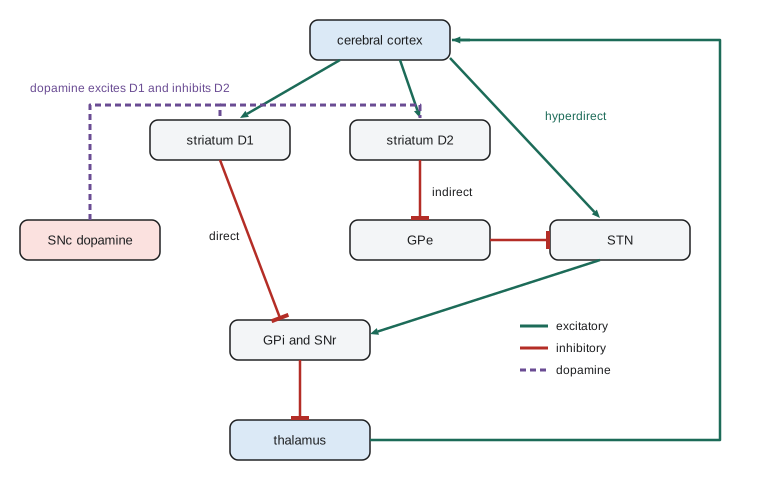
The basal ganglia are a set of interconnected nuclei: the striatum (caudate and putamen), the globus pallidus with external (GPe) and internal (GPi) segments, the subthalamic nucleus (STN) and the substantia nigra, with its dopamine-producing pars compacta (SNc) and GABAergic pars reticulata (SNr). They form loops: cortex projects to the striatum, the basal ganglia output nuclei (GPi and SNr) project to the thalamus, and the thalamus projects back to cortex. The output nuclei are tonically active and inhibitory, holding thalamic targets under constant restraint.
In the classic model, two pathways leave the striatum. The direct pathway, from striatal neurons expressing D1 dopamine receptors, inhibits GPi and SNr, releasing the thalamus from inhibition and facilitating movement. The indirect pathway, from neurons expressing D2 receptors, inhibits GPe, which disinhibits the STN, which excites GPi and SNr, increasing inhibition of the thalamus and suppressing movement. A third, hyperdirect pathway runs straight from cortex to the STN and can quickly halt actions. Dopamine from the SNc excites D1 neurons and inhibits D2 neurons, so it promotes movement through both pathways. A useful summary is that the basal ganglia select one action and suppress competitors, with dopamine biasing the choice toward actions that have been rewarded (lesson 10). Recent work shows the two pathways are co-active during movement, consistent with selecting the wanted action while suppressing others, rather than one simply acting as an accelerator and the other as a brake.
Parkinson's disease
Parkinson's disease, which affects more than 100,000 people in Canada and about a million in the United States, results largely from the death of dopamine neurons in the SNc; symptoms appear after roughly half or more of these neurons are lost. The cardinal motor signs are slowness of movement (bradykinesia), rigidity, a resting tremor and, later, postural instability; non-motor features such as loss of smell, constipation, sleep disorders and depression often precede them by years. In the basal ganglia model, dopamine loss weakens the direct pathway and strengthens the indirect pathway, increasing inhibitory output from GPi and SNr and suppressing movement. Recordings show excessive synchronized oscillations in the beta band (13 to 30 hertz) in the STN and GPi that correlate with bradykinesia and rigidity. Most cases are sporadic, but mutations in genes such as SNCA (alpha-synuclein), LRRK2 and GBA raise risk, and clumps of misfolded alpha-synuclein called Lewy bodies are the pathological hallmark. In 2023 a seed amplification assay that detects misfolded alpha-synuclein in spinal fluid showed high accuracy in diagnosing the disease, a step toward biological diagnosis.
Treatment began with levodopa in the 1960s, after Arvid Carlsson showed that dopamine is a transmitter and Oleh Hornykiewicz found it depleted in Parkinsonian brains. Levodopa remains the most effective drug, but over years its benefit fluctuates and involuntary movements (dyskinesias) appear. Deep brain stimulation (DBS), developed by Alim-Louis Benabid and colleagues in Grenoble in the late 1980s, delivers continuous high-frequency electrical pulses, around 130 hertz, through electrodes implanted in the STN or GPi. It reduces tremor, rigidity and bradykinesia and allows lower drug doses; well over 200,000 people worldwide have received DBS. How it works is still debated: it may disrupt pathological beta oscillations, effectively replacing an abnormal pattern with a regular one that the circuit ignores. Adaptive or closed-loop DBS systems that sense beta activity and adjust stimulation in response were approved by the US Food and Drug Administration in 2025. DBS is also used for essential tremor and dystonia and is being studied for depression and obsessive–compulsive disorder. Huntington's disease, by contrast, involves early loss of indirect-pathway striatal neurons and produces excessive, involuntary movements (chorea), the opposite pattern to Parkinson's disease.
The cerebellum
The cerebellum contains more than half of all the neurons in the brain, most of them tiny granule cells, yet damage to it does not paralyze. Instead movements become uncoordinated: reaches overshoot and undershoot (dysmetria), tremor appears as the hand approaches a target, alternating movements become clumsy, gait becomes broad-based and unsteady, and speech is slurred. The cerebellum compares intended movements with actual sensory feedback and corrects errors, and it learns.
Its circuit is strikingly regular. Mossy fibers, carrying information from the cortex (through pontine nuclei), spinal cord and vestibular system, excite granule cells, whose axons ascend and split into parallel fibers running along the folia. Each Purkinje cell, the sole output of cerebellar cortex, receives about 200,000 parallel fiber synapses, each weak. In contrast, each Purkinje cell receives a single climbing fiber from the inferior olive, which wraps around its dendrites and makes hundreds of synapses, producing a powerful complex spike. Purkinje cells inhibit the deep cerebellar nuclei, which send the cerebellum's output to motor cortex (through the thalamus) and brainstem.
In 1969 David Marr, and in 1971 James Albus, proposed that the cerebellar cortex works as a learning machine: the climbing fiber signals an error, and parallel fiber synapses active at the same time as the climbing fiber are weakened, so the Purkinje cell learns to change its output in that context. Masao Ito later demonstrated this form of LTD at parallel fiber synapses. The theory explains how adaptation of the vestibulo-ocular reflex, which keeps the eyes steady when the head moves, recalibrates after wearing magnifying glasses, and how eyeblink conditioning, in which a tone comes to elicit a protective blink before an air puff, depends on the cerebellum. Computationally, the cerebellum appears to build internal models that predict the sensory consequences of movements, allowing smooth control despite feedback delays. It also contributes to cognition and emotion through loops with prefrontal cortex.
Worked problem
Interpreting a clinical motor examination
An invented patient has weakness of the left arm and leg, brisk reflexes and increased tone on the left, and an upgoing toe on the left when the sole is stroked (Babinski sign). Coordination of the right limbs is normal. A second patient has no weakness but shows overshooting reaches and tremor that increases near the target in the right arm, with a broad-based gait. Where are the lesions likely to be?
Show solution
- Weakness with spasticity, brisk reflexes and a Babinski sign are upper motor neuron signs, from damage to motor cortex or the corticospinal tract.
- The corticospinal tract crosses in the medulla, so left-sided signs point to a lesion in the right hemisphere above the decussation, for example a stroke affecting the right internal capsule.
- The second patient has ataxia and intention tremor without weakness, cerebellar signs.
- The cerebellum controls the same side of the body, because its pathways cross twice, so right-sided signs point to the right cerebellar hemisphere.
Answer: Patient 1: right hemisphere corticospinal pathway (upper motor neuron lesion). Patient 2: right cerebellar hemisphere.
Common mistakes
MistakeAssuming the brain chooses which motor units to recruit for each task.
InsteadRecruitment follows the size principle automatically, from small to large motor neurons.
MistakeThinking rhythmic walking requires continuous commands from the brain.
InsteadSpinal central pattern generators produce the basic rhythm; the brain starts, stops and adjusts it.
MistakeExpecting single motor cortex neurons to specify movement direction precisely.
InsteadIndividual neurons are broadly tuned; direction is read out from the population.
MistakeDescribing the direct and indirect pathways as simply "go" and "stop" acting in turn.
InsteadBoth are active during movement, selecting desired actions and suppressing competitors.
MistakeExpecting cerebellar damage to cause paralysis.
InsteadIt causes incoordination, intention tremor and impaired motor learning, not weakness.
Summary and key terms
- A motor unit is a motor neuron and its fibers; units are recruited from small to large by the size principle.
- Spindles and tendon organs drive spinal reflexes; central pattern generators produce rhythmic movements.
- Motor cortex neurons are broadly tuned to direction; population vectors decode movement precisely.
- Basal ganglia direct, indirect and hyperdirect pathways select actions, with dopamine favoring movement.
- Parkinson's disease from dopamine neuron loss is treated with levodopa and deep brain stimulation, now including adaptive DBS.
- The cerebellum corrects and learns movements; climbing fiber error signals drive parallel fiber LTD.
- motor unit
- A motor neuron together with all the muscle fibers it innervates.
- size principle
- Recruitment of motor units in order of increasing motor neuron size.
- muscle spindle
- A stretch receptor in muscle that signals length and its rate of change.
- central pattern generator
- A neural circuit that produces rhythmic output without rhythmic input.
- corticospinal tract
- The pathway from motor cortex to the spinal cord, crossing in the medulla.
- population vector
- The sum of neurons' preferred directions weighted by their activity.
- direct pathway
- Striatal D1 neurons that inhibit basal ganglia output and facilitate movement.
- indirect pathway
- Striatal D2 neurons whose net effect increases basal ganglia output and suppresses movement.
- deep brain stimulation
- Therapeutic high-frequency stimulation through electrodes implanted in deep brain nuclei.
- climbing fiber
- An input from the inferior olive that powerfully excites a Purkinje cell and signals errors.
Practice problems
Work each problem on paper first, then reveal the answer to check.
Why do small motor neurons reach threshold first?
Show answer
Their high input resistance means a given synaptic current produces a larger depolarization.
Describe the circuit of the patellar stretch reflex.
Show answer
Spindle Ia afferents excite quadriceps motor neurons monosynaptically and inhibit hamstring motor neurons through an interneuron.
What is a half-center oscillator?
Show answer
Two mutually inhibitory groups of neurons that alternate activity, producing rhythm.
Why does a population vector become more accurate with more neurons?
Show answer
Independent noise in individual neurons averages out.
In the classic model, how does dopamine loss reduce movement?
Show answer
It weakens the direct pathway and strengthens the indirect pathway, increasing inhibitory output to the thalamus.
Which two nuclei are the usual DBS targets for Parkinson's disease?
Show answer
The subthalamic nucleus and the internal globus pallidus.
What signal is thought to be carried by climbing fibers?
Show answer
An error or teaching signal that drives plasticity at parallel fiber synapses.
A patient overshoots targets with the left hand. Which side of the cerebellum is affected?
Show answer
The left cerebellar hemisphere.
Lesson quiz · 5 questions
Score 60% or more to complete this lesson.
Create a free account to save your progress, take quizzes and earn a certificate.