
BIOL-420 · Lesson 8 of 12 · 26 min
Hearing, touch, pain and the chemical senses
Each sensory system faces the same basic tasks: transduce a physical or chemical stimulus into an electrical signal, encode its quality, intensity, timing and location, and send that code to the brain through dedicated pathways. The systems in this lesson solve those tasks with very different machinery. Hearing uses mechanical amplification and microsecond timing; touch uses specialized endings tuned to vibration, pressure and stretch; pain uses channels that detect heat, cold, chemicals and tissue damage and can be turned up or down by the brain; and smell and taste use large receptor families to recognize molecules. BIOL-350 introduced sensory transduction in general terms; here we look at how each system works and what goes wrong.
Sound and the cochlea
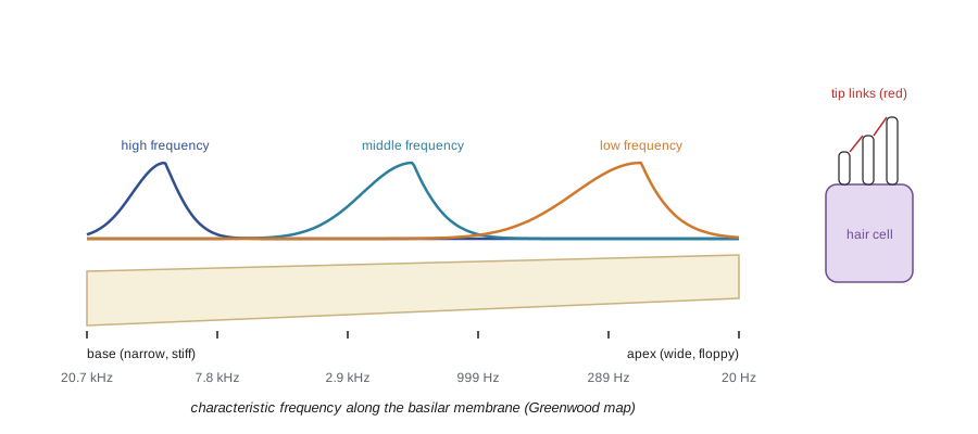
Sound is a pressure wave. The outer ear funnels it to the eardrum, and the three middle-ear bones, malleus, incus and stapes, transfer vibrations to the oval window of the fluid-filled cochlea, matching the low impedance of air to the high impedance of fluid; without this, about 99.9 percent of the energy would be reflected. Inside the cochlea, a spiral about 35 millimeters long if unrolled, the basilar membrane varies systematically: narrow and stiff at the base near the oval window, wide and floppy at the apex. Georg von Békésy showed, in work that earned the 1961 Nobel Prize, that sound produces a traveling wave along the membrane that peaks at a place determined by frequency: high frequencies near the base, low frequencies near the apex. This place code, tonotopy, is preserved from the cochlea through the brainstem and thalamus to auditory cortex.
The place–frequency map of the human cochlea is described by Greenwood's function, \(f = A(10^{a x} - k)\), where \(x\) is the fractional distance from the apex. For humans, with \(A\) = 165.4 Hz, \(a\) = 2.1 and \(k\) = 0.88, the map runs from about 20 Hz at the apex to about 20,000 Hz at the base, roughly logarithmic over most of its length.
Hair cells and the cochlear amplifier
The receptors are hair cells, so called for the bundle of 50 to 100 stereocilia on their apical surface, arranged in rows of increasing height like a staircase. Fine filaments called tip links connect each stereocilium to its taller neighbor. When sound deflects the bundle toward the tallest row, tip links tighten and pull open mechanotransduction channels, now known to be formed by the proteins TMC1 and TMC2, letting potassium flow in from the potassium-rich endolymph. The channels open within microseconds, far faster than any second-messenger pathway could, which is essential for hearing frequencies up to 20 kilohertz. Movement of just a few nanometers, about the diameter of the channel, produces a detectable response.
The human cochlea has about 3,500 inner hair cells, which send signals to the brain through about 30,000 auditory nerve fibers, each inner hair cell contacted by 10 to 30 fibers, and about 12,000 outer hair cells, which amplify. Outer hair cells contain the motor protein prestin, which makes the cell lengthen and shorten with changes in membrane potential at acoustic frequencies, pumping energy into the basilar membrane's vibration at the right place. This cochlear amplifier sharpens frequency tuning and increases sensitivity about a hundredfold. Its activity even produces sounds, called otoacoustic emissions, that can be recorded in the ear canal and are used to screen newborns for hearing loss.
Hearing loss affects a large share of the population. Outer hair cells are especially vulnerable to loud noise, ototoxic drugs such as aminoglycoside antibiotics and cisplatin, and aging, and mammalian hair cells do not regenerate. Noise can also destroy the synapses between inner hair cells and nerve fibers while leaving hearing thresholds normal, so-called hidden hearing loss, which may explain difficulty hearing speech in noisy places. Cochlear implants bypass lost hair cells by stimulating auditory nerve fibers directly with electrodes placed along the tonotopic axis, a successful neural prosthesis used by more than a million people. Gene therapy for one form of congenital deafness, caused by mutations in the otoferlin gene, restored hearing in children in clinical trials reported from 2024.
Occupational noise is a major preventable cause of hearing loss. OSHA's permissible exposure limit is 90 dBA averaged over eight hours with a 5 dB exchange rate, with a hearing conservation program required from an action level of 85 dBA; NIOSH recommends a stricter limit of 85 dBA with a 3 dB exchange rate.
Localizing sound
Unlike vision and touch, hearing has no map of space on its receptor surface; location must be computed. For horizontal position, the brain uses two cues. Interaural level differences arise because the head shadows high-frequency sound, making it louder at the nearer ear; they are processed in the lateral superior olive. Interaural time differences (ITDs) arise because sound reaches the nearer ear earlier, by up to about 700 microseconds for a sound directly to one side of a human head; humans can detect ITDs of about 10 microseconds, far shorter than the duration of a single action potential. ITDs are processed in the medial superior olive in mammals and in the nucleus laminaris in birds.
In 1948 Lloyd Jeffress proposed a model for ITD detection: axons from the two ears run in opposite directions past a row of coincidence-detector neurons, forming delay lines, so that each neuron fires best when the extra travel time along one axon exactly compensates for the acoustic delay at the other ear. The array of neurons then forms a place map of ITD, and hence of azimuth. The barn owl's nucleus laminaris fits the model remarkably well. In mammals the mechanism appears to rely more on precisely timed inhibition than on axonal delay lines, and ITD may be encoded by the relative activity of two broadly tuned populations in each hemisphere rather than a place map.
# Jeffress model: coincidence detectors with axonal delay lines estimate interaural time difference.
import numpy as np
rng = np.random.default_rng(2)
true_itd_us = 300.0                       # sound arrives 300 microseconds earlier at the left ear
freq = 500.0                              # Hz tone
period_us = 1e6 / freq
n_cycles = 200
# phase-locked spike times in each auditory nerve (one spike per cycle, with timing jitter)
left = np.arange(n_cycles) * period_us + rng.normal(0, 60, n_cycles)
right = left + true_itd_us + rng.normal(0, 60, n_cycles)
# detectors: internal delay applied to the left input (left signal travels farther to some detectors)
delays = np.arange(-600, 601, 100)        # microseconds of extra delay added to the left input
window = 80.0                             # coincidence window, microseconds
print("internal delay (us)   coincidences")
counts = []
for d in delays:
    shifted = left + d
    c = sum(np.any(np.abs(right - s) < window) for s in shifted)
    counts.append(c)
    print(f"   {d:+5d}               {c:4d} {'#' * (c // 8)}")
best = delays[int(np.argmax(counts))]
print(f"best detector compensates a delay of {best} us -> estimated ITD {best} us (true {true_itd_us:.0f})")
az = np.degrees(np.arcsin(best / 690.0))
print(f"approximate azimuth: {az:.0f} degrees toward the left")internal delay (us) coincidences
-600 0
-500 0
-400 0
-300 0
-200 0
-100 0
+0 0
+100 3
+200 83 ##########
+300 162 ####################
+400 75 #########
+500 5
+600 0
best detector compensates a delay of 300 us -> estimated ITD 300 us (true 300)
approximate azimuth: 26 degrees toward the leftTouch and proprioception
The skin contains several types of mechanoreceptor, each paired with a class of fast-conducting, myelinated A-beta fiber. In glabrous (hairless) skin, Merkel cell complexes signal sustained pressure and fine spatial detail, letting you read Braille; Meissner corpuscles signal light touch and low-frequency flutter, detecting slip when you grip an object; Pacinian corpuscles, onion-like capsules deep in the skin, respond to high-frequency vibration around 250 hertz, letting you feel texture through a tool; and Ruffini endings signal skin stretch. They differ in receptive field size and adaptation: rapidly adapting receptors report changes, slowly adapting ones report sustained stimuli. The mechanotransduction channel Piezo2, identified by Ardem Patapoutian's group in 2014, is essential for light touch and for proprioception, the sense of body position from muscle spindles and tendon organs; people with loss-of-function mutations in PIEZO2 have severely impaired touch discrimination and body awareness.
Touch information ascends in the dorsal column–medial lemniscus pathway: primary afferents enter the spinal cord and travel up the dorsal columns to the medulla, where second-order neurons cross to the opposite side and project to the thalamus and then to primary somatosensory cortex in the postcentral gyrus. There the body is mapped as a distorted homunculus in which the lips, tongue and fingertips occupy much more area than the back, reflecting receptor density. The map changes with experience: after amputation, neighboring representations can expand into the deprived area, and phantom limb sensations are common.
Pain
Pain is detected by nociceptors, free nerve endings of thinly myelinated A-delta fibers, which carry sharp first pain, and unmyelinated C fibers, which carry slower, burning second pain. They express receptors for noxious stimuli. TRPV1, cloned by David Julius's group in 1997, is activated by heat above about 43 °C, by acid and by capsaicin, the pungent ingredient of chili peppers, which is why chili tastes hot. TRPM8 detects cool temperatures and menthol; TRPA1 detects irritants such as mustard oil and is activated in inflammation. Julius and Patapoutian shared the 2021 Nobel Prize in Physiology or Medicine for discovering receptors for temperature and touch. Nociceptors also depend on particular sodium channels, Nav1.7 and Nav1.8, which explains why people lacking Nav1.7 feel no pain (lesson 2) and why Nav1.8 blockers such as suzetrigine are being developed as non-opioid analgesics.
Pain pathways differ from touch pathways. Nociceptive fibers synapse in the dorsal horn of the spinal cord; second-order neurons cross immediately and ascend in the spinothalamic tract to the thalamus and then to somatosensory cortex, which localizes pain, and to the insula and anterior cingulate cortex, which contribute its unpleasantness. A spinal lesion affecting one side therefore impairs touch on the same side below the lesion but pain and temperature on the opposite side.
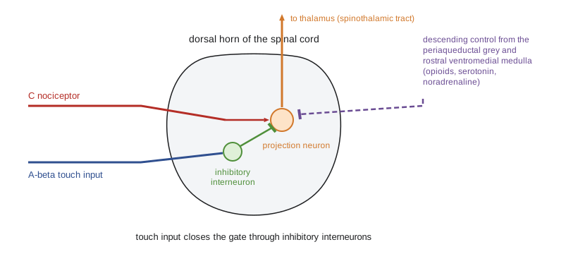
Pain is strongly modulated. In 1965 Ronald Melzack, at McGill, and Patrick Wall proposed the gate control theory: activity in large touch fibers excites inhibitory interneurons in the dorsal horn that reduce transmission from nociceptors, which is why rubbing an injured spot helps and the basis of transcutaneous electrical nerve stimulation. Descending pathways from the periaqueductal gray of the midbrain, through the rostral ventromedial medulla, inhibit dorsal horn transmission using endogenous opioids, serotonin and noradrenaline; this system underlies stress-induced analgesia and placebo analgesia, which can be partly blocked by naloxone.
After injury, pain systems become sensitized. Inflammatory mediators lower nociceptor thresholds (peripheral sensitization), and intense nociceptive input triggers NMDA receptor-dependent plasticity in the dorsal horn (central sensitization), so that touch becomes painful (allodynia) and painful stimuli more painful (hyperalgesia). Usually this resolves as tissue heals. When it persists, pain becomes chronic, a disease in its own right that involves changes in spinal, brainstem and cortical circuits and in microglia. Neuropathic pain from nerve damage, in diabetes, shingles or chemotherapy, responds poorly to opioids and is often treated with drugs such as gabapentinoids and certain antidepressants that enhance descending inhibition.
Chronic pain affects roughly one in five US adults, according to National Health Interview Survey estimates published by the CDC, and its treatment became entangled with the overdose crisis when opioid prescribing rose in the 1990s and 2000s; the CDC's 2022 prescribing guideline emphasizes individualized care and non-opioid options.
Smell and taste
Smell begins with olfactory sensory neurons in the nasal epithelium, each expressing just one of about 400 functional odorant receptor genes in humans (about 1,000 in mice), out of the largest gene family in the genome, discovered by Linda Buck and Richard Axel in 1991; they shared the 2004 Nobel Prize. Odorant receptors are GPCRs that signal through cyclic AMP. Each receptor binds many odorants and each odorant binds several receptors, so odors are encoded combinatorially. All neurons expressing the same receptor send their axons to one or two glomeruli in the olfactory bulb, creating a map in which each odorant activates a characteristic pattern of glomeruli. From the bulb, signals go directly to olfactory cortex without a thalamic relay, and to the amygdala and entorhinal cortex, which may explain the strong link between smell, emotion and memory. Olfactory sensory neurons are replaced throughout life. Loss of smell, anosmia, was a prominent symptom of COVID-19, caused mainly by infection of supporting cells in the nasal epithelium; it is also an early sign of Parkinson's and Alzheimer's diseases.
Taste is detected by taste receptor cells in taste buds on the tongue and palate. Five basic qualities are well established: sweet, umami and bitter are detected by GPCRs (T1R2 plus T1R3 for sweet, T1R1 plus T1R3 for umami, and about 25 T2R receptors for bitter); salty taste involves the epithelial sodium channel; and sour taste is detected by the proton channel OTOP1. Each taste cell type is tuned to one quality, and the labeled-line view holds that each quality is carried by dedicated pathways, although flavor depends heavily on smell, texture and temperature.
| System | Receptor or channel | Transduction speed | Coding strategy |
|---|---|---|---|
| Hearing | TMC1/2 channels in hair cells, opened by tip links | Microseconds | Place (tonotopy) and timing (phase locking) |
| Touch | Piezo2 in Merkel cells and A-beta endings | Milliseconds | Receptor type, adaptation and somatotopy |
| Pain and temperature | TRPV1, TRPM8, TRPA1; Nav1.7 and Nav1.8 | Milliseconds to seconds | Dedicated fibers, modulated by gates and descending control |
| Smell | About 400 odorant GPCRs in humans | Hundreds of ms | Combinatorial patterns of glomeruli |
| Taste | T1R and T2R GPCRs, ENaC, OTOP1 | Hundreds of ms | Largely labeled lines for five qualities |
Worked problem
Using Greenwood's map to locate damage
An invented patient with noise-induced hearing loss has a notch of reduced sensitivity centered at 4,000 Hz. Using Greenwood's function f = 165.4(10^(2.1x) − 0.88), where x is the fractional distance from the apex, estimate where along a 35 mm cochlea the damage lies.
Show solution
- Solve for x: 10^(2.1x) = f/165.4 + 0.88 = 4000/165.4 + 0.88 ≈ 25.06.
- 2.1x = log10(25.06) ≈ 1.40, so x ≈ 0.666.
- The damage lies about two-thirds of the way from the apex, or about one-third of the way from the base.
- On a 35 mm cochlea that is about 0.666 × 35 ≈ 23 mm from the apex, or about 12 mm from the base, consistent with the 4 kHz notch typical of noise damage.
Answer: About 23 mm from the apex (12 mm from the base).
Common mistakes
MistakeThinking the cochlea analyses frequency only by timing.
InsteadPlace coding along the basilar membrane is primary; phase locking adds timing information at low frequencies.
MistakeAssuming inner hair cells amplify sound.
InsteadOuter hair cells, through prestin, amplify; inner hair cells send most signals to the brain.
MistakeTreating pain as a simple readout of tissue damage.
InsteadPain is modulated by spinal gates, descending pathways, expectation and context, and can persist without ongoing damage.
MistakeExpecting a one-sided spinal lesion to affect touch and pain on the same side.
InsteadPain pathways cross at the spinal level and touch pathways in the medulla, so a one-sided lesion affects pain on the opposite side and touch on the same side.
MistakeSaying each odor has its own receptor.
InsteadOdors are encoded combinatorially by patterns across many receptors.
Summary and key terms
- The basilar membrane separates frequencies by place; tonotopy is preserved up to auditory cortex.
- Hair cell tip links open TMC channels within microseconds; outer hair cells amplify through prestin.
- Sound location is computed from interaural level and time differences; Jeffress proposed delay lines and coincidence detectors.
- Distinct mechanoreceptors and Piezo2 serve touch and proprioception, mapped somatotopically in cortex.
- Nociceptors use TRP channels and Nav1.7 and Nav1.8; gate control, descending opioid pathways and sensitization shape pain.
- Smell uses a large odorant receptor family with combinatorial coding; taste uses receptors for five basic qualities.
- tonotopy
- Orderly mapping of sound frequency along the cochlea and auditory pathway.
- hair cell
- A mechanosensory cell with a bundle of stereocilia whose deflection opens channels.
- tip link
- A filament linking adjacent stereocilia that gates mechanotransduction channels.
- cochlear amplifier
- Active amplification of basilar membrane motion by outer hair cells.
- interaural time difference
- The difference in arrival time of a sound at the two ears.
- nociceptor
- A sensory neuron that detects potentially damaging stimuli.
- TRPV1
- A channel activated by noxious heat, acid and capsaicin.
- gate control theory
- The idea that touch-fiber activity inhibits pain transmission in the dorsal horn.
- central sensitization
- Increased responsiveness of central pain pathways after intense or prolonged input.
- glomerulus
- A cluster of synapses in the olfactory bulb receiving input from neurons with the same receptor.
Practice problems
Work each problem on paper first, then reveal the answer to check.
Why does the middle ear improve hearing?
Show answer
It matches the impedance of air to the cochlear fluid, preventing most sound energy from being reflected.
Where on the basilar membrane do high frequencies peak?
Show answer
Near the base, close to the oval window.
What protein lets outer hair cells change length?
Show answer
Prestin.
A sound arrives 400 µs earlier at the right ear. Which Jeffress detector fires most?
Show answer
The one whose internal delay on the right-ear input is about 400 µs, compensating for the acoustic delay.
Which receptor detects sustained pressure and fine spatial detail?
Show answer
Merkel cell complexes.
Why does rubbing an injury reduce pain, according to gate control theory?
Show answer
Large touch fibers excite inhibitory interneurons that reduce nociceptive transmission in the dorsal horn.
How does capsaicin produce a burning sensation?
Show answer
It activates TRPV1 on nociceptors, the same channel activated by noxious heat.
Why can one odorant receptor contribute to many smells?
Show answer
Each receptor binds many odorants, and odors are coded by combinations of activated receptors.
Lesson quiz · 5 questions
Score 60% or more to complete this lesson.
Create a free account to save your progress, take quizzes and earn a certificate.