
BIOL-380 · Lesson 5 of 12 · 29 min
Antibodies and B cells
Antibodies were the first products of the adaptive immune system to be identified and remain its most versatile tools. A single person can make antibodies against an almost unlimited range of molecules, including synthetic chemicals that no organism has ever met, and antibodies made in the laboratory have become some of the best-selling medicines in the world. This lesson explains how antibodies are built, how their diversity is generated by rearranging genes, how B cells are activated and how their antibodies improve during a response, and how the different antibody classes protect the body.
Antibody structure
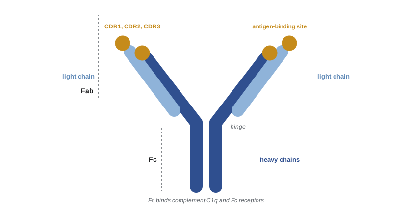
Antibodies, also called immunoglobulins (Ig), are Y-shaped glycoproteins built from two identical heavy chains and two identical light chains, held together by disulfide bonds. Rodney Porter and Gerald Edelman worked out this structure in the late 1950s and early 1960s, Porter by cleaving antibodies with the enzyme papain and Edelman by separating the chains, and shared the 1972 Nobel Prize. Papain cuts an IgG molecule into two identical antigen-binding fragments (Fab) and one fragment that crystallizes (Fc). The two Fab arms bind antigen; the Fc stem interacts with complement and with Fc receptors on immune cells, determining what happens to the antigen once it is bound.
Each chain is built from immunoglobulin domains of about 110 amino acids, folded into a sandwich of two β sheets, a structure that recurs in hundreds of immune and other proteins, the immunoglobulin superfamily, including the T cell receptor, MHC molecules, CD4 and CD8. The amino-terminal domain of each chain is the variable (V) domain, which differs greatly between antibodies; the rest are constant (C) domains. Within each V domain, variability is concentrated in three short loops called hypervariable regions or complementarity-determining regions (CDR1, CDR2 and CDR3), which together form the antigen-binding site at the tip of each arm. The third CDR of the heavy chain, CDRH3, is the most variable and usually makes the most contact with antigen.
Antibodies bind antigens through many weak noncovalent interactions, hydrogen bonds, electrostatic attractions, van der Waals forces and hydrophobic effects, across a contact surface of roughly 700 to 900 square ångströms. The strength of binding of a single site to a single epitope is its affinity, measured by the dissociation constant \(K_d\); high-affinity antibodies have \(K_d\) values of nanomolar to picomolar. Because antibodies have two or more binding sites, their overall binding to a multivalent antigen such as a bacterial surface, called avidity, can be far stronger than the affinity of a single site, because both arms must let go at once for the antibody to detach. IgM, with ten binding sites, can bind avidly even when each site has modest affinity.
Antibody classes
Humans make five classes, or isotypes, of antibody, defined by the constant region of the heavy chain: IgM, IgD, IgG, IgA and IgE, with heavy chains μ, δ, γ, α and ε. Light chains are of two types, κ and λ, which do not affect function. IgG is subdivided into four subclasses, IgG1 to IgG4, and IgA into two. Each class has a characteristic structure, location and set of effector functions.
| Class | Form | Share of serum Ig | Main location | Key functions |
|---|---|---|---|---|
| IgM | Pentamer (with J chain) | About 5–10% | Blood; B cell receptor on naive cells | First antibody in a primary response; strong complement activation; high avidity |
| IgD | Monomer | Under 1% | B cell surface | Antigen receptor on naive B cells; roles in mucosal defense |
| IgG | Monomer | About 75–80% | Blood and tissues | Opsonization, complement activation, neutralization, ADCC; crosses the placenta; half-life about 3 weeks |
| IgA | Monomer in blood; dimer in secretions | About 10–15% of serum (most IgA is in secretions) | Mucosal secretions, breast milk | Neutralization at mucosal surfaces without inflammation |
| IgE | Monomer | Trace | Bound to mast cells and basophils | Defense against worms; allergy |
IgG is unusual in having a long half-life, about three weeks for most subclasses, because the neonatal Fc receptor (FcRn) on endothelial cells rescues it from degradation in lysosomes and returns it to the blood. FcRn also carries IgG across the placenta, so newborns are protected by their mothers' IgG for the first months of life, and this is why maternal vaccination against pertussis, influenza and RSV in pregnancy protects infants. Therapeutic antibodies are almost always IgG for the same reason, and drugs that block FcRn are now used to lower harmful autoantibodies in diseases such as myasthenia gravis.
IgA is the most abundant antibody made by the body as a whole, though not in blood: plasma cells beneath mucosal surfaces secrete dimeric IgA joined by a J chain, which binds the polymeric immunoglobulin receptor on the basal side of epithelial cells and is carried across them, a process called transcytosis. At the lumenal surface the receptor is cleaved, releasing secretory IgA with a fragment of the receptor, the secretory component, attached, which protects it from digestion. Secretory IgA neutralizes toxins and microbes and traps them in mucus, a process called immune exclusion, without triggering inflammation. Breast milk is rich in secretory IgA, which protects breastfed infants' guts.
Effector functions of antibodies
Antibodies protect in several ways. Neutralization is binding that blocks a microbe or toxin from attaching to host cells: antibodies against the receptor-binding domain of a virus's surface protein prevent infection, and antitoxins against diphtheria and tetanus toxins block toxin binding. Opsonization coats microbes with IgG, whose Fc regions bind Fcγ receptors on phagocytes, greatly increasing phagocytosis. Complement activation by IgM and IgG1 and IgG3 through C1q leads to further opsonization, inflammation and lysis. Antibody-dependent cellular cytotoxicity (ADCC) occurs when NK cells, through the Fc receptor CD16, recognize and kill antibody-coated cells. Finally, IgE bound to mast cells triggers their degranulation when antigen cross-links it, which helps expel worms but also causes allergy.
Generating diversity: V(D)J recombination
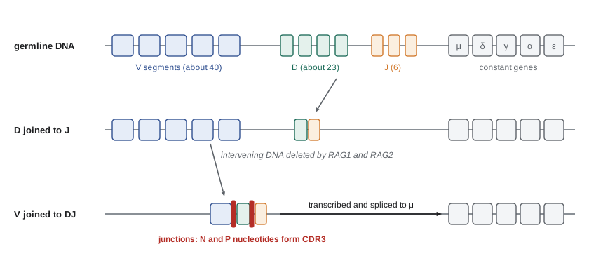
The human genome contains about 20,000 protein-coding genes, yet a person can make billions of different antibodies. How is this possible? In 1976 Susumu Tonegawa and Nobumichi Hozumi showed that the DNA encoding an antibody light chain was arranged differently in antibody-producing cells than in embryonic cells: separate gene segments were brought together by DNA rearrangement. Tonegawa received the 1987 Nobel Prize. The variable domain of the heavy chain is encoded by three kinds of segment, variable (V), diversity (D) and joining (J); the light chain uses only V and J. In humans the heavy chain locus has roughly 40 functional V, 23 D and 6 J segments; the κ locus about 40 V and 5 J; and the λ locus about 30 V and 4 to 5 functional J segments.
During B cell development in the bone marrow, the recombinase enzymes RAG1 and RAG2 cut the DNA at recombination signal sequences flanking each segment, and the cell's DNA repair machinery joins one D to one J, then one V to the DJ unit, deleting the DNA in between. The light chain then rearranges V to J. This process is called V(D)J recombination. Combining segments at random generates combinatorial diversity, but most diversity comes from imprecision at the joints: nucleotides are lost by trimming and added by the enzyme terminal deoxynucleotidyl transferase (TdT), which inserts random N nucleotides, and by the formation of hairpins that add palindromic P nucleotides. This junctional diversity is concentrated in CDR3. Pairing of any heavy chain with any light chain multiplies the total again.
import random, math
# Part 1: combinatorial diversity from gene segments (approximate functional human segment numbers).
V_H, D_H, J_H = 40, 23, 6
V_K, J_K = 40, 5
V_L, J_L = 30, 5
heavy = V_H * D_H * J_H
light = V_K * J_K + V_L * J_L
print(f"heavy chain V-D-J combinations: {heavy:,}")
print(f"light chain V-J combinations (kappa + lambda): {light:,}")
print(f"heavy x light pairings: {heavy * light:,}")
print("junctional diversity (N and P additions, trimming) raises the potential repertoire to over 1e13")
# Part 2: a toy model of affinity maturation (mutation plus selection).
random.seed(380)
cells = [1.0] * 50                      # starting affinity score of 50 B cells (arbitrary units)
print("\nround  mean affinity  best affinity")
for rnd in range(1, 9):
    new = []
    for a in cells:
        for _ in range(2):              # each cell divides into two daughters
            r = random.random()
            if r < 0.5:   a2 = a        # silent or neutral mutation
            elif r < 0.85: a2 = a * random.uniform(0.3, 0.95)   # damaging
            else:          a2 = a * random.uniform(1.05, 1.6)   # improving
            new.append(a2)
    new.sort(reverse=True)
    cells = new[:50]                    # selection: only the 50 best compete for T cell help
    if rnd in (1, 2, 4, 6, 8):
print(f"{rnd:5d} {sum(cells) / len(cells):13.2f} {max(cells):13.2f}")heavy chain V-D-J combinations: 5,520
light chain V-J combinations (kappa + lambda): 350
heavy x light pairings: 1,932,000
junctional diversity (N and P additions, trimming) raises the potential repertoire to over 1e13
round mean affinity best affinity
1 1.11 1.55
2 1.30 2.13
4 1.73 2.54
6 2.40 4.24
8 3.20 4.49The first part of the code shows that combinatorial joining of heavy and light chain segments alone gives a few million antibodies, and the second part shows why affinity rises during a response. Mutations are mostly neutral or harmful, but selection of the best binders in each round raises the average affinity steadily, much as artificial selection improves a crop. Real germinal centers select B cells through competition for antigen and T cell help rather than by explicit ranking, and the improvement is often tenfold to a thousandfold or more over weeks.
Because rearrangement is random, two-thirds of joints shift the reading frame and produce no functional protein; a B cell then tries the second chromosome. Once a functional heavy chain is made, it pairs with a surrogate light chain to form a pre-B cell receptor, which signals the cell to stop rearranging the other heavy chain allele, a process called allelic exclusion, so that each B cell makes antibody of only one specificity, as clonal selection requires. B cells whose receptors bind self antigens strongly in the bone marrow can edit their receptors by further light chain rearrangement or are deleted, part of central tolerance (lesson 8). Mutations that inactivate RAG1 or RAG2 prevent any B or T cells from forming and cause a form of severe combined immunodeficiency (lesson 11).
B cell activation
Mature naive B cells leave the bone marrow carrying IgM and IgD on their surface as their B cell receptor, together with the signaling proteins Igα and Igβ (CD79a and CD79b). They circulate through the follicles of lymph nodes and the spleen. When a B cell receptor binds antigen, especially multivalent antigen that cross-links several receptors, signaling through kinases such as Syk and Bruton's tyrosine kinase (BTK) activates the cell. Complement fragment C3d bound to the antigen engages the coreceptor CR2 (CD21) at the same time and lowers the threshold for activation, linking innate recognition to the B cell response. Mutations in BTK cause X-linked agammaglobulinemia, in which boys have almost no B cells or antibodies, and BTK inhibitors such as ibrutinib are used to treat B cell cancers.
Some antigens, particularly bacterial polysaccharides with many repeating units, can activate B cells without T cell help. These T-independent responses, carried out largely by marginal zone B cells in the spleen and B-1 cells, produce mainly IgM of modest affinity and little memory. Young children respond poorly to them, which is why plain polysaccharide vaccines against pneumococcus and Haemophilus influenzae type b work poorly in infants. Conjugate vaccines solve the problem by linking the polysaccharide to a carrier protein, so that B cells specific for the polysaccharide take up the conjugate, present carrier peptides to helper T cells and receive help, converting the response into a T-dependent one with class switching and memory. The introduction of Hib conjugate vaccines in the late 1980s and 1990s nearly eliminated Hib meningitis in children in Canada and the United States.
Most protein antigens elicit T-dependent responses. A B cell that has bound and internalized a protein antigen processes it and displays peptides on MHC class II. It then moves to the border between the follicle and the T cell zone, where it meets helper T cells that were activated earlier by dendritic cells presenting peptides from the same antigen. The T cell recognizes peptide–MHC on the B cell and provides help through the CD40 ligand (CD154), which binds CD40 on the B cell, and through cytokines. Because the B cell and T cell must recognize the same molecule, though not necessarily the same epitope, this linked recognition ensures help goes only to B cells binding the relevant antigen. Boys with mutations in CD40 ligand have hyper-IgM syndrome: they make IgM but cannot switch to other classes and are susceptible to opportunistic infections.
Germinal centers, affinity maturation and class switching
Some activated B cells become short-lived plasma cells that quickly secrete IgM. Others, with T follicular helper cells, enter the follicle and form a germinal center, a structure that appears about a week after immunization and can persist for weeks to months. In the dark zone of the germinal center, B cells proliferate rapidly and their V region genes undergo somatic hypermutation, introducing point mutations at a rate of about one per thousand base pairs per cell division, roughly a million times the normal background rate. The mutations are introduced by the enzyme activation-induced cytidine deaminase (AID), which converts cytosine to uracil in the DNA, triggering error-prone repair.
The mutated B cells then move to the light zone, where they compete for antigen held on follicular dendritic cells and for help from T follicular helper cells. Cells whose mutations improve affinity capture more antigen, present more peptide, receive more help and return to the dark zone for further rounds; cells whose affinity has fallen die by apoptosis. Repeated cycles of mutation and selection produce affinity maturation, the progressive increase in antibody affinity during a response and after boosting. Selected cells leave the germinal center as long-lived plasma cells, which migrate to the bone marrow and can secrete antibody for decades, or as memory B cells.
AID also drives class switch recombination. The constant region genes lie downstream of the rearranged VDJ unit in the order μ, δ, γ3, γ1, α1, γ2, γ4, ε and α2 in humans. Each is preceded by a repetitive switch region. AID-initiated breaks in two switch regions are joined, deleting the intervening DNA and placing a new constant gene next to the VDJ unit, so that the B cell keeps its specificity but changes the class of antibody it makes. Which class is chosen depends on cytokines from helper T cells: interferon-γ favors certain IgG subclasses, interleukin-4 favors IgE and IgG4, and TGF-β favors IgA. Because the deleted DNA is lost, switching is irreversible. Mutations in AID cause another form of hyper-IgM syndrome, with no class switching and no somatic hypermutation.
Monoclonal antibodies
An immune response normally produces a polyclonal mixture of antibodies from many B cell clones. In 1975 César Milstein and Georges Köhler, working in Cambridge, fused antibody-producing B cells from an immunized mouse with myeloma cells, a cancer of plasma cells, creating immortal hybridoma cells, each secreting a single defined antibody: a monoclonal antibody. They shared the 1984 Nobel Prize and deliberately did not patent the technique. Mouse antibodies provoke immune responses in humans, so later methods produced chimeric, humanized and then fully human antibodies, using transgenic mice with human antibody genes, phage display libraries, a method for which Gregory Winter and George Smith shared the 2018 Nobel Prize in Chemistry, and single B cells cloned from people who have recovered from infection.
Therapeutic monoclonal antibodies now treat cancers, autoimmune diseases, asthma, migraine, high cholesterol, osteoporosis and infections, and their international nonproprietary names carry the stem -mab; older names show the source with infixes such as -xi- for chimeric, -zu- for humanized and -u- for fully human (as in rituximab, trastuzumab and adalimumab), a scheme the World Health Organization simplified in 2017 by dropping the source infixes and, in 2021, decided to replace for new antibodies with four new stems (-tug, -bart, -mig and -ment). Long-acting antibodies such as nirsevimab, given once to infants to prevent RSV disease, show how antibodies can also provide passive immunization. Examples recur throughout the course: anti-TNF and anti-CD20 antibodies in lesson 10, anti-IgE in lesson 10, and checkpoint inhibitors in lesson 12.
Worked problem
Interpreting an antibody response
A researcher measures antibodies after two doses of a protein vaccine given four weeks apart. After the first dose, specific IgM peaks at day 10 and IgG is low. After the second dose, specific IgG rises within five days to 20 times its first peak, and its binding affinity is about 50 times higher than after the first dose. Explain these observations.
Show solution
- The early IgM after the first dose comes from short-lived plasma cells formed before germinal centers mature, so it has low affinity.
- Germinal centers formed after the first dose generate memory B cells that have already undergone class switching to IgG and somatic hypermutation.
- On the second dose, these memory B cells, more numerous and more easily activated than naive cells, quickly differentiate into plasma cells secreting IgG.
- Further rounds of germinal center selection after the boost raise affinity again, which is why booster doses spaced weeks or months apart improve the quality as well as the quantity of antibody.
Answer: The secondary response is faster, larger, switched to IgG and of higher affinity because memory B cells generated by germinal center reactions respond to the booster dose.
Common mistakes
MistakeThinking each antibody gene is inherited as a complete unit.
InsteadVariable domains are assembled by V(D)J recombination of separate gene segments in each B cell.
MistakeConfusing affinity with avidity.
InsteadAffinity is the strength of one binding site; avidity is the overall strength of multivalent binding.
MistakeBelieving class switching changes antibody specificity.
InsteadSwitching replaces the constant region; the variable region and specificity are kept.
MistakeAssuming somatic hypermutation happens during B cell development in the bone marrow.
InsteadIt occurs in germinal centers after activation, driven by AID.
MistakeExpecting plain polysaccharide vaccines to work well in infants.
InsteadT-independent responses are weak in young children; conjugate vaccines recruit T cell help.
Summary and key terms
- Antibodies have two heavy and two light chains; variable domains with three CDRs form the antigen-binding site, and the Fc region determines effector function.
- IgM, IgD, IgG, IgA and IgE differ in structure, location and function; IgG crosses the placenta and lasts weeks thanks to FcRn.
- Antibodies neutralize, opsonize, activate complement and trigger ADCC and mast cell degranulation.
- V(D)J recombination by RAG1/RAG2 plus junctional diversity from TdT generates a vast repertoire; allelic exclusion ensures one specificity per cell.
- T-independent responses to polysaccharides are weak in infants; conjugate vaccines recruit T cell help.
- In germinal centers, AID drives somatic hypermutation and class switching; selection produces affinity maturation, plasma cells and memory B cells.
- Monoclonal antibodies, from hybridomas to fully human antibodies, are major therapeutics.
- immunoglobulin
- An antibody molecule or the B cell receptor.
- complementarity-determining region
- A hypervariable loop of an antibody variable domain that contacts antigen.
- isotype
- An antibody class defined by its heavy chain constant region.
- affinity
- The binding strength of a single antigen-binding site for its epitope.
- avidity
- The overall binding strength of a multivalent antibody to a multivalent antigen.
- V(D)J recombination
- Assembly of variable region genes from V, D and J segments by RAG1 and RAG2.
- allelic exclusion
- Expression of only one rearranged allele so each B cell has one specificity.
- germinal center
- A site in a follicle where B cells mutate, are selected and switch class.
- somatic hypermutation
- Introduction of point mutations into antibody variable genes during a response.
- class switch recombination
- DNA recombination that changes the antibody constant region.
- monoclonal antibody
- An antibody of a single specificity produced by one B cell clone.
Practice problems
Work each problem on paper first, then reveal the answer to check.
What do the Fab and Fc fragments of an antibody do?
Show answer
Fab binds antigen; Fc binds complement and Fc receptors to trigger effector functions.
Why is IgM effective despite modest affinity?
Show answer
Its ten binding sites give it high avidity, and it activates complement strongly.
How does IgG reach the fetus?
Show answer
By FcRn-mediated transport across the placenta.
Name three sources of junctional diversity.
Show answer
Nucleotide trimming, N nucleotides added by TdT and P nucleotides from hairpin opening.
Why do conjugate vaccines work in infants?
Show answer
Linking polysaccharide to a carrier protein recruits helper T cells, giving class switching and memory.
What enzyme drives both somatic hypermutation and class switching?
Show answer
Activation-induced cytidine deaminase (AID).
What does CD40 ligand deficiency cause, and why?
Show answer
Hyper-IgM syndrome, because B cells cannot receive help to switch class.
What did Köhler and Milstein invent?
Show answer
Hybridomas that produce monoclonal antibodies.
Lesson quiz · 5 questions
Score 60% or more to complete this lesson.
Create a free account to save your progress, take quizzes and earn a certificate.