
BIOL-380 · Lesson 12 of 12 · 28 min
Capstone: tumor immunology, immunotherapy and immunology in public health
This final lesson applies the whole course to two areas where immunology has changed practice most dramatically in recent years. The first is cancer: how the immune system recognizes and controls tumors, how tumors escape, and how checkpoint inhibitors, engineered T cells and cancer vaccines turn the immune system against them. The second is the use of immunological tools in public health, from antibody tests and their interpretation to population serosurveys. The lesson ends with an integrated case that draws on every earlier lesson.
Immunosurveillance and immunoediting
Paul Ehrlich suggested in 1909 that the immune system might suppress tumors, and in the 1950s Burnet and Lewis Thomas proposed immunosurveillance: lymphocytes patrol the body and eliminate transformed cells. The idea fell out of favor when early studies found that immunodeficient nude mice did not develop more spontaneous tumors, but those mice still had NK cells and some T cells. From the late 1990s Robert Schreiber and colleagues showed that mice lacking interferon-γ signaling or RAG2 developed more chemically induced and spontaneous tumors, and that tumors arising in immunodeficient mice were often rejected when transplanted into normal mice, whereas tumors from normal mice were not. Humans with immunodeficiency, such as transplant recipients and people with untreated HIV, have greatly increased rates of cancers caused by viruses, such as Kaposi sarcoma, Epstein–Barr virus lymphoma and HPV-related cancers, and modestly increased rates of some others.
Schreiber, Lloyd Old and Gavin Dunn summarized these findings in 2002 as cancer immunoediting, with three phases. In elimination, innate and adaptive immunity destroy nascent tumor cells. In equilibrium, some tumor cells survive but are held in check, sometimes for years; immunity both controls them and selects variants that are less immunogenic. In escape, edited tumor cells that evade immunity grow into clinically apparent cancers. This framework explains why established tumors are, by definition, those that have defeated immune control, and why therapies must overcome the escape mechanisms they have acquired.
Tumor antigens
T cells can recognize several kinds of tumor antigen. Neoantigens are peptides created by mutations in tumor cells, unique to each tumor and not present in normal tissue, so T cells that recognize them have not been deleted by central tolerance. Tumors with many mutations, such as melanomas caused by ultraviolet light, lung cancers caused by smoking and tumors with defective DNA mismatch repair, have more neoantigens and respond better to immunotherapy. Cancer–testis antigens, such as NY-ESO-1 and the MAGE family, are normally expressed only in germ cells, which lack MHC class I, but are reactivated in some tumors. Differentiation antigens, such as tyrosinase and gp100 in melanocytes, are shared by tumors and their normal tissue of origin, so attacking them can damage normal cells, as seen in the vitiligo that sometimes accompanies successful melanoma immunotherapy. Viral antigens are expressed by virus-induced cancers.
Viral cancers can be prevented by vaccines. The hepatitis B vaccine, introduced in the 1980s, has reduced liver cancer in vaccinated cohorts in Taiwan and elsewhere. The human papillomavirus vaccine, based on virus-like particles of the L1 protein, prevents infection with the high-risk HPV types that cause nearly all cervical cancers and many anal, oropharyngeal and other cancers. Scottish data published in 2024 found no cases of invasive cervical cancer among women fully vaccinated at age 12 or 13, Both Canada and the United States include HPV vaccination in adolescent schedules, and in 2024 NACI recommended a single-dose schedule for most young people, which provinces have been adopting.
How tumors escape
Tumors evade immunity in many ways. They lose or reduce MHC class I, through mutations in β2-microglobulin, HLA genes or the antigen-processing machinery, becoming invisible to CD8 T cells, although this makes them vulnerable to NK cells. They express PD-L1, often in response to interferon-γ released by attacking T cells, a form of adaptive resistance. They recruit regulatory T cells and myeloid-derived suppressor cells, immature myeloid cells that inhibit T cells, and polarize macrophages toward tumor-promoting states. They secrete TGF-β, interleukin-10 and prostaglandin E2, consume nutrients such as glucose and tryptophan that T cells need, and produce adenosine and lactate that suppress them. Their abnormal blood vessels and dense stroma can physically exclude T cells. Tumors are often classified as hot, infiltrated by T cells and more likely to respond to checkpoint inhibitors, or cold, with few T cells.
Checkpoint blockade
The insight that inhibitory receptors limit antitumour T cells (lesson 8) led to checkpoint blockade. In 1996 James Allison's group showed that antibodies blocking CTLA-4 caused tumor rejection in mice. Ipilimumab, an anti-CTLA-4 antibody, became in 2011 the first drug shown to prolong survival in metastatic melanoma, and a fraction of patients lived for a decade or more. Antibodies against PD-1, such as pembrolizumab and nivolumab, approved from 2014, and against PD-L1, proved more effective and less toxic, and are now used in more than 20 cancer types, including lung, kidney, bladder, head and neck cancers, Hodgkin lymphoma and any tumor with mismatch repair deficiency. Former US President Jimmy Carter's melanoma, which had spread to his brain, went into remission in 2015 after treatment that included pembrolizumab; he lived until December 2024.
Responses vary greatly. In melanoma, combining anti-CTLA-4 and anti-PD-1 produces durable responses in roughly half of patients, and ten-year follow-up of the CheckMate 067 trial, published in 2024, found that more than 40 percent of patients receiving the combination were alive, compared with a median survival of well under a year for metastatic melanoma before these drugs. In other cancers, most patients do not respond. Predictors of response include tumor mutational burden, PD-L1 expression, T cell infiltration, mismatch repair deficiency and, intriguingly, the gut microbiota: studies in 2018 found that the composition of patients' gut bacteria correlated with response, and that antibiotics before treatment reduced benefit. Because checkpoint inhibitors release brakes on all T cells, they cause immune-related adverse events, autoimmune inflammation of the colon, skin, liver, lungs, thyroid, pituitary and other organs, in many patients, usually manageable with corticosteroids but occasionally fatal; type 1 diabetes caused by anti-PD-1 is often permanent.
Engineered T cells and other cell therapies
Chimeric antigen receptor (CAR) T cells are a patient's own T cells genetically modified to express a synthetic receptor that combines an antibody-derived binding domain with the signaling domains of CD3ζ and a costimulatory molecule such as CD28 or 4-1BB. A CAR recognizes intact surface antigen without needing MHC, so tumor loss of MHC does not matter. The first CAR-T therapies target CD19, present on B cells and on the cancer cells of most B cell leukemia and lymphoma. In 2012 Emily Whitehead, a six-year-old with relapsed acute lymphoblastic leukemia, became the first child treated with CD19 CAR-T cells, at the Children's Hospital of Philadelphia, and remains in remission more than a decade later. CAR-T cells targeting BCMA are used in multiple myeloma.
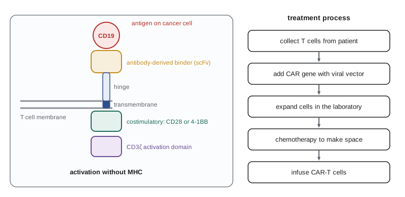
CAR-T therapy can cause cytokine release syndrome, fever and low blood pressure from massive T cell activation, treated with the anti-interleukin-6 receptor antibody tocilizumab, and neurotoxicity. Its high cost, manufacturing time of several weeks and limited effectiveness against solid tumors remain challenges, and researchers are developing CAR-T cells for solid tumors, off-the-shelf CAR cells from donors, CAR-NK cells and in vivo engineering in which T cells are modified inside the patient. Other cell therapies include tumor-infiltrating lymphocyte therapy, in which T cells grown from a patient's tumor are expanded and reinfused; lifileucel, the first such product, was approved in the United States for melanoma in 2024. Bispecific antibodies, such as blinatumomab, which binds CD19 on leukemia cells and CD3 on T cells, bring T cells into contact with tumor cells without genetic engineering.
In the United States, the Food and Drug Administration approved the first CAR-T therapy, tisagenlecleucel, in 2017, and seven CAR-T products had been approved by 2024 for leukemia, lymphoma and myeloma. In 2024 the FDA required boxed warnings about rare secondary T cell cancers after CAR-T therapy, and in 2025 it removed requirements for patients to stay near the treatment center for as long as previously required, easing access. Costs of several hundred thousand dollars per treatment are covered variably by insurers and Medicare.
Cancer vaccines
Therapeutic cancer vaccines aim to expand T cells against tumor antigens. Early vaccines against shared antigens had limited success; sipuleucel-T, a dendritic cell vaccine approved in the United States in 2010 for prostate cancer, extended survival by about four months. Personalized neoantigen vaccines are more promising. Tumor and normal DNA are sequenced, mutations predicted to produce neoantigens that bind the patient's HLA molecules are identified by algorithms, and a vaccine encoding up to dozens of them is manufactured for that patient within weeks. In a phase 2 trial in resected melanoma, the mRNA vaccine intismeran autogene, developed by Moderna and Merck, combined with pembrolizumab reduced the risk of recurrence or death by about 44 percent compared with pembrolizumab alone, and in August 2026 the companies reported that the phase 3 INTerpath-001 trial had met its primary endpoint of recurrence-free survival. A personalized mRNA vaccine for pancreatic cancer, developed by BioNTech and Memorial Sloan Kettering, induced T cell responses associated with delayed recurrence in an early trial published in 2023. Regulatory decisions on these vaccines were pending at the time of writing (October 2026).
Immunology in diagnosis and public health
Many diagnostic tests use antibodies as reagents. In an enzyme-linked immunosorbent assay (ELISA), an antigen or capture antibody is attached to a plastic well; the sample is added, and bound antibody or antigen is detected with an enzyme-linked antibody that converts a colorless substrate into a colored product. Concentrations are read from a standard curve, usually fitted with a four-parameter logistic equation. Lateral flow tests, such as home pregnancy and rapid antigen tests, use the same principle on a paper strip with antibodies conjugated to colored particles. Neutralization assays measure the ability of serum to block infection of cells in culture and are closer to function than binding assays.
import math
import numpy as np
from scipy.optimize import curve_fit
# Fit a four-parameter logistic standard curve to ELISA readings and read an unknown (invented data).
def four_pl(x, a, b, c, d):
    return d + (a - d) / (1.0 + (x / c) ** b)
conc = np.array([0.5, 1, 2, 4, 8, 16, 32, 64, 128])               # standards, ng/mL
od = np.array([0.08, 0.11, 0.17, 0.30, 0.55, 0.95, 1.42, 1.80, 2.02])  # optical density
params, _ = curve_fit(four_pl, conc, od, p0=[0.05, 1.2, 15, 2.2], maxfev=10000)
a, b, c, d = params
print(f"fitted: bottom a = {a:.3f}, slope b = {b:.2f}, midpoint c = {c:.1f} ng/mL, top d = {d:.3f}")
def inverse(y):
    return c * ((a - d) / (y - d) - 1.0) ** (1.0 / b)
for sample_od in (0.42, 1.10, 1.95):
    est = inverse(sample_od)
    flag = "  (near top of curve: dilute and retest)" if sample_od > 0.85 * d else ""
print(f"sample OD {sample_od:.2f} -> {est:6.1f} ng/mL{flag}")fitted: bottom a = 0.065, slope b = 1.27, midpoint c = 21.2 ng/mL, top d = 2.222 sample OD 0.42 -> 5.9 ng/mL sample OD 1.10 -> 19.9 ng/mL sample OD 1.95 -> 97.4 ng/mL (near top of curve: dilute and retest)
The fitted curve is sigmoidal: near the bottom and top, small changes in signal correspond to large changes in concentration, so readings there are imprecise. Laboratories therefore report results only within the curve's reliable range and dilute samples that fall near the top, as the code flags for the third sample.
Even an accurate test can mislead if its results are interpreted without considering how common the condition is. Sensitivity is the probability that a test is positive in someone with the condition, and specificity the probability that it is negative in someone without it. The quantity a patient or clinician wants, the probability of having the condition given a positive result, called the positive predictive value (PPV), depends strongly on prevalence.
# Positive and negative predictive values of an antibody test at different prevalences.
sensitivity, specificity = 0.95, 0.98
print("prevalence   PPV     NPV    false positives per 10,000 tested")
for prev in (0.001, 0.01, 0.05, 0.20, 0.50):
    tp = sensitivity * prev
    fp = (1 - specificity) * (1 - prev)
    tn = specificity * (1 - prev)
    fn = (1 - sensitivity) * prev
    ppv = tp / (tp + fp)
    npv = tn / (tn + fn)
print(f"{prev:9.1%} {ppv:5.1%} {npv:6.2%} {fp * 10000:8.0f}")prevalence PPV NPV false positives per 10,000 tested
0.1% 4.5% 99.99% 200
1.0% 32.4% 99.95% 198
5.0% 71.4% 99.73% 190
20.0% 92.2% 98.74% 160
50.0% 97.9% 95.15% 100When a condition is rare, most positive results from even a good test are false positives: at a prevalence of one in a thousand, a test with 98 percent specificity gives about 20 false positives for every true positive. This is why screening tests for rare conditions are followed by confirmatory tests, why early antibody surveys during a new epidemic can overestimate how many people have been infected, and why testing is most informative when targeted at people with a reasonable prior probability of the condition.
Serosurveys, studies that measure antibodies in samples of a population, estimate how many people have been infected or vaccinated, including those who never had symptoms or were never tested. During the COVID-19 pandemic, serosurveys of blood donors by Canadian Blood Services and Héma-Québec, the COVID-19 Immunity Task Force and national surveys in the United States tracked the proportion of people with antibodies from infection, distinguishing them from vaccine-induced antibodies by testing for antibodies to the nucleocapsid protein, which is not in most vaccines; by early 2023 a large majority of adults in both countries had antibodies from infection. Serosurveys also guide measles and rubella programs by identifying age groups with gaps in immunity, and are used to verify elimination.
An integrated case
Consider an invented patient, a 58-year-old woman diagnosed with melanoma that has spread to her lungs. Her tumor has a high mutational burden and is infiltrated with CD8 T cells. She receives combined anti-CTLA-4 and anti-PD-1 antibodies, and her lung lesions shrink over three months. Almost every lesson of the course helps explain what happens.
Her tumor's mutations created neoantigens, processed by the proteasome and loaded on MHC class I through TAP (lesson 4), recognized by CD8 T cells that escaped central tolerance because the mutated peptides were never present in the thymus (lessons 6 and 8). Those T cells had been primed in lymph nodes by cDC1 dendritic cells cross-presenting tumor antigens (lesson 4), with CD4 help licensing the dendritic cells (lesson 6), but had become exhausted, expressing PD-1, as the tumor upregulated PD-L1 in response to their interferon-γ (lessons 8 and 12). Anti-PD-1 reinvigorated progenitor exhausted T cells, and anti-CTLA-4 enhanced priming of new clones and may have depleted intratumoral regulatory T cells through Fc-mediated mechanisms (lessons 5, 7 and 8). The expanded cytotoxic T cells killed tumor cells with perforin and granzymes (lesson 7).
Six weeks into treatment, she develops severe diarrhea. A colonoscopy shows colitis infiltrated with CD8 T cells, an immune-related adverse event caused by the same release of brakes on T cells that recognize gut antigens or commensal bacteria, normally restrained by regulatory T cells and CTLA-4 (lessons 8 and 10). She is treated with corticosteroids and, when these are not enough, with infliximab, an anti-TNF antibody (lesson 10), and the colitis resolves. Before infliximab is given, she is screened for latent tuberculosis, because blocking TNF can allow granulomas to break down (lesson 9). Her melanoma remains controlled. Later, during an influenza season, her oncologist recommends the inactivated influenza vaccine, safe during immunotherapy, rather than a live attenuated vaccine (lesson 9).
Worked problem
Interpreting a positive screening test
A new antibody test for an infection has 95 percent sensitivity and 98 percent specificity. In a community where 1 percent of people have been infected, a randomly selected person tests positive. What is the probability that the person has truly been infected? How would the answer change in a group of close contacts of known cases, where 30 percent have been infected?
Show solution
- In 10,000 people at 1 percent prevalence, 100 are infected; the test detects 95 of them (true positives).
- Of 9,900 uninfected people, 2 percent, or 198, test falsely positive.
- PPV = 95 / (95 + 198) ≈ 0.32, so only about a third of positives are true.
- Among 10,000 close contacts at 30 percent prevalence: 2,850 true positives and 0.02 × 7,000 = 140 false positives, so PPV = 2,850 / 2,990 ≈ 0.95.
Answer: About 32 percent in the general community but about 95 percent among close contacts; the same test is far more informative when prior probability is higher.
Common mistakes
MistakeAssuming tumors are invisible to the immune system because they are self.
InsteadNeoantigens, cancer–testis antigens and viral antigens allow T cells to recognize tumors.
MistakeThinking checkpoint inhibitors attack cancer cells directly.
InsteadThey block inhibitory receptors so that the patient's T cells can attack the tumor.
MistakeExpecting CAR-T cells to need MHC presentation.
InsteadCARs recognize intact surface antigen directly, independent of MHC.
MistakeReading a positive result from a highly specific test as almost certainly true.
InsteadWhen prevalence is low, most positives can be false; predictive value depends on prevalence.
MistakeTreating immune-related adverse events as unrelated to treatment efficacy.
InsteadThey arise from the same release of T cell brakes that drives tumor responses.
Summary and key terms
- Immunosurveillance and immunoediting describe elimination, equilibrium and escape of tumors under immune pressure.
- Neoantigens, cancer–testis, differentiation and viral antigens allow T cells to recognize tumors; vaccines against HBV and HPV prevent cancers.
- Tumors escape by losing MHC class I, expressing PD-L1, recruiting suppressive cells and secreting inhibitory factors.
- Checkpoint inhibitors against CTLA-4, PD-1 and PD-L1 produce durable responses in some cancers but cause immune-related adverse events.
- CAR-T cells, TIL therapy, bispecific antibodies and personalized neoantigen vaccines extend immunotherapy.
- ELISA and lateral flow tests use antibodies as reagents; predictive values depend on prevalence.
- Serosurveys measure population immunity, and an integrated case shows how all parts of the immune system interact in treatment.
- immunosurveillance
- Detection and elimination of transformed cells by the immune system.
- cancer immunoediting
- The elimination, equilibrium and escape phases of tumor–immune interaction.
- neoantigen
- A tumor antigen created by a mutation and absent from normal tissue.
- myeloid-derived suppressor cell
- An immature myeloid cell that suppresses T cells in tumors.
- checkpoint inhibitor
- An antibody blocking an inhibitory receptor such as CTLA-4 or PD-1.
- immune-related adverse event
- Autoimmune inflammation caused by checkpoint inhibitors.
- chimeric antigen receptor
- A synthetic receptor combining an antibody binding domain with T cell signaling domains.
- cytokine release syndrome
- A systemic inflammatory reaction caused by massive T cell activation.
- ELISA
- Enzyme-linked immunosorbent assay, an antibody-based test that measures antigens or antibodies.
- positive predictive value
- The probability that a person with a positive test truly has the condition.
- serosurvey
- A study measuring antibodies in a population sample to estimate past infection or vaccination.
Practice problems
Work each problem on paper first, then reveal the answer to check.
What evidence supports immunosurveillance in humans?
Show answer
Immunodeficient people, such as transplant recipients, have increased rates of virus-driven and some other cancers.
Why do tumors with many mutations respond better to checkpoint inhibitors?
Show answer
They carry more neoantigens that T cells can recognize.
Name three ways tumors evade T cells.
Show answer
Loss of MHC class I, PD-L1 expression and recruitment of regulatory T cells or suppressor cells.
Why is vitiligo sometimes seen in patients responding to melanoma immunotherapy?
Show answer
T cells against shared melanocyte differentiation antigens also attack normal melanocytes.
What are the parts of a chimeric antigen receptor?
Show answer
An antibody-derived binding domain, hinge and transmembrane region, a costimulatory domain and CD3ζ.
What is the PPV of a test with 95 percent sensitivity and 98 percent specificity at 5 percent prevalence?
Show answer
About 71 percent.
How did COVID-19 serosurveys distinguish infection from vaccination?
Show answer
By testing for antibodies to the nucleocapsid protein, which most vaccines lack.
Why must patients be screened for tuberculosis before anti-TNF therapy?
Show answer
TNF maintains granulomas; blocking it can reactivate latent infection.
Lesson quiz · 5 questions
Score 60% or more to complete this lesson.
Create a free account to save your progress, take quizzes and earn a certificate.